Eye spots are any externally visible change that may be present on the surface of the eye. Those. this term does not mean the spots that you may sometimes notice before your eyes, such as so-called “floaters.” Therefore, it is the formations that are noticeable from the outside that will be discussed in the article.
The appearance of dots or spots on the eyeball can be caused by various diseases and conditions. Some of them may be harmless, others may be serious. The spots may be yellow, brown, white or pink, depending on the causative factor.
For example, a strong sneeze can destroy a small superficial blood vessel, which then creates a red spot. However, there are cases where spots could indicate a serious problem, such as inflammation in the eye that could threaten your vision, or even the formation of cancer. You should immediately seek help from an ophthalmologist if you notice:
- Loss of vision
- Unexpected increase in the number of spots
- Pimples appearing before the eyes
Stains inside
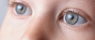
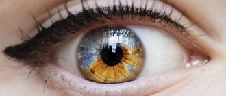
Several factors are known to cause pigmentation changes in human eyes. The most common pigmented formation in this case is a mole (nevus). It is a collection of pigmented cells known as melanocytes. They may be located at the front of the eye, around the iris, or under the retina at the back.
Moles (nevi) around the sclera (left) and on the iris (right)
Moles in the eye are usually benign, although there is always a chance that they can develop into melanoma. Melanoma is a serious type of cancer.
Therefore, it is important to have spots in the eyeball checked by a good eye doctor.
Other possible causes of discolored areas of the eyeball include:
Minor bruising that occurs after a minor injury to the eye
A ruptured blood vessel is very common in young children who are physically active.
Red spot due to hemorrhage
Are they dangerous?
In general, spots on the iris cannot be considered dangerous because the danger is entirely related to the cause of the injury.
Although most spots on the eye are benign lesions that do not pose a health risk, there are malignant spots, such as melanoma, and others that are symptomatic, such as those caused by macular degeneration.
For this reason, it is important to see an ophthalmologist to check the structure of the eye and suggest a diagnosis.
Examining a site in the eye can be key to identifying important eye conditions such as macular degeneration or cataracts.
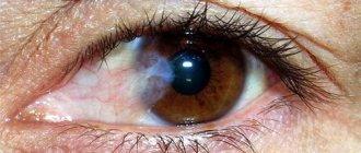
Dark, black spots on the eyeball
When a mole appears on the white part of the eye, it tends to attract more attention and also raise concerns about whether it could be a health risk. They are harmless in most cases. However, special attention should be paid to sudden darkening, as this may be a sign of malignancy.
Nevi that appear on your eyeball are collectively known as pigmented neoplasms. According to the Eye Cancer Network website, congenital nevi are the most common and are mostly harmless. A biopsy should be performed to identify the neoplasm.
Diagnostics
Diagnosis of the patient’s condition consists of several stages:
- Anamnesis collection. This is data obtained from the words of the patient or his close relatives. Based on these, the doctor may prescribe further examination.
- General examination of the patient's condition. The doctor evaluates the extent to which the superficial tissues of the eyes are affected. The skin of the eyelids and surrounding tissues may be involved in the process.
- Submission of laboratory tests. These include general blood and urine analysis, blood biochemistry, glucose determination, lipid profile. The presence of an inflammatory focus is detected using indicators of leukocytes and ESR, the amount of glucose and fat deposits.
- Fundus examination. A solution is first instilled into the patient’s eyes, which temporarily eliminates eye accommodation. The doctor looks at the condition of the lens, vitreous body, retina, and eye cameras. Evaluates the transparency or opacity of these areas, the presence of damage, hemorrhages.
- MRI, . Not only the structures of the eyeball, but also the brain are assessed. The doctor can reveal the condition of these structures layer by layer, since many tissues are visible through fundus examination.
Based on the data obtained, the doctor can make a reliable diagnosis. Only after diagnosis is treatment prescribed individually for each patient.
Stains outside
Spots and dots may form on the conjunctivae and the area near your iris. Their growth should not be ignored as they can spread to the outer layer called the cornea, causing vision impairment
Common reasons include:
Pterygium
A pterygium is a triangular white formation with blood vessels, consisting of conjunctival tissue.
This may be the reason for the appearance of a white spot on your eyeball. The pathology is also known as pterygoid hymen. And in English the term “surfer's eye” is also used, which often affects people who regularly surf on a board. The problem is quite common and mainly affects people who spend most of their time outdoors.
Externally, this disease is expressed in the accumulation of white tissue with blood vessels. In some cases, this may be accompanied by burning or itching. Severe cases can lead to vision impairment.
Risk factors
The underlying cause of this disease is unknown, but experts believe risk factors include:
- Prolonged exposure to ultraviolet rays
- The presence of a large number of external irritants, for example, smoke, pollen, wind
- Prolonged stay outdoors
Pinguecula
Pinguecula is a yellow-white raised formation on the conjunctiva.
It is characterized by the appearance of small light tubercles, which are located in the area of contact of the conjunctiva with the cornea. This condition does not cause pain, but it appears as if there is a white bump on your cornea that is clearly visible. It does not cause any additional symptoms. It is known that its appearance is greatly influenced by exposure to ultraviolet sunlight.
UV radiation
Exposure to ultraviolet rays is also associated with the appearance of spots on the eyeball. Prolonged sun exposure causes damage to the thin collagen fibers found in your conjunctiva. Then a color change occurs. The fibers that have been damaged will then appear as lumps.
Environmental irritants
Additional factors that can lead to the formation of spots on the eyeball are wind, dust and sand. Any person who is frequently exposed to such elements will have an increased risk of developing these spots. Examples would be people who spend a lot of time gardening, playing golf, and construction workers.
Eye damage
Trauma caused directly to your eyeball will certainly cause a spot to appear. The area may be blood-stained or white. For example, welders suffer from eyeball stains, especially if they do not wear safety glasses.
Prevention
To prevent problems with the visual system, you need to monitor your health, do not smoke, do not drink alcohol, and eat food rich in vitamins. Try to exercise at least minimally, don’t overstrain your eyes, get plenty of rest, be sure to periodically walk outside, and don’t spend a lot of time looking at monitors of computer devices.
If you are nearsighted or farsighted, follow all doctor's instructions. To avoid vision problems, periodically relieve your eyes and do not allow them to become too strained. If you have black spots flying before your eyes or other symptoms, consult a doctor immediately. Timely help from a specialist will allow you to quickly get rid of the disease and prevent the condition from worsening.
Gray spots on the eyeball
It is important to have an ophthalmologist examine any abnormal or unusual types of pigmentation that may be present on your eyeball. The exam is designed to determine whether your eyes require immediate treatment.
There are various formations, such as apigmented conjunctival nevus or ocular melanocytosis, which may appear as gray spots.
Ocular melanocytosis
Yellowish (pinguecula)
The pinguecula is a yellowish formation that rises slightly above the conjunctiva. Most often it is located at the junction of the cornea and conjunctiva. Does not cause discomfort or pain.
Occurs under the influence of unfavorable environmental factors:
- dust;
- wind;
- infrared radiation;
- ultraviolet radiation.
People who, due to their profession, spend a lot of time outdoors are at risk of developing pinguecula. For such patients, ophthalmologists recommend protecting their vision with contact lenses and safety glasses with an ultraviolet filter.
Red spot on the white of the eye
Subconjunctival hemorrhage is the term used for red spots that cover the white part of your eyeball (sclera). These spots occur when thin blood vessels burst inside the eye. This is a benign condition that is not known to cause any eye or vision problems and goes away on its own over time.
Although the exact cause of their occurrence is known, medical experts believe that the following factors may contribute to their occurrence:
- Eye injury
- Sudden increase in blood pressure due to sneezing, laughing, weight lifting, and constipation
- Taking blood thinners or aspirin
- Vitamin K deficiency
- Eye surgeries
Treatment of floating spots
| You can get rid of this problem with conservative treatment or choose laser therapy, a more modern method. The specialist decides which method is suitable. |
Homeopathic treatment
In addition to traditional drug treatment, homeopathic medicines can be used. They should not be confused with dietary supplements, so they can only be taken after the approval of a doctor. Among such remedies are the following: Agaricus, Calcarea Carbonica, Iodum, Argentum Nitricum, Cocculus, Thuja and Lithium Carbonicum.
Therapeutic approach
There are plenty of pharmaceutical products that can remove blackheads that interfere with reading and writing. However, you need to understand that in order for dark spots in the eyes to disappear forever, it is necessary to treat exactly the reasons that caused their appearance. The main rule is that treatment must begin with a correct diagnosis.
If no serious pathology is detected, drops can be used that will promote the resorption of insoluble deposits and restore the functionality of the vitreous body itself. Among such funds are the following:
- Wobenzym;
- Emokipin;
- Taufon;
- Quinax.
Laser therapy for floating spots
Vitreolysis is a complex but effective procedure using a YAG laser. The advantages of the method are that vitreolysis occurs without surgical intervention.
The disadvantage is that the particles that need to be removed are mobile, and this complicates the work of the specialist. That is why vitreolysis is used when the clarity of vision is reduced due to the destruction of the vitreous body.
The essence of this technique is that a laser with an accuracy of at least 6 microns is directed at particles that interfere with vision, and it breaks them into small grains. Due to their new size, they become invisible to the human eye.
| Laser therapy is performed only in hospital settings. The patient remains in the hospital for some time after the procedure. |
Return to contents
Surgical treatment of floating spots
If you need to remove a dark spot in the eye that appears when you look at a white wall, vitrectomy is used. This is a complex surgical operation in which the vitreous humor is removed through a small incision and replaced with a special solution.
There are often cases when vitrectomy does not bring the desired results and after a while the patient again begins to complain about dark spots. Most often this happens if the operation itself was accompanied by complications.
White dot on the iris: what is it?
There may be several reasons:
- keratitis of any etiology (viral, tuberculous, bacterial, etc.), the most common cause of cataract);
- eye injury: exposure to chemicals, burns, injuries, surgeries, etc.;
- trachoma (inflammation of the conjunctiva and cornea of the eye caused by chlamydia, leading to blindness due to scarring in the mucous membrane and destruction of the cartilaginous structures of the eyelids);
- congenital corneal opacity.
Whether white cloudiness in the eye due to leukoma is dangerous depends on the size, location and intensity.
Treating eyesores
Small cloud-like opacities and defects in the periphery do not require surgical intervention, but they need to be monitored over time.
Conservative therapy is indicated to stabilize the condition and includes:
instillation of Dionin in increasing concentrations during the initial stages of the disease);- corticosteroids: Hydrocortisone ointment, Dexamethasone eye drops, etc.;
- vitamins B12, A, C, E;
- physiotherapy: electrophoresis with potassium iodide, Papain, Lidase, Aloe extract or Vitreous body, course intramuscular injections are possible;
- for ulceration - ointments that enhance reparation processes, for example, with Solcoseryl, Actovegin, etc.
- drugs for the prevention of secondary glaucoma (Pilocarpine hydrochloride);
- pathogenetic therapy (depending on the etiological factor: antituberculosis, antiviral, antibiotics, etc.).
- diet: an excess of fatty foods under certain conditions can lead to an increase in cholesterol levels and its deposition on the scar tissue of the cataract, which will aggravate the condition. On the contrary, food containing large amounts of vitamins A, , , B, gives the body strength to fight the disease.
Surgical treatment of ocular leukoma
Keratoplasty is an operation that is performed if conservative therapy is not indicated and the patient’s quality of life suffers. Using a laser, the affected segment is excised layer by layer, followed by rehabilitation therapy.
, corneal keratoprosthesis helps restore vision in case of leukoma . The biomaterial is removed from a deceased donor, and keratoprosthesis with an artificial corneal implant is also possible.
note
There are special banks in which ophthalmic transplants are stored. All of them have been tested for infections and are safe for humans.
Progress of the operation:
- Under anesthesia, the affected area of the cornea is removed using a special laser.
- A donor graft is placed in its place and secured with sutures.
- Full or partial prosthetics are possible, which is decided on an individual basis.
- After the operation, all physical activity is excluded, the sutures are removed after 6 months, and final scarring occurs after 12 months.
- Hospitalization takes 24-48 hours.
The precision of the excimer laser avoids bleeding, astigmatism and keratoconus.
Goals of surgery for eyesores:
- increasing visual acuity by restoring the shape and transparency of the cornea;
- slowing down the progression of pathology in the future.
If visual acuity is high enough (0.2-0.4), you should refrain from surgery, since there remains a danger of clouding of the graft due to incompatibility and loss of vision.
A cataract complicated by glaucoma or impaired light perception is a contraindication for corneal transplantation.
The prognosis for recovery after surgery is positive; for leukomas caused by burns or extensive trauma, it is doubtful.
Mishina Victoria, doctor, medical columnist
32, total, today
( 62 votes, average: 4.13 out of 5)
Keratitis: symptoms and treatment
Boil on the eye: treatment