Description of the pathology
Retinitis is an inflammation of any nature that affects the photosensitive membrane of the eyeball. This layer of the eye is a delicate tissue that is responsible for light perception and is about 1/6 of a millimeter thick. The retina is the inner membrane adjacent to the choroid. A large amount of nutrients necessary for the normal functioning of cells located in this layer is provided precisely by the choroid.
The retina does not contain nerve endings that perceive pain, therefore all inflammatory and infectious processes developing in this layer are painless.
Most often, when the retina is damaged, inflammation involves the choroid, which is called the choroid. This progression of pathology leads to the formation of chorioretinitis in the patient.
Pigmentation with retinitis
As the disease develops, a characteristic ophthalmoscopic picture is observed. In this case, it is impossible to determine exactly in which area of the photosensitive layer of tissue the primary focus of inflammation occurs.
Experts refer to retinitis as damage to the retina caused by prolonged exposure to sunlight. Such damage to the structures of the eye is not inflammatory in nature.
The disease is characterized by the appearance of destructive changes in the affected area and inflammatory infiltration on the periphery of the formed lesion. Simultaneously with this process, infiltration also occurs in the inner layers of the choroid. The result is the formation of scar tissue.
Retinitis pigmentosis. Retinal pigmentary dystrophy - video
Causes of the disease
The cause of this disease is infection with harmful microorganisms, such as:
- staphylococci;
- pneumococci;
- streptococci;
- Treponema pallidum;
- toxoplasma;
- herpes virus;
- tuberculosis bacillus and others.
The cause of the pathology may be ionizing radiation or eye injury, which leads to damage to the retina. The development of retinitis is also provoked by prolonged exposure to direct sunlight on the organ of vision.
At risk are:
- patients diagnosed with high myopia;
- pregnant women;
- workers in hazardous industries, for example, welders;
- elderly people.
Causes of development and types of inflammation of the retina
The main reasons for the formation of retinitis are the following:
- diseases that develop as a result of infection of the body by various pathogenic flora;
- collagenosis;
- strengthening of allergic processes in the body;
- blood diseases;
- diseases associated with the endocrine system;
- degenerative pathological changes in the structure of the retina;
- eye injuries;
- intoxication of the body;
- high energy radiation.
The appearance and progression of the disease is provoked by microbes, protozoa, parasites or viruses. The most common causes of the development of pathology are infections such as syphilis, tuberculosis and toxoplasmosis. In addition, some other purulent and viral infections can provoke the disease.
The pathogen penetrates the organ of vision through the bloodstream. Very often, the development of retinitis begins simultaneously with the first exacerbations of pyelonephritis or when pathogenic microflora enters the body. In addition, the onset of the disease can be triggered by general blood poisoning, as well as meningitis and endocarditis.
Most often, a person is diagnosed with retinitis caused by pathogenic bacterial flora; much less often, the disease develops as a result of viral infection.
Bacteria that can cause disease are most often specific and nonspecific microorganisms:
- treponema pallidum;
- Mycobacterium tuberculosis;
- staphylococcal infection;
- streptococcal infection.
Among viral infections, the most common are:
- HSV types 1 and 2;
- chickenpox virus;
- influenza virus, measles;
- adenoviruses and some others.
In very rare cases, the development of retinitis can be triggered by brucellosis, mycoplasmosis, chlamydia and typhus.
Often the cause of a pathological disorder is injury to the organ of vision and damage to the photosensitive membrane of the eye. Intense exposure to ionizing radiation can also provoke retinitis.
Classification: viral, tuberculous, pigmentary, cytomegalovirus, exudative and other types of retinitis
Depending on the cause, experts distinguish several types of retinitis:
- viral retinitis;
- tuberculosis;
- septic;
- syphilitic;
- rheumatic;
- toxoplasmosis;
- cytomegalovirus;
- brucellosis;
- pseudoalbuminuretic;
- diabetic;
- leukemic;
- retinitis pigmentosa;
- exudative (Coats disease).
Depending on the origin, the pathology is divided into two types:
- exogenous;
- endogenous.
Exogenous types of disease arise as a result of the influence of external factors, endogenous types of pathology appear as a consequence of the influence of internal factors on the body.
Brief characteristics of viral and bacterial retinitis
Viral retinitis develops under the influence of pathogens such as:
- herpes virus;
- adenovirus;
- influenza virus.
Pathological disorders in the structure of the photosensitive membrane of the eye are very often detected in the event of infection of the body with the influenza virus. In this case, there is a change in the degree of transparency of the retina from slight clouding to the formation of white spots of different shapes and sizes.
Cytomegalovirus retinitis is a disease that develops against the background of an immunodeficiency condition, such as HIV. This type of pathology is characterized by the formation of a slow-growing retinal lesion in the fundus, which, with long-term progression, involves all layers of the retina. During the development of the disease, hematogenous detachment of the photosensitive layer can form.
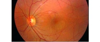
Fundus of the eye with CMV retinitis
Syphilitic retinitis occurs due to the penetration of Treponema pallidum into the human body. This type of disease can develop as a result of the birth of a child with congenital syphilis.
Congenital syphilitic retinitis is characterized by the formation of a large number of white lesions, which alternate with dark ones. This symptom of the disease persists throughout life.
If an acquired form of syphilitic retinitis is detected in a person, the disease is characterized by the appearance of swelling of the retina and optic nerve head. In the vitreous body, as this form progresses, the appearance of a diffuse suspension in combination with floating opacities is noted; in addition, hemorrhage is observed in the retinal layer.
Toxoplasmosis retinitis develops as a result of intrauterine infection. The disease is characterized by the presence of atrophic and cicatricial foci in which hypertrophy of the pigment layer of the photosensitive membrane is observed. Foci of hypertrophy most often form single ones.
As the pathology progresses, one should be wary of the occurrence of exudative retinal detachment and retinal hemorrhages.
Toxoplasmosis retinitis develops as a result of intrauterine infection
Tuberculous retinitis develops in the organ of vision when the eye is damaged by mycobacteria. As it progresses, hemophthalmitis occurs (penetration of blood into the vitreous), which can provoke tractional retinal detachment.
Rheumatic retinovasculitis is characterized by the appearance of whitish couplings, which have jagged edges located along the vessels. With prolonged progression, an exudative focus may appear in the area of the optic nerve head, which has a star-shaped shape.
Pathologies caused by metabolic disorders and diseases of the circulatory system
Diabetic retinitis occurs when, during the development of diabetes, there is a deterioration in the condition of the blood vessels that supply nutrition to the retina. Their walls begin to let blood and other fluids through. Vessels can increase in size and become brittle. The progression of pathology leads to an increase in the incidence of cataracts.
A characteristic sign of diabetic retinitis is the appearance of capillary microaneurysms and fusiform dilations of veins, and pinpoint hemorrhages in the photosensitive membrane also appear. With the further development of the disease, the formation of fibrous growths occurs.
Leukemic retinitis occurs in the acute form of leukemia. This type of disease is characterized by discoloration of the fundus of the eye, which is accompanied by the appearance of multiple hemorrhages in the retinal layer. In addition, the formation of small round lesions is observed on the retina, which in some cases have a border in the form of a red rim.
Heredity - retinitis pigmentosa
Retinitis pigmentosa develops as a result of dystrophy and not as a consequence of the progress of the inflammatory-infectious process. This type of disease appears under the influence of hereditary factors that provoke the course of degenerative processes in the retina, which are associated with the presence of abnormalities of photoreceptors and pigment epithelium in the body. The progression of the pathology leads to the loss of a large number of photoreceptors, which leads to the development of varying degrees of blindness.
Retinitis pigmentosa appears under the influence of hereditary factors
The main sign of the pathological disorder is the formation of deposits in the form of bone bodies, a decrease in the number and thickening of blood vessels, in addition, the disease is characterized by the appearance of waxy pallor of the optic nerve head.
Retinitis of unknown etiology
Coates' retinitis is a very rare congenital disorder that can cause partial or complete blindness. The reasons for the progression of this disease have not been studied. It is characterized by the appearance of anomalies in the development of blood vessels in the circulatory system that supplies the retina. The formation of massive exudation and hemorrhage is observed in the superficial layers of the retina. Between the photosensitive layer and the choroid there is a concentration of exudate and blood, which provokes retinal detachment.
Coates' retinitis is a very rare congenital disorder.
According to medical statistics, pathology is most often detected among the male population in childhood or adolescence. The most likely option is that one eye is affected.
As the disease progresses, the patient experiences blurred vision and deterioration.
An interesting fact is that when photographing with a flash, the patient’s eye appears yellow, not red, in the pictures. This is due to the reflection of light from deposits formed by cholesterol.
The lesions that appear as the disease progresses do not have a clear boundary and are most often located near the macula or optic nerve head. As the pathology develops, they increase in size and merge with each other. A characteristic sign of the disease is miliary aneurysms (small abnormal formations on the venous vessels of the eye).
Solar and traumatic retinitis
Solar retinitis is characterized by the appearance of a blind spot in the visual field, not associated with its peripheral boundaries, which is formed after the organ of vision receives a dose of radiation, chromatopsia (distortion of color perception). A distinctive feature is the formation of yellowish-white spots, which are surrounded by a gray rim. Their color changes; after 10–14 days after receiving irradiation, they acquire a reddish color.
Traumatic retinitis is formed as a result of a person receiving an injury to the organ of vision. The disease develops when the retina of the eye is damaged.
Diagnostics
If specific signs are detected, you should consult an ophthalmologist. The doctor will survey the patient regarding complaints and the presence of factors predisposing to retinitis (injuries, recent illnesses, sunbathing, etc.), and will also conduct:
- ophthalmoscopy - examination of eye structures using an ophthalmoscope;
- biomicroscopy - examination of the fundus using a slit lamp;
Biomicroscopy is performed using a slit lamp
- visometry - determination of visual acuity;
- computer perimetry - study of visual fields;
- color testing - a study to exclude color blindness;
- diaphanoscopy (transillumination, transillumination) - identifying mainly tumors in the eyeball;
- optical coherence tomography (OCT) - assessment of the condition of retinal tissue using infrared radiation;
- fluorescein angiography - examination of the vessels of the retina.
In addition, to identify the causative agent of the disease (if retinitis is infectious), the doctor will refer the patient for blood tests.
Differential diagnosis
As a rule, differential diagnosis is aimed at distinguishing one form of retinitis from another. Each type of retinal inflammation has its own clinical signs.
Clinical signs depending on the type of retinitis - table
| Septic retinitis | Viral | Tuberculous retinitis | Syphilitic retinitis | Toxoplasmosis retinitis | Pseudoalbuminuric retinitis | Solar retinitis |
|
|
| In the congenital form:
For acquired syphilis (4 options):
|
|
| yellowish-white spots with a gray rim on the fundus, subsequently turning into red lesions with clear boundaries |
Symptoms and signs of the disease
As retinitis progresses, the patient does not feel pain. Therefore, the presence of the disease can only be detected by a decrease in the acuity and quality of vision. In the case of the formation of a focus of inflammation in the macular zone, a change in the function of central vision and the correct perception of the color spectrum of images occurs.
If the inflammation spreads to the periphery of the retina, the formation of so-called tunnel vision occurs, which limits visibility to the sides; in addition, twilight vision is impaired.
Quite often, as the disease progresses, objects appear to split into two, the contours of objects become distorted, and sparks appear in the eyes. If inflammation subsides, the formation of scar tissue is observed on the retina of the eye, which subsequently leads to a severe decrease in visual acuity.
With the further development of the disease, a large number of disorders may occur, among them the most common are:
- uveitis;
- endophthalmiitis;
- panophthalmeitis.
All these pathologies increase the risk of vision loss.
Symptoms
Symptoms that should alert you and prompt the patient to see a doctor:
- The appearance before the eyes of such optical phenomena as “flies”, “worms”, as well as bright flashes or sparks.
- Difficulty in perceiving the colors of surrounding objects.
- The development of “night blindness”, that is, a decrease in twilight vision, when a person stops seeing after sunset.
- The appearance of “blind spots”, that is, large dim spots that make it difficult to fully see the objects in front of the eyes.
- Reduced field of vision, “tunnel vision,” when a person sees only what is in front, but stops noticing objects on the sides.
- Blurred outlines of objects, so-called metamorphopsia.
Diagnostic methods
The main methods for diagnosing the disease are:
- examination of the organ of vision by a specialist. Examination of the fundus plays a vital role in diagnosing pathology. When retinitis occurs, which has a tuberculous etiology, damage to the retina and the presence of a large number of foci of inflammation are detected in it. If necessary, the attending physician may prescribe an X-ray examination, computed tomography, magnetic resonance imaging;
- performing a standard visual acuity test;
- viziometry. The use of viziometry allows one to study one of the main functions of the eye - visual acuity, that is, the physiological ability of the eye to see two different points at a minimum angle. When performing viziometry, it is necessary to recognize objects that are called optotypes;
- conducting a color test to determine visual fields. Used to recognize intervals of vision loss in the general range. Testing can help identify eye diseases or innervation problems that may limit the ability to see visual objects clearly. Most often, to obtain a clear picture of the pathology, several tests are required during the examination: a test to determine the focusing of the eye;
- Amsler grid. With its help, age-related macular degeneration is checked;
- test to determine perimeters. It is carried out using a special apparatus;
- tangential screen vision test. The examination uses a black screen with concentric circles and lines that emanate from the center. The test provides a complete diagram of the patient's visual field;
- color discrimination test;
Computer perimetry is one of the most modern methods of studying visual fields
When diagnosing pathology, the most important data are ophthalmoscopic examination of the fundus. In addition, the etiology of the disease is established based on an assessment of the patient’s condition and data from a general examination of the body.
Septic retinitis
When pyogenic microbes from internal organs affected by inflammation enter the eye and directly into the retina, there is a risk of developing septic retinitis, in which the condition of the vessels of the retina worsens. Near them there is a focus of inflammation, which looks like whitish spots. The development of the disease leads to swelling of the optic nerve, which transmits signals from the retina to the brain. The vitreous body also subsequently becomes cloudy.
Ultimately, all this can result in panophthalmitis - severe purulent inflammation and melting of almost all parts of the eyeball.
Treatment
Treatment of the disease is complex. It must be carried out in a hospital setting at a medical institution. The choice of treatment method is determined by the type of pathology and the accuracy of diagnosing the causes of its occurrence. Depending on the results of the examination, surgical or medicinal treatment may be used.
Conservative treatment methods
Carrying out drug treatment of the disease involves the use of various means:
- Broad-spectrum antibiotics when identifying the bacterial nature of the disease (Tetracycline).
- Local glucocorticosteroids in the form of gels, drops and ointments (Dexamethasone and Methylprednisolone). In order to ensure resorption of the inflammatory focus, Gemaza, Lidaza, Fibrinolysin are used.
- Antiviral drugs in the form of instillations, injections. Such agents are, for example, the interferon inducer Poludan, which is used in the form of subconjunctival and parabulbar injections (injections of the drug through the skin into the lower eyelid area). Acyclovir is taken orally.
- Special anti-tuberculosis drugs and other drugs.
- Medicines that increase metabolism in the retina to improve the condition of the vascular system. Such drugs are vitamins A, E, B vitamins, Solcoseryl and some others.
- To ensure the strengthening of the vascular wall, doctors prescribe the use of Ascorutin, Dicynone, and Ascorbic acid.
Additionally, during drug treatment, agents are prescribed to stabilize the state of the vascular system, antispasmodics, vitamin complexes and metabolic drugs, and agents to accelerate tissue recovery. For specific retinitis, therapy is carried out for the underlying disease that provoked the development of the pathology.
Physiotherapeutic methods are also effective: electrophoresis and phonophoresis of medicinal substances, the use of high-frequency currents, magnetic fields, and ultrasound.
Medications - photo gallery
Solcoseryl increases metabolism in the retina
Ascorutin strengthens the walls of blood vessels Dexamethasone is a corticosteroid drug that has an anti-inflammatory effect. Poludan is an antiviral drug.
Tetracycline is a broad-spectrum antibiotic, necessary for the bacterial nature of the disease
Types of retinitis: solar, pigmentary, exudative, cytomegalovirus and others
Depending on the route of infection of the retina of the eye, retinitis is distinguished:
- External (exogenous).
- Internal (endogenous) - in the presence of a focus of infection in another organ, when the pathogen is transferred with the movement of blood.
Most often, the infection enters the eyes through the endogenous (hematogenous) route. The source of the disease may be in an organ far from the eyes, for example, in the kidneys. Sometimes, especially in weakened people with reduced immunity, the cause of retinitis can be ordinary caries or sore throat.
Among external retinitis there are:
- solar - retinitis of exogenous origin, which occurs in people who spend a long time in the sun;
- traumatic - retinitis, which appears as a result of injury or chemical burn;
- post-operative, etc.
Endogenous can be:
- infectious (syphilitic, tuberculosis, staphylococcal, leprosy, cytomegalovirus);
- viral, which occurs in diseases of measles, scarlet fever, herpes and influenza;
- leukemic, diabetic - accompany systemic diseases (diabetes mellitus, leukemia);
- hereditary. For example, retinitis pigmentosa, which often leads to severe vision impairment and blindness;
- unknown etiology (shingles, exudative, etc.)
This type of pathology also includes Coats disease, which is characterized by abnormal development of blood vessels behind the retina. It is also called external exudative Coats retinitis.
If retinitis appears as a concomitant disease, then treatment is carried out comprehensively, together with the underlying disease.
Retinitis pigmentosis - video
Surgical treatment
If complications are detected during retinitis, surgical intervention is performed, which is aimed at removing the vitreous body. The procedure is called vitrectomy. Indications for the use of this method of surgical treatment are:
- destruction of the vitreous body;
- retinal detachment;
- macular fold;
- diabetic retinitis;
- macular damage;
- hemorrhage in the vitreous region.
In case of retinal detachment, emergency surgical intervention is required using the laser coagulation method.
Laser coagulation of the retina is a surgical method for treating areas of the retina that are subject to pathological changes.
Laser coagulation is an operation to strengthen the inner lining of the eye
The procedure helps strengthen the retina and secure its fixation to the underlying layer of tissue. Laser exposure leads to cauterization of the retina and proliferation of connective tissue at the site of exposure, which restores blood circulation and increases visual acuity. In addition, laser coagulation prevents further detachment.
Performance
Coats disease usually affects only one eye (unilateral) and occurs predominantly in 1/100,000 young males, with symptoms usually appearing in the first decade of life. Peak age of onset is between 6–8 years, but onset can range from 5 months to 71 years.[2][3]
Coats disease causes gradual loss of vision. Blood leaks from abnormal vessels at the back of the eye, leaving behind cholesterol deposits and damaging the retina. Coats disease usually progresses slowly. In later stages, retinal detachment may occur. Glaucoma, atrophy, and cataracts may develop as secondary processes to Coats disease. In some cases, removal of the eye (enucleation) may be necessary.
Consequences and complications
The formation of a scar on the retina during the development of pathology leads to such unpleasant consequences as deterioration or loss of vision, loss of certain fields. If the macula is damaged, a person may lose the ability to see in the affected eye. As a result of inflammation, hemorrhages occur, which provoke retinal detachment, and this contributes to the occurrence of severe blindness.
The main and most serious complication is the formation of chorioretinal scars with a large affected area, which remain even after the source of inflammation is localized.
If left untreated, atrophy or neuritis of the optic nerve occurs, uveitis develops and retinal detachment occurs with complete loss of vision.
Coats disease. Similar disorders
- Retinoblastomas are extremely rare malignant tumors that develop in the retina. Patients are usually diagnosed with retinoblastoma before the age of three. The main features of retinoblastomas include leukocoria and strabismus. These clinical features of retinoblastoma may be identical to Coats' disease, so it is important to properly distinguish between these conditions, since treatment for retinoblastoma can lead to the development of life-threatening complications.
- Norrie disease is a rare inherited disorder characterized by the early onset of blindness in both eyes shortly after birth. Some children with this disorder may experience developmental delays. Additional symptoms associated with Norrie's disease include hearing loss and other eye abnormalities. Cataracts can develop in early infancy.
- Other disorders that may be confused with Coats disease include familial exudative vitreoretinopathy and macular telangiectasia type I.
Viral retinitis
They become a consequence of the development of herpes, influenza, and adenovirus infection. These diseases are accompanied by retinitis quite often, although to varying degrees. Sometimes the retina simply becomes less transparent. In some cases, white spots form on it. Under a microscope, the white areas look like cotton wool. They are located mainly in the area of the macula - the central point of the retina - or near the optic nerve head. If the cause of the pathology is eliminated, that is, the infectious disease is cured, the white spots become smaller and disappear completely over time. Visual functions return after this. There are also unfavorable outcomes when retinal detachment occurs or degenerative processes begin.
Cytomegalovirus retinitis
CMV retinitis is an inflammatory lesion of the retina caused by cytomegalovirus.
Most often occurs in people with weakened immune systems, incl. against the background of HIV infection. Typically acute or gradual loss of vision. At the same time, blurred contours of the objects in question appear, flickering of spots before the eyes and the presence of floating spots. Cytomegalovirus is characterized by sequential involvement of one and then the other eye. Retinitis of different nature is clinically similar to each other, so it is difficult to distinguish them. But there are certain features that are very rarely encountered with cytomegalovirus infection. So, it is not typical for her to experience pain and a burning sensation in the eye, increased tearing and irritation of the conjunctival membrane.