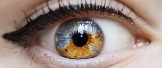
What is exophthalmos?
Protrusion in humans is accompanied by protrusion of the eyeballs from the orbital cavity.
However, this happens not due to correction of their sizes, but due to displacement of the visual apparatus. Women most often encounter pathology when they have problems with the thyroid gland. In men, the disease develops as a result of injury to the eye. With exophthalmos, the eyes protrude forward or move slightly to the side, depending on the location of the destructive processes occurring inside the organ of vision. Another characteristic manifestation of the disease is the formation of a white gap between the iris and the upper eyelid.
| The disease can develop over several years or appear in a couple of weeks. A severe form of the disease is visible to the naked eye. With ophthalmological abnormalities, exophthalmos affects one eye, with general pathologies in both. |
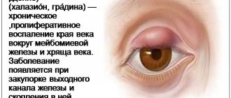
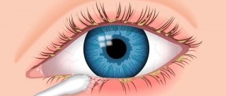
What is ulcerative blepharitis?
The staphylococcal/ulcerative type of the disease, as a rule, manifests itself at an early age in children, and later it can continue throughout a person’s life. This type of blepharitis is expressed by very noticeable redness of the eyelids and the formation of purulent crusts. In addition, in its severe forms, bleeding ulcers may appear on the eyelids, which then turn into scars on the affected areas.
According to statistics, ulcerative or staphylococcal blepharitis occurs more often in women and can be accompanied by seborrheic dermatitis, recurrent stye and rosacea. This disease is also often observed in patients with atopic eczema.
Prevention
Preventive measures to prevent the occurrence of mydriasis are advisory in nature and do not guarantee a person’s protection from its possible occurrence. To reduce the risk of developing the disease, you should undergo an annual examination by an ophthalmologist (including ophthalmoscopy under mydriasis), promptly treat identified eye diseases, and not use any medications without prior medical consultation. Patients with diabetes should adjust their blood sugar levels. It is recommended to avoid places and activities where injury to the eyes and head is possible.
Parasitic diseases
Eyelash mite
Opportunistic organisms may be present on human skin and multiply when immune defenses are weakened. Hairy parts of the body, including eyelashes, are the favorite habitats of demodex mites.
You can become infected through direct contact, as well as through towels and clothing. The waste products of ticks are toxic and cause allergic reactions. Microorganisms cannot be seen with the naked eye - their size is no more than 0.4 mm.
Experts determine demodicosis by external signs and on the basis of microscopic analysis of eyelashes and skin scrapings. The existence of a mite on the eyelashes should be suspected if there is itching, swelling and redness of the eyelids, the eyelashes are glued together with purulent discharge, the edges of the eyelids are covered with scales and dried crusts.
In treating the disease you will have to show persistence and patience. In addition to antiparasitic drugs, antibacterial and antihistamine drugs are prescribed. A big role is given to hygienic procedures: scales must be regularly removed with alcohol-containing solutions and ointment (Blefarogel, Demalon) applied to the edges of the eyelids. To strengthen general immunity, vitamin complexes and supplements are prescribed.
Pediculosis
accumulation of lice and nits on eyelashes
Eyelashes can be affected by lice, which are usually infected through close contact and through the personal belongings of the sick person. Insects crawl onto the eyelashes from other parts of the body, or nits get on the skin of the eyelids when the patient scratches the bites and touches the face.
Parasite secretions cause itching, irritation and inflammation of the skin. The eyelids swell and look puffy. Eyelashes take on an untidy appearance due to eggs laid by lice: black or brown spots are visible between the hairs, formations similar to broken eyelashes are characteristic.
Treatment of eyelash lice requires special care due to the close proximity of the eye mucosa. Vaseline oil is applied to the eyelashes and the insects are combed out with a fine comb or removed with tweezers. In some cases, for example in children, eyelashes should be cut to the root. Permitrin group products are used as prescribed by a doctor, which should be diluted with water first to avoid burning the eyes.
Symptoms of the disease
With trichiasis, eyelashes grow inward, and an additional row of hairs may be located behind the normal eyelashes. As a rule, they are thinner and shorter, lack pigmentation, are arranged in a chaotic manner, and are very difficult to see even with a slit lamp.
Touching the eyeball, eyelashes lead to irritation of its surface and the appearance of erosions on the cornea. Other symptoms may include:
- photophobia;
- involuntary spasm of the eye muscles (blepharospasm);
- pain when blinking and closing your eyes;
- sensation of a foreign object in the eye;
- burning, stinging, itching;
- redness and inflammation of the proteins, sometimes their bleeding;
- headache;
- absence of eyelashes (madarosis).
Trichiasis usually develops gradually. The intensity of symptoms depends on the number of abnormally growing hairs and the stage of the disease.
Trichiasis. Diagnosis and treatment
Consultation with an ophthalmologist is free!
Consultation with an ophthalmologist is FREE for removal of chalazion, neoplasms (papillomas) of the eyelids, foreign bodies of the cornea and conjunctiva! (price .
Discounts for friends from social networks!
This promotion is for our friends on Facebook, Twitter, VKontakte and Instagram! If you are a friend or subscriber of the clinic’s page &laqu.
This month, residents of the Savelovsky, Begovoy, Airport, and Khoroshevsky districts.
Ardamakova Alesya Valerievna
Ophthalmologist, laser surgeon
Candidate of Medical Sciences
Do you experience pain in your eyes from bright light, blink frequently, suffer from increased lacrimation, and do you have a persistent sensation of a foreign body in your eyes? Perhaps these problems are associated with improper growth of eyelashes on the eyelids, i.e. trichiasis.
Incorrect growth of eyelashes is not just a cosmetic defect, but a medical problem that needs to be solved so that the situation is not complicated by deterioration of vision and inflammatory eye diseases.
Trichiasis is an abnormal growth of eyelashes in which they are directed towards the cornea, which leads to irritation and injury to the eye. Trichiasis can be congenital or acquired.
In its normal state, the eyelid with normally growing eyelashes protects the eye from dirt and dust, too bright lighting and other aggressive environmental factors. But when the ciliary edge turns inward of the eye, the eyelashes begin to scratch the cornea, which, if the problem is not solved, can lead to numerous ulcerations.
Diagnosis and treatment of trichiasis
Diagnosis and treatment of trichiasis
Eyelash diseases and their symptoms
Diseases of the eyelashes can be divided into 2 groups - parasitic lesions and changes in the physiological characteristics of the structure of the organ.
The first group includes:
- pediculosis;
- demodicosis
In the first case, the disease is caused by pubic lice. Their cephalic relatives do not live on eyelashes and other areas covered with hair. Infection occurs through contact.
Signs of the disease:
- itching;
- conjunctivitis;
- gray coating on hairs;
- traces of blood.
In the second case, the infectious agent is a microscopic mite. This insect lives in the ducts of hair follicles and sebaceous glands and is considered a representative of opportunistic microflora.
Most often, this disease is diagnosed in veterinary practice, but it also occurs in humans.
Signs of pathology:
- discharge from the eyes;
- hyperemia of the eyelid margin;
- hypertrophy of the eyelid margin;
- decreased production of tear fluid;
- development of dry eye syndrome;
- hair loss.
The second group of diseases is associated with impaired eyelash growth and their number. Pathologies in this group include:
- trichiasis - in this case, eyelashes grow in the wrong direction towards the eyeball. There may be a single hair, or there may be many - along the entire length of the edge of the eyelid;
- distichiasis is a congenital pathology in which 2 strips of hairs grow on each eyelid. That is, doubling the number of hairs;
- hypertrichosis is an acquired disease. An increase in the number of hairs with a normal number of ciliary rows;
- madarosis or hypotrichosis - you can find photos on the Internet - complete or partial absence of hairs. Often this is an acquired pathology, which is a consequence of a serious illness with aggressive treatment.
All these pathologies can be accompanied by conjunctivitis, blepharitis, and in severe cases, an ulcer of the cornea or sclera develops.
Prevention
Not a single woman is immune from the occurrence of background diseases of the cervix. Therefore, the relevance of preventive measures remains for all representatives of the fairer sex. To reduce the likelihood of developing cervical pathologies, it is recommended:
- undergo regular preventive examinations with a gynecologist in order to suspect and treat pathological processes in time;
- avoid casual sexual intercourse, and in case of a change of partner, use barrier contraceptives;
- use contraception to avoid abortion;
- if infectious diseases occur, strange discharge from the genital tract or other disturbing signs appear, immediately seek medical help;
- Maintain personal hygiene and use vaginal products less often unless recommended by a doctor.
Women are often interested in various issues related to cervical diseases. The most common is sexual activity. Doctors say that intimacy with ectopia is not contraindicated if the disease is not at the stage of treatment. However, the eroded area is an open path for infection
Therefore, it is especially important for women with erosion to use protective equipment
Prevention of madarosis of eyelashes
In addition to pharmaceutical products, following simple rules will help prevent or speed up recovery. They improve immunity.
Prevention methods:
- Proper nutrition, rich in healthy fats and protein. This is the basis for restoring skin with hair roots.
- Partial refusal of cosmetics during treatment.
- Avoid tearing eyelashes.
- Do not wipe the inflamed eyelids, do not remove hairs - they will fall off on their own, without harm to the hair follicles.
- Use high-quality, proven cosmetics.
- Do not overload your eyes with continuous work on the computer.
Madarosis is a symptom indicating infection, blepharitis, barley, conjunctivitis. You can get rid of it by curing the underlying disease. Oils, cosmetic gels and vitamins A, E and B will help speed up recovery and the growth of new hairs.
Eyelashes – long, thick – are every girl’s dream. But like any human organ, they are susceptible to various diseases. Some of them are the result of thoughtless and careless actions.
What are the types of eyelash diseases? What are the symptoms of the pathological process? Is there adequate treatment or is there only one way out - plucking the affected hairs?
Symptoms of microphthalmos
Microphthalmos is characterized by a small eye that causes minor complaints. Usually we are talking about a congenital form of the disease, so the diagnosis is made immediately after birth.
Sometimes the eyeball and cornea are formed correctly, but are reduced in size.
People with microphthalmos may have coloboma. Coloboma is the absence of part of the eye membrane. Appears as cuts or gaps in:
- the colored part of the eye called the iris;
- the retina, which is the specialized light-sensitive tissue that lines the back of the eye;
- a layer of blood vessel under the retina called the choroid;
- optic nerves, which carry information from the eyes to the brain.
Colobomas may be present in one or both eyes. Affect visual perception.
Children with microphthalmos may also have other vision problems, including clouding of the lens of the eye (cataract) and a narrowed opening to the eye (narrowed palpebral fissure). In addition, the presence of this pathology increases the risk of developing glaucoma.
Microphthalmos refers to Lenz syndrome and sometimes causes anomalies in other systems. Other signs:
- impaired visual acuity;
- Lenticonus;
- ecstasy.
Treatment of enophthalmos
The choice of treatment depends on the origin of the disease. If the disease is caused by contraction of retrobulbar tissues, an injection is made from the patient's own fat cells taken from the anterior abdominal wall. This way you can bring the eyeball into the correct position, avoiding complications. During surgical treatment, implants made of polymers or titanium are installed in the retrobulbar space.
Conservative treatment of enophthalmos after injury is carried out only in patients with minor manifestations (posterior displacement less than 2 mm) without interposition of the inferior rectus extraocular muscle and in the absence of double vision. Treatment tactics boil down to prescribing a course of antibacterial drugs and corticosteroids. After traumatic enophthalmos, bone fragments are repositioned. If the damage is minimal, endoscopic surgery is performed.
Pain syndrome is relieved with non-narcotic analgesics. Edema is removed by instillation of hypertonic saline solutions. For inflammation of the retrobulbar tissue, a course of conservative therapy is carried out.
Treatment
Once the ophthalmologist determines that the patient has blepharitis (symptoms and treatment are directly related), he needs to carry out a set of hygienic and therapeutic procedures.
First, tampons soaked in a specialized ointment, furatsilin solution or fish oil are applied to the eyelids to soften the purulent crusts. Then the eyes are thoroughly cleaned, treated with an antiseptic and the ointment recommended by the doctor is applied.
Etiology
Many etiological factors have been described that lead to impaired growth of eyelashes and eyebrows. Madarosis can be a consequence of local changes or systemic pathologies. Leading causes of the disorder:
- Injuries.
In the area of traumatic injuries, keloid scars may form, on the surface of which there is no hair. Similar deviations occur with burns. Damage to the hair follicles is observed in obsessive-compulsive disorder (trichotillomania, trichoteiromania, trichothemamania). - Blepharitis.
Chronic inflammation of the eyelids, accompanied by periods of remissions and exacerbations, leads to eyelash loss. Blepharitis of staphylococcal nature is often observed with rosacea. Trachoma is a rarer cause, but leads to irreversible consequences. - Dermatological diseases.
Madarosis occurs as a result of a number of skin diseases that cause itching and dryness. The most common among them: atopic dermatitis, seborrhea, ichthyosis. In women over 50 years of age, the main cause is postmenopausal frontal fibrosing alopecia. - Taking medications.
Hair growth is directly affected by prolonged use of heparin, anticonvulsants, and androgens. Generalized changes in hair growth occur with the use of chemotherapy drugs. - Intoxication.
Thallium and mercury poisoning leads to alopecia areata, dysfunction of the nervous system and gastrointestinal tract. With hypervitaminosis A, retinoids have a toxic effect on the skin and its appendages. - Neoplasms.
Hair regrowth is difficult with malignant tumors: sclerosing sweat gland cancer, basal cell and squamous cell carcinoma. Madarosis occurs in systemic mastocytosis and cutaneous T-cell lymphoma. - Endocrine diseases.
Hair follicles become affected in response to thyroid dysfunction. With hypo- and hyperthyroidism, hair growth is disrupted at the cellular level, so the entire hairline suffers. - Nutrient deficiency.
The disease occurs with nutritional deficiency, manifested by hypoproteinemia and deficiency of vitamins and microelements (zinc, iron, vitamin B7).
- Ophthalmologic: blepharitis. Anterior blepharitis is either staphylococcal or seborrheic, and posterior blepharitis is caused by a blockage of the meibomian glands.
- Dermatological conditions: There are several types of dermatological conditions that can lead to madarosis. These include atopic dermatitis, seborrheic dermatitis, psoriasis. Also: frontal fibrosing alopecia, keratosis pilaris, rosacea, telogen effluvium, mycosis fungoides and sarcoidosis.
- Nutritional disorders: Severe malnutrition can cause chronic hair loss. Hypoproteinemia, lack of biotin and iron causes degeneration of hair follicles. Zinc deficiency, such as in acrodermatitis enteropathica, can lead to eyebrow/eyelash hair loss. Also leads to hair loss.
- Infections: There are many bodily infections that can cause loss of eyelashes/eyebrows. Lepromatous leprosy, syphilis or other viral infections such as herpes or HIV can also cause eyelash loss, as well as fungal infections (paracoccidioidomycosis, trichophyton or microsporum).
- Trauma: Most traumas are caused by madarosis from a psychological point of view, known as trichotillomania.
- Drugs/Medications: Crack cocaine or chemotherapy drugs. Other drugs include: propranolol, valproic acid, barbiturates, MMR vaccine, botulinum toxin, epinephrine, antithyroid drugs, anticoagulants and lipid-lowering drugs.
- Genetics
- Autoimmune disorders: alopecia areata, discoid lupus erythematosus, chronic cutaneous lupus erythematosus, Graham-Little syndrome and Parry Romberg syndrome
- Other diseases: hypothyroidism, hyperthyroidism, hypoparathyroidism, hypopituitarism, amyloidosis and congenital erythropoietic porphyria (Gunther's disease)
Causes and symptoms
Hair growth depends on the condition of the hair follicle and the skin around it. The causes of madarosis are pathologies that interfere with the normal metabolism of cells in the area of the roots of the hairs of the eyebrows and eyelashes:
- blepharitis;
- barley;
- chemotherapy;
- damage to the bulbs during depilation;
- skin burn near the eyebrows and eyes;
- demodicosis (subcutaneous mite activity);
- syphilis;
- leprosy.
Madarosis is aggravated by bad cosmetics. Its frequent use clogs the pores in the skin, disrupting the flow of oxygen and metabolic processes. This is an auxiliary factor; cosmetics cannot cause loss of eyebrows and eyelashes.
Need advice from a beauty expert? Get advice from a beauty expert online. Ask your question right now.
ask a free question
The key factor in the manifestation of the disease is genetic predisposition. Naturally brittle, sparse hairs are easier to damage; they are likely to begin to fall out at the age of 50-70.
In most cases, madarosis is caused by ophthalmic diseases. Especially blepharitis, barley - inflammatory diseases. They develop and become chronic, causing permanent baldness of eyelashes and, less commonly, eyebrows.
Madarosis can be recognized by bald spots in the eyelash growth area. They are more often localized in the center and are easy to notice. Then the hairs fall out along the edges. There are primary symptoms by which the disease can be recognized before hair loss:
- Tearfulness associated with inflammatory processes is also observed with blepharitis and barley.
- Long lasting swelling of the eyelid.
- Hyperemia and redness of the skin.
In a child, eyelash loss can be associated with neurological conditions when children independently pull out hairs or play with them.
What sizes can there be?
When about 15% of the mucous membrane of an organ is covered with ulcers, they speak of a small lesion. If 45% is affected, the erosion is considered to be medium; a large lesion is considered to be a lesion that affects more than 60% of the mucosa.
False
False erosion is nothing more than pseudo-erosion. It can also be called ectopia or endocervicosis.
This mucosal condition develops after the period of true erosion ends and the velvety epithelial layer begins to be gradually replaced by columnar epithelial cells. False erosion can occur due to a combination of acquired and congenital factors.
If even in utero a woman develops a certain feature of the distribution of epithelial cells in the genital organs, ectopia may appear at different periods of life.
Under the influence of sex hormones, the area of false erosion can fluctuate, since the cervical epithelium is extremely sensitive to the influence of hormones and responds to any change in their concentration with active growth. As soon as the concentration of hormones is normalized, the stratified epithelium is restored due to an increase in growth cells.
In this regard, at different periods of a woman’s life, ectopia can either not appear at all or be pronounced.
In modern medicine, there is no clear line that would separate the pathological condition of the cervical mucosa from a physiological feature that does not require medical intervention.
There are several subtypes of ectopia, which differ histologically:
- Papillary erosion. In this case, the epithelium forms growths in the form of papillae, which are inflammatory infiltrates. This form of erosion occurs under the influence of various factors that can provoke an inflammatory process.
- Glandular erosion. It is characterized by the formation of cysts and follicles, which are covered with stratified epithelial cells. For what reasons this pathology occurs is not known to science; there is an assumption that cellular changes or the influence of hormones are involved.
- Mixed erosion. It is manifested by the formation of both papillary growths and the formation of cystic changes.
In addition, false erosion can occur in a healing form. In this case, the multi-layered covering of the vaginal part of the cervix is restored to its normal state without treatment.
Some experts do not agree with the existing classification, since absolutely all forms of erosive lesions are considered pathological and require treatment.
Glandular
With glandular erosion, cylindrical cells leave the cervical canal, as a result of which the transformation zone shifts. As already mentioned, the causes of glandular erosion have not been studied; this pathology is not accompanied by a clinical picture, so this type is diagnosed by chance.
If an infection joins the process, the symptoms become more pronounced as inflammation develops.
Glandular erosion is characterized by highly developed erosion glands. If the pathology is accompanied by the development of cystic formations that contain mucous contents, atypical cells are replaced by squamous epithelial cells, as a result of which the erosive glands are blocked.
In this case, the inflammatory process begins to develop, and the cervical region becomes extremely vulnerable to various infections.
IMPORTANT! As for symptoms, non-inflammatory glandular erosion is not accompanied by a clear clinical picture, but if cysts and follicular formations occur, a woman may experience pain and discomfort during intimacy.
Papillary
Papillary erosion is characterized by the formation of growths that look like papillae. From above these formations are covered with columnar epithelial cells.
Some doctors do not classify this pathology as a disease, but consider it as an individual anatomical feature or a pathology of tissue formation.
Symptoms of papillary erosion may be as follows:
- slight pain in the lower abdomen;
- vaginal itching;
- spotting after sexual intercourse;
- the volume of leucorrhoea increases significantly.
Glandular, papillary and cystic erosion in their pure form are extremely rarely diagnosed.
As a rule, they are combined with each other. Most often, patients are diagnosed with glandular-cystic pseudoerosion.
This is a rather serious pathology, because the secretion that blocks the glandular channels provokes inflammation, and this is a gateway for various pathogens.
Treatment of madarosis
If the disease has just begun with profuse eyelash loss, you just need to give them a “rest.” That is, for two weeks, give up makeup or at least stop dyeing your eyelashes. During this time, they will become noticeably stronger and will be able to recover.
Vegetable oils are effective means of restoring the growth of eyelash hair and strengthening existing ones. The best results are shown by using castor oil and flaxseed oil. Before using them, you need to comb your eyelashes with a special brush and only then apply oil along the hairline with a regular cotton swab. It is better to perform this procedure at night before going to bed.
An excellent means of restoring hair and improving its growth is vitamin E. This is an oily substance that must be applied to the eyelashes with a brush from roots to tips. To improve the effect, vitamin E capsules should be taken orally according to the instructions.
The use of traditional medicine recipes shows very good results in the treatment of madarosa. To restore damaged eyelash hair follicles and improve the appearance of eyelashes, infusions of medicinal herbs are excellent: cornflower, chamomile, calendula, eucalyptus, sage, etc. They are used in the form of lotions or compresses on the eyes twice a day. The exposure time of compresses with herbal infusions is 10-15 minutes, the best time is morning and evening.
Fragility and loss of eyelashes are very often associated with a deficiency in the body of vitamin E or A, as well as B vitamins. In this regard, to combat madarosis, it is very important to pay attention to the foods that make up the diet. The condition of your eyelashes will definitely improve if you supplement your daily menu with portions of tomatoes, sweet peppers, carrots, leafy vegetables, fruits, herbs, fatty fish, and liver.
Modern pharmacology also offers an extensive line of medicinal cosmetics, the use of which gives quick results in treating eyelash loss and improving their appearance.
If there are no results from treating madarosis at home. Be sure to seek advice from a specialist. Perhaps the cause of eyelash loss is a systemic disease, the treatment of which, in order to avoid serious consequences, should be handled exclusively by a doctor.
Classification
The disease is divided into imaginary and true:
- In the first case, the disease manifests itself with congenital asymmetry of the visual apparatus, abnormal development of the skull, or an increase in the palpebral fissure;
- True is diagnosed in case of inflammation and general pathologies, as well as in case of injury to the eye and the appearance of neoplasms.
According to the type of occurrence, exophthalmos is divided into the following categories:
- Constant. Most often it is diagnosed when malignant or benign tumors form in the orbit;
- Intermittent. Develops against the background of pathological processes occurring in the orbital veins. This form of anomaly is characterized by the fact that protrusion occurs only at a moment of strong physical stress, when the vessels are filled with blood. The pathology is most clearly visible if you tilt your head;
- Pulsating. Diagnosed with aneurysm and injuries of the visual apparatus. The main symptom of the disease is pronounced pulsation in the eye. A severe headache is also observed; when the eyelid is closed, a noise is recorded in the upper part of the eye. As the disease progresses, the veins on the forehead and temples become enlarged;
- Hypothalamic-pituitary. It is characterized by rapid development and manifests itself under the influence of increased production of the pituitary hormone. Within a few days, the eyelids swell and conjunctival chemosis begins.
In the absence of deviations, the eyeball extends beyond the orbit by a maximum of twenty millimeters.
| Depending on this parameter, mild, moderate and severe forms of exophthalmos are distinguished. In the first case, the eyes protrude by 21 - 23 mm, in the second the figure reaches twenty-seven millimeters, the severe stage is characterized by a deviation of up to 28 mm. |