If intrauterine development is disrupted, retinoblastoma can form in children.
It is most common in children under 5 years of age. This is a condition in which a benign tumor forms in the eye.
Its cells can migrate through the optic nerve into the brain. If tissue malignancy has formed, metastases spread to the bones and bone marrow. The disease is dangerous with many complications and therefore requires treatment.
We recommend reading: Retinoblastoma
Reasons for development
The exact causes of retinal cancer in children have not yet been proven, but in the majority of cases the disease is hereditary.
Retinoblastoma in children can occur due to the following factors:
- harmful environmental conditions, which are observed in large industrial cities,
- poor quality food containing many carcinogens,
- professional activities of parents related to chemicals or other harmful substances,
- age of the parents – the risk of retinoblastoma increases in children if the parents are over 40 years old at the time of the child’s birth.
Most often, retinoblastoma is considered hereditary, but it has a clear connection with the environmental impact on a woman during pregnancy, which affects the fetus. As retinoblastoma grows, it grows beyond the retina and affects the eye socket and optic nerve. Treatment of retinoblastoma in children must be carried out without delay, otherwise the tumor will gradually involve the orbital tissue, and distant metastases will begin to affect vital organs. Parents who have already encountered this pathology are obliged to carefully monitor the development of their children's eyes and regularly visit an ophthalmologist with them.
Causes
The main cause of the pathology is a violation of embryonic development, as a result of which a defective neural tube is formed. This condition can occur with the following negative effects on a woman during pregnancy:
- bacterial or viral infection;
- woman's use of drugs, nicotine, alcohol;
- pathology of the placenta;
- fetal hypoxia;
- hereditary manifestations;
- gene mutation.
Therefore, during pregnancy, a woman should maintain a healthy lifestyle, eat right, visit doctors and undergo laboratory tests.
What is retinoblastoma?
Retinoblastoma is a malignant tumor that develops in the intraocular space from the retinal tissue. This disease occurs in the vast majority of cases in children under 5 years of age, regardless of the gender of the child. This happens for the reason that the neoplasm originates from embryonic tissues. We can say that the disease begins during the period of intrauterine formation of the child. Such children are born in more than 50% of cases from parents who suffered the disease in childhood, i.e. this pathology is of a genetic nature.
Retinoblastoma is located on the retina in the intraocular space
Does retinoblastoma occur in adults? Rarely. Specialists with many years of clinical practice note that over the entire period of work they encounter isolated cases of histologically proven disease in adult patients. It can be assumed that the cause of the development of retinoblastoma in this case is a tumor node that was formed in childhood, but due to unclear circumstances did not develop.
Treatment of retinoblastoma
To treat retinoblastoma in modern oncology, surgical removal, chemotherapy, and radiation therapy are used. To obtain satisfactory results, a combined approach is recommended. When choosing a tactic, the following parameters of retinoblastoma should be taken into account:
- Is vision preserved?
- Pair of lesions;
- Extension of the process beyond the eye;
- Involvement of the orbit and central nervous system in the tumor process, formation of distant metastases.
Doctors try in every possible way to limit themselves to conservative methods. Among them, photocoagulation and cryotherapy have become very popular. These techniques rarely cause complications and help preserve vision. If the disease recurs, this minimally invasive procedure can be repeated. Cryotherapy is used when retinoblastoma is located in the anterior retina; photocoagulation is more suitable for tumors of the posterior retina.
Classification of pathology
First of all, retinoblastomas are divided into hereditary and acquired. The hereditary form of the disease begins to manifest itself at the age of 2-3 years after birth, and its characteristic feature is that both eyes are affected. Acquired cases of the disease are characterized by damage to only one eye.
When classifying retinoblastoma, the direction of growth is of great importance:
- Endophytic growth - retinoblastoma grows from cells that are located on the inner surface of the retina. The tumor develops inward, affecting all layers of the retina and destroying the vitreous body.
- Exophytic - retinoblastoma affects the retina, behind which exudate begins to accumulate, and also involves the optic nerve and elements of the orbit.
Tumor cells can be either differentiated or undifferentiated, which determines the course of the disease. Undifferentiated tumors are characterized by more aggressive development with intense clinical manifestations. Deposits of calcium salts may accumulate in the tissues of the formation, and necrosis may develop.
Radiation treatment
To try to preserve vision in patients with retinoblastoma, radiation is given to the tumor. The goal of this technique is to cure the disease and preserve vision. In this case, the doctor must assess all the risks of immediate and long-term results.
The technique of radiation therapy can be different, but external irradiation (two lateral fields) is most often used. Due to the frequent occurrence of bilateral retinal involvement and multifocal tumor growth, the training field should cover all these areas. In addition to the retina, radiation therapy irradiates the vitreous body and 10 mm of the anterior segment of the optic nerve. Doses depend on the stage of the disease (at stages 1-3 - 3500 Gy, at stages 4-5 - 4500 Gy). Special blocks are used to protect the lens. This helps prevent the formation of post-radiation cataracts. If, after lateral irradiation, tumor recurrences occur in the anterior segment, cryotherapy is additionally prescribed.
During irradiation, young children are put under anesthesia or their head is firmly fixed. With this treatment, about 75% of patients are cured, which is due to the high sensitivity of the tumor to radiation therapy. If cryotherapy is additionally performed, the percentage of cured patients increases even more.
Sometimes radioactive plates are used, but this technique is limited. This is due to the uneven distribution of the radiation dose. Vascular damage and hemorrhage may occur at the site of greatest activity.
Radiation therapy is most often used for tumor processes affecting the central zones of the retina, as well as for solid retinoblastomas.
Local (local) chemotherapy
This chemotherapy is indicated for patients from groups C and D in combination with systemic chemotherapy. The technique involves using a superthin catheter to inject a cytostatic drug called Melphalan through the internal carotid and femoral arteries directly into the mouth of the ophthalmic artery. Before this, carotid angiography of the internal carotid artery is performed to visualize the structure of the vessels.
Chemotherapy with Melphalan (selective intra-arterial) is carried out 3-4 weeks after the 1st course of systemic chemotherapy. The dosage is 5-7.5 mg/m2:
- a dosage of 5 mg/m2 is indicated when both eyes are treated simultaneously;
- if only one is affected, then 7.5 mg/m2 should be administered.
16 mcg of Melphalan (this is 0.05 ml) is injected into the vitreous body (med. – “intravitreal”). This concentration is effective against tumor screenings and is safe.
Treatment
The main goal of treatment for retinoblastoma is to preserve the child’s life, and only then to preserve vision. The treatment method for retinal cancer is selected taking into account a number of factors:
- type of lesion
- stage of tumor development,
- the degree of spread of pathology throughout the body,
- the likelihood of maintaining vision.
Modern ophthalmology offers the following methods of treating retinoblastoma in children:
- Enucleation. The most effective method of treating unilateral retinoblastoma, the essence of which is to remove the tumor along with the diseased eye. Sometimes it is possible to save the eyeball.
- Brachytherapy. A radioactive plate is sutured to the eye, providing maximum radiation exposure directly to the tumor.
- Cryodestruction. The malignant tumor is exposed to cold, which destroys the pathological focus.
- Thermotherapy. With the help of infrared radiation, the tumor warms up to high temperatures for a long time, resulting in the death of pathological cells.
- Intra-arterial chemotherapy . An antitumor drug is injected into the artery supplying blood to the eye tumor, destroying the cancer focus.
- Intravitreal chemotherapy. An antitumor drug is injected into the vitreous body affected by the tumor.
Small tumors can be eliminated using photocoagulation. Surgical methods for treating retinal cancer are used for serious damage to the visual organs and significant loss of vision. In most cases, doctors try to eliminate the tumor using chemotherapy (Etoposide, Vincristine) and radiation therapy. To achieve a positive result, several methods of therapy are usually used at once.
By following the clinical recommendations of specialists, you can save your vision.
Stages
When retinoblastoma appears in children, the TNM system distinguishes the following stages of disease development:
- T1 - tumor size does not exceed more than 25% of the fundus,
- T2 - the neoplasm grows throughout the retina, but does not occupy more than 50% of its surface,
- T3 - retinoblastoma affects more than half of the retina and can even extend beyond it,
- T4 - neoplastic cells spread beyond the orbital boundaries,
- N1 - single metastasis occurs in regional lymph nodes,
- MI - the tumor metastasizes to distant organs.
In the acute course of the disease and the risk of metastases, the life of the sick child arises. The most dangerous period is when retinoblastoma reaches the soft shell of the brain, after which it begins to grow over its surface
Stages of retinoblastoma
Treatment for retinoblastoma is determined by the size of the tumor and the extent of the pathological process. In this regard, it is very important to correctly stage the tumor.
Among the classifications of retinoblastoma, the one most often used is Reese-Ellsworth, created back in 1969. However, it only includes intraocular tumors. This classification is based on the location of retinoblastoma, the extent of organ damage and the impact of this on the risk of vision loss. All patients with retinal cancer are divided into five groups, which differ in the likelihood of maintaining visual function.
Enucleation for retinoblastoma
The most effective treatment for eye cancer is enucleation - removal of the eyeball. During the operation, the optic nerve is cut as far as possible, so this method shows the best results in the treatment of retinoblastoma. 6 weeks after surgery, prosthetics can be performed to eliminate the visual defect. Because patients with retinoblastoma are often children, enucleation poses some challenges. After removal of the eyeball, the bones of the orbit begin to grow more actively, causing a severe defect.
Indications for enucleation:
- extensive damage and inability to restore vision,
- massive seeding of the vitreous body,
- the tumor affects the posterior chamber of the eye and reaches the posterior capsule of the lens,
- localized near the ciliary body or occupies most of the anterior part of the eye,
- glaucoma due to vascular proliferation,
- advanced secondary complications (uveitis, cataracts, hemophthalmos, hyphema),
- the onset of blindness with the inability to restore vision.
After enucleation, the eyes are sent for histological analysis. If the tumor has affected the optic nerve and choroid or has extended beyond the sclera, this indicates a poor prognosis for the patient. When retinoblastoma extends beyond the orbit, orbital exenteration (removal of orbital contents) is recommended.
In what cases are enucleation and conservative treatment combined:
- retinoblastoma grows into the optic nerve,
- scleral damage,
- extrabulbar tumor spread,
- location in the peripapillary zone,
- the presence of large and multiple tumor nodes,
- penetration of lesions into the vitreous body, iris, anterior segment of the eye,
- choroidal involvement.
Course of retinoblastoma
If the retinal tumor is located intraocularly, there is a threat to vision. With ophthalmoscopy, cancer can be determined if the node enlarges to 0.25 mm. Externally, such a node looks like a translucent hemispherical mass, which is located within the fundus of the eye and covers the choroid.
As the tumor node grows, it becomes impenetrable and acquires a pinkish color. Sometimes the growth of retinoblastoma is directed into the vitreous body (endophytic tumor). This type of retinal cancer is usually friable and spreads to the vitreous substance and even to the structures of the anterior chamber of the eye. During intraocular tumor growth, secondary glaucoma occurs due to a large tumor size or retinal detachment. At the same time, increased vascular growth occurs within the iris, which leads to an increase in intraocular pressure. In a child, intraocular hypertension can cause an increase in the size of the eyeball, which gradually loses visual function. Later pain, anorexia, and vomiting develop. When the tumor grows beyond the retina, an exophytic variant of retinoblastoma occurs.
When the process spreads beyond the orbit, there is a threat not only to the patient’s vision, but also to his life. This is due to the immediate proximity of the central nervous system. Most often, the tumor penetrates into brain structures through the pia mater or along the fibers of the optic nerve. when the choroid is damaged, we are talking about hematogenous metastasis of retinoblastoma. Sometimes the tumor grows into the sclera. Lymph nodes are also affected, and in later stages distant metastases occur.
Symptoms
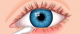
The symptoms of retinoblastoma are largely determined by its current stage of development. It is often very difficult to clarify all the symptoms, since young children are not yet able to fully speak and describe what they are feeling. At the first stage, no pronounced manifestations occur, but a change in the diameter of the pupil may be observed. Loss of binocular and central vision is an early symptom of this pathology, which is why children often experience strabismus.
The second stage of retinoblastoma growth is characterized by the appearance of photophobia, hyperemia, and the development of iridocyclitis and uveitis. Due to tumor invasion, children experience pain. The pressure inside the eyeball increases, leading to the appearance of secondary glaucoma. In the presence of a tumor dissection of the retina, protrusion of the neoplasm occurs.
During the third stage, children often develop displacement of the eyeball. The intensity of this shift may vary. Orbital tissue and its walls may also be damaged. The neoplasm grows into the paranasal sinuses and fills the subarachnoid space.
In stage four, retinoblastoma destroys the optic nerve and involves the pia mater of the brain. The neoplasm spreads metastases throughout the body, initially affecting the brain, and then the liver and other important organs. The spread of malignant cells occurs through the bloodstream and lymph. The patient is in critical condition, he experiences severe weakness, nausea with vomiting, headaches, and intoxication develops.
What are the symptoms of the disease?
While retinoblastoma is very small, children usually do not complain about anything. For quite a long time the disease does not manifest itself in any way; children do not have any symptoms. The first complaints appear when the tumor becomes larger or begins to grow into other parts of the eye. The child begins to lose vision, sometimes to the point of complete blindness. The visual acuity of both eyes becomes different and therefore strabismus may appear in children (occurs in approximately 25 to 30% of cases).
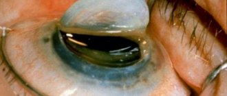
The most common first symptom, according to which retinoblastoma is found in approximately one third of children, is a white glow of the pupil in certain lighting (in the language of specialists, this symptom is called leukocoria). For example, in bright light from a camera flash, a healthy pupil becomes red or black, and a white reflex appears in a sick pupil. Such a white pupil is called a “cat’s eye,” and the “cat’s eye” effect itself indicates that the tumor has already grown beyond the lens. Less often, it happens that a child's eye hurts, turns red or swells when intraocular pressure increases.
Alarming symptoms in children are:
- when the pupil (or both pupils) becomes whitish-yellow (leukocoria)
- the child develops strabismus or deteriorates vision/visual acuity
- redness or swelling of the eyes, the eyes begin to hurt
If a child has one or several of the listed symptoms at once, this does not mean that he has contracted retinoblastoma or some other type of cancer. Some of these symptoms appear for completely harmless reasons and have nothing to do with cancer. However, we recommend that you consult a doctor as soon as possible to find out the exact cause. If it really is retinoblastoma (or some other malignant disease), then timely diagnosis is the best condition for a good treatment outcome.
Children from families with an increased risk of a hereditary disease should undergo regular examinations by an eye doctor, even if they do not have any symptoms or complain of anything. Only then can a specialist diagnose retinoblastoma at an early stage and the child will be treated on time.
Postthrombotic form of the disease
This type of eye damage is associated with the formation of a blood clot in the central retinal vein. Due to occlusion, blood microcirculation is disrupted, vascular permeability increases, which leads to an outpouring of blood and plasma into the retinal space. Edema of the macula leads to rapid ( within a week ) loss of vision.
In ICD-10, the disease is assigned code H.34 - “Occlusion of retinal vessels.”
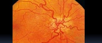
Photo 2. Fundus of the eye with postthrombotic retinopathy. There is extensive hemorrhage in the retina.
Pathogenesis occurs in three stages:
- Prethrombosis. Blood circulation is disrupted. Along with the expansion of capillaries, local ocular hemorrhages occur. Characteristic symptoms of this condition: blurred, foggy vision, slight loss of visual acuity.
- Thrombosis. Against the background of narrowing of the arteries, the veins expand, the macula swells, and the boundaries of the optic nerve head blur. Visual acuity drops noticeably, blind spots appear, and a veil covers the eyes.
- Actually postthrombotic retinopathy. Degenerative changes in the vascular system of the visual organs, the formation of fibrosis in the fundus. Acute ischemia can lead to retinal detachment.
Possible complications
Retinoblastoma is a dangerous disease that rapidly affects the visual system, causing vision loss. Lack of timely treatment leads to the rapid spread of tumor metastases throughout the body, which in most cases causes death. Often, even after successful treatment of the tumor, there is a relapse of the malignant neoplasm in adulthood. If a child is diagnosed with a congenital form of pathology, then there is a high risk of developing other types of cancer.
Diagnostics
When a child experiences symptoms indicating retinal cancer, an urgent consultation with an ophthalmologist is necessary. Oncologists and neurologists also take part in the diagnosis of retinoblastoma. First of all, the doctor conducts an external examination and checks vision. The presence of retinoblastoma may be indicated by strabismus, a weak reaction to light and dilation, as well as leukocoria.
If a child has these signs of the disease, additional tests are prescribed to make an accurate diagnosis:
- ophthalmoscopy,
- fluorescein angiography,
- radiography,
- computed tomography (CT),
- magnetic resonance imaging (MRI),
- ultrasound examination (ultrasound),
- biopsy.
Carrying out tomographic studies is necessary to assess the extent of the tumor. An intraocular biopsy is prescribed to a child only if there is an urgent need for this study, since this procedure may lead to the dissemination of malignant cells in the eye. Only after receiving the results of all the studies performed, the doctor can prescribe a further course of therapy.
Signs of retinoblastoma of the eye
Although retinal tumors have several characteristic warning signs, they are very difficult to spot in children. So,
Loss of vision will be one of the first signs, which is difficult to diagnose. Children under 2 years of age may not notice a decrease in visual acuity. This period is characterized by loss of visual fields and blurred images.
Cat's eye syndrome (white pupillary reflex) is one of the first symptoms. It is most often noticed by parents. From a certain angle, it seems that the baby’s eye (or eyes) sparkled “like a cat’s.” Sometimes there is a feeling that the eye has turned white; the swelling is noticeable through the pupil. This symptom appears quite early, the tumor is clearly visible during an ophthalmological examination. Timely diagnosis allows us to give good prognosis for vision preservation.
Strabismus is also one of the first symptoms of a retinal tumor. This sign is easy to notice and often detection of retinoblastoma occurs during examination regarding complaints of childhood strabismus.
Other symptoms. In later stages, the tumor affects the ENT organs and the nervous system. These specialists issue a referral to an ophthalmologist. In the later stages of the disease, the child becomes lethargic, gets tired quickly (a sign of intoxication), and complains of severe headaches.
Differential diagnosis
It is very important to distinguish retinoblastoma from other diseases that can cause retinal detachment in children. The clinical manifestations of retinoblastoma are similar to many other pathologies, including retinal dysplasia, cicatricial retinopathy, and soft tissue sarcoma.
In addition, it is necessary to distinguish this tumor from vitreous granulomas and retinal hamartomas. The latter can also be combined with neurofibromatosis and tuberculous sclerosis. The effectiveness of further treatment depends on how accurately the diagnosis is made, because in each individual case the methods of therapy are radically different.
Symptoms of manifestations
The primary symptom of the pathology is redness around the organs of vision.
Bilateral retinoblastoma develops gradually. First, parents notice redness around the eyes, swelling and a subtle white spot on the retina. Over time, it increases and, at the same time, vision deteriorates, strabismus, glaucoma, lacrimation, and photophobia develop. In infants, this is less noticeable, since the peak of clinical manifestations occurs at the age of 2-3 years.
General symptoms of malignancy are also inherent in binocular retinoblastoma. The child often gets sick due to underdevelopment of the immune system and intoxication of the body with tumor metabolic products. There is a decrease in appetite and indigestion, which leads to a slowdown in physical and mental development and low activity. Children complain of headaches and discomfort in the eyes.
Treatment methods
With timely detection of retinoblastoma of the eye, with adequate treatment, in 90% of cases the prognosis for children is favorable. Treatment methods for retinoblastoma vary, and when choosing the most optimal method, ophthalmologists take into account the following factors:
- preservation of vision, as well as the chances of its recovery after treatment,
- number of affected eyes,
- retinoblastoma size,
- the tumor is located on or behind the equator,
- involvement of the optic nerve, spread of the process to the tissues of the orbit,
- the presence or absence of distant metastasis.
During treatment, not only the ability to preserve vision, but also the safety of the eye itself is of particular importance. After surgery to remove an intraocular tumor, there may be a significant disruption in the growth of the skull in the facial area, which can cause serious cosmetic defects. Maximum preservation of eye tissue is possible with drug treatment of retinoblastoma, but it is rational only at the beginning of the development of the disease. For bilateral lesions, a separate treatment method is selected for each eye.
Cryotherapy and photocoagulation
These methods can be rationally used in the first stages of development, while they allow preserving not only the organ, but also visual function. After several sessions of cryotherapy, small tumors located on the anterior periphery can be eliminated. Photocoagulation allows you to remove small tumors that form on the retina. After such treatment, there is still a risk of retinoblastoma recurrence, but in such a situation, you can undergo another course of treatment.
Surgery
During surgical procedures, not only the eye, but also the orbital tissue can be removed. This operation is prescribed if the following indications exist:
- extensive damage to the organ, when the use of conservative methods does not produce results,
- development of glaucoma due to tumor growth,
- large tumor size,
- loss of vision without the possibility of its restoration.
During surgical procedures, it is very important that the optic nerve is cut off as far as possible from the affected area. This is necessary to prevent the spread of neoplasia. Some time after the operation, it is possible to install a prosthesis.
Radiation therapy
Radiation therapy can be carried out remotely and using applications of radioactive substances. It is rational to use irradiation for small tumors in children located outside the optic nerve. If a child suffers from congenital retinoblastoma, he is prescribed external irradiation, which allows one to influence several foci of pathology at once.
In approximately 70% of cases, irradiation can achieve a positive result and even preserve vision. Radiation therapy is also combined with surgery and drug treatment. The radiation dose is usually about 4500 Gy. In addition to the retina, the vitreous body and 10 mm of the anterior part of the optic nerve are irradiated.
Chemotherapy
Indications for a course of chemotherapy are:
- extensive damage to the tissues of the orbit,
- optic nerve damage
- tumor metastasis.
During treatment, patients are prescribed special medications that can be administered intravenously or orally. This method is quite effective for treating children with diagnosed retinoblastoma, but chemotherapy has a large number of side effects, including hair loss, nausea, apathy and general malaise.
Folk remedies
It is impossible to get rid of retinoblastoma using traditional methods. Folk remedies include the use of decoctions and teas that help relieve tension and weaken the clinical manifestations of the disease. Also, after a course of chemotherapy, acupuncture is recommended, which can improve the general condition of the patient. Before using any alternative medicine, you should always consult a doctor.
Reese-Ellsworth classification
Group 1 is the most favorable:
A. A solitary tumor is located at or behind the equator. Has a diameter less than four times the size of the optic disc. B. Multiple nodes, the diameter of which does not exceed 4 disc diameters. Located in the same area.
Group 2 also considers it favorable:
A. A single tumor measuring 4-10 disc diameters, located at or behind the equator. B. Multiple tumors of the same size and location.
Group 3 has a questionable prognosis:
A. A tumor of any size located in front of the equator of the eye. B. A single tumor measuring more than 10 discs behind the equator.
Group 4 has an unfavorable prognosis for vision:
A. Multiple nodes larger than 10 disc diameters. B. Any swelling in the area anterior to the dentate line.
Group 5 has the most unfavorable prognosis:
A. The tumor affects more than half of the retina. B. Contamination of the vitreous body has occurred.
To determine the size of the tumor node, the diameter of the optic nerve (1.5 mm) is used.
There is no uniform staging classification for common retinoblastomas. According to the classification proposed by scientists from the USA, there are:
- The first stage is characterized by the location of retinoblastoma only within the retina.
- The second stage is accompanied by the spread of the tumor to other structures of the eyeball.
- The third stage has extraocular spread of the tumor.
- At the fourth stage, distant metastases of retinoblastoma are present.
Because this classification does not include important characteristics of retinal cancer, some doctors prefer to use a different classification to guide their treatment of the disease. There are several variants of tumor growth that have prognostic significance:
- Intraocular location of retinoblastoma.
- Tumor damage to the optic nerve fibers.
- Involvement of orbital tissue in the process.
- Formation of distant metastases.
Prognosis and prevention
It is very good when retinoblastoma can be detected in children at the very beginning of their growth. Then, in addition to surgery, it is possible to carry out other treatment methods to preserve the organ of vision: cryotherapy, photocoagulation and radiation therapy. When metastases appear, retinoblastoma is removed by enucleation of the eye, which allows the child to be completely cured. However, the loss of the eye leaves a significant cosmetic defect. If, as retinoblastoma grows, secondary lesions appear in the brain, the chances of saving the child’s life are significantly reduced.
The main reason for the development of a disease such as retinoblastoma in children is genetic mutations that are transmitted hereditarily. Therefore, if one of the parents has already had retinal cancer, the family is required to undergo medical genetic counseling. Children at risk should undergo a full medical examination from an early age.
What protocols and registries are used to treat children?
In order to choose for each specific case the most optimal treatment method, which is recommended for each risk group (in the language of specialists - risk-adapted treatment), there must be a base that is based on reliable statistical data. But retinoblastoma is an extremely rare disease (about 4 children get sick every year in Germany and Austria). Therefore, to date, little data has been accumulated on this disease.
Therefore, unlike other forms of cancer that occur in children and adolescents, there is no single standard protocol for the treatment of retinoblastoma, which in Germany is called therapy optimization studies. (German protocols, or therapy optimization studies, are clinical studies, they are strictly controlled. They treat children and at the same time study a specific form of cancer).
RB-Registry was opened in 2013 . This registry will collect data on the epidemiology [epidemiology] of retinoblastoma and how this disease progresses for several years. The goal is to gather more information about this form of cancer and how different types of therapy respond to treatment.
This registry accepts all children and adolescents from Germany and Austria under 18 years of age who are newly diagnosed with retinoblastoma and/or who have a germline mutation of the RB1 gene (mutation in the germline [germline]) and have not yet received any treatment. The central research office is located at the University Hospital Essen. Head – Ph.D. Petra Temming.
In addition, for patients with a hereditary form of retinoblastoma, there is an opportunity to be included in a European research protocol that investigates the causes of the appearance of secondary tumors after retinoblastoma has been successfully treated (research protocol “ Screening of secondary tumors in children with hereditary retinoblastoma” ). As part of this screening study, children undergo an MRI of the head once a year. The conditions under which children are included in this protocol are: a hereditary form of retinoblastoma, the children received radiation therapy during treatment, and the child’s age is between 8 and 18 years.
A research protocol on the effectiveness of chemotherapy is currently being planned. But it will be able to start only when funding is received.
Causes of retinoblastoma
Tumor risk factors include:
- Heredity and genetic mutations (usually in the 13th pair of chromosomes);
- Age of parents;
- Adverse environmental influences and occupational hazards (metallurgical industry, in particular) experienced by future parents or a woman during pregnancy.
The causes of retinoblastoma usually lie in genetic abnormalities, more often inherited from parents to children. The hereditary form of retinoblastoma is characterized in the vast majority of cases by bilateral involvement, while retinoblastoma caused by other causes usually affects only one eye. A hereditary tumor is detected early, in the first two to three years of a child’s life. As a rule, parents who know about such a pathology or have experienced it carefully monitor the development of their children’s eyes and promptly visit an ophthalmologist.
Retinoblastoma, not associated with heredity, is usually unilateral and can be detected at any age, although this pathology is extremely rare in adults. Such cases are not familial, but their cause still lies in spontaneous mutations of individual genes.
The older the future parents are and the more external influences they experience from the environment or from working in hazardous or hazardous industries, the higher the likelihood of retinoblastoma developing in their offspring.
The tumor can form one growth focus or several at once (with a hereditary variant of retinoblastoma), grow inside the eye, affecting the retina and vitreous body, or outward (exophytic), causing retinal detachment and accumulation of exudate behind it. Infiltrative growth with damage to all layers of the organ is also possible.
The cells that make up the tumor are polymorphic and divide intensively, as evidenced by a significant number of pathological mitoses. Necrosis and deposition of calcium salts are common in neoplasm tissue. The degree of differentiation of retinoblastoma cells implies the separation of differentiated and undifferentiated forms of the tumor, which affects the course of the disease.
Gradually increasing in size, retinoblastoma can extend beyond the retina and eye tissue and invade the optic nerve and elements of the orbit. Like any other malignant tumor, retinoblastoma can metastasize to the brain, bones, and liver.
Classification
Forms depending on the prevalence on the eyes:
- monocular – only one organ of vision is affected;
- binocular - both eyes are affected.
Forms depending on the cause of occurrence:
- hereditary - the cause of pathology in the transmission of mutated genes from parents to children;
- sporadic - occurring spontaneously, under the influence of maternal disease, genetic damage.
Based on the type of formation, tumors are divided into the following types:
- endophytic - forms at some point of the eye, then grows inside, resulting in damage to the intraocular elements;
- exophytic – spread occurs only to the retina;
- infiltrative - grows throughout the entire eye, so it completely stops functioning; this type is rare.
Based on cell type, tumors are divided into:
- differentiated - this is a benign tumor, which is the least dangerous for the patient;
- undifferentiated are malignant cells that can metastasize to various tissues and organs.
It is the doctor's responsibility to classify the tumor to determine which treatment is appropriate for the patient.
Video about what methods are best to cure retinoblastoma
The video talks about innovative ways to treat the disease. Firstly, it is a malignant tumor of the retina that can appear in children between 2 and 5 years old. Secondly, treatment approaches in different countries differ from each other, and very seriously.
In the CIS countries, intravenous chemotherapy is carried out, which has a systemic effect not only on the formation itself, but also on the entire growing organism. The second method is enucliation - this is the complete removal of the organ.
In European countries, this disease is treated with cryotherapy (used for anterior lesions of the retina, and when the formation is frozen using special equipment) and photocoagulation (this method is based on the use of laser beams used for posterior lesions of the retina).
Regardless of the choice of therapy, I recommend adhering to preventive measures so that the disease goes away.
Chemotherapy
In combination with surgery, chemotherapy treatment can be carried out, which guarantees 80-90% cure. Chemotherapy is required for:
- significant intraocular damage;
- optic nerve invasion;
- orbital lesions;
- regional metastases.
Retinoblastoma is very sensitive to chemotherapy. Several cytostatics can be used for treatment at once. The best results can be obtained by combining drugs such as Carboplatin, Vepesid, Vincristine.
Chemotherapy can reduce the size of the tumor, which is why this technique is recommended before surgery. In addition, this technique is intended for systemic therapy in advanced cases.