State Description
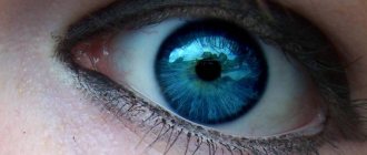
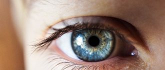
When enophthalmos occurs, an abnormal, that is, anatomically incorrect and incorrect from a physiological point of view, localization of the eyeball is observed. It is located in the orbit, but it moves deeper into the orbital cavity. The eye seems sunken and deep-set, but this condition not only represents an external defect, but also provokes visual impairment, and is also accompanied by other abnormalities, sometimes dangerous and serious.
Despite the fact that enophthalmos sometimes only has manifestations that affect a person’s appearance, sometimes the reasons for its development are hidden inside the body, are provoked by serious pathologies and even threaten the health and life of the patient. That is why the condition requires full diagnosis and timely treatment.
Good to know! The reasons why enophthalmos develops can be very different, and they do not always directly affect the organs of vision. An experienced ophthalmologist will help to identify and eliminate negative factors and triggers.
Enophthalmos is not an independent disease: in ophthalmology it refers to abnormal conditions that accompany certain diseases or pathologies. This phenomenon was first discovered in 1889 by British-born surgeon V. Lang. He treated a patient after a serious injury and, among other injuries, observed a deepening of the eyeball into the socket by 8 millimeters.
Enophthalmos and exophthalmos: what is the difference
In ophthalmology there is another concept - exophthalmos. Uninformed people confuse it with enophthalmos; this condition also affects the eyeball, but in a slightly different way. With enophthalmos, the eye moves deeper into the orbital cavity, as if sunken. With exophthalmos, the visual organ, on the contrary, partially extends beyond the orbit, that is, it protrudes.
In medical practice, exophthalmos is often called protrusion of the eyeball, bulging eyes or proptosis. This condition is more noticeable and is usually detected earlier than enophthalmos, as it has pronounced characteristic external manifestations.
The causes of enophthalmos and exophthalmos are different in most cases. Bug-eye develops, as a rule, after traumatic brain injuries (especially with fractures of the base of the skull), damage to the orbital cavity, the parasitic disease trichinosis, against the background of Graves' disease. Exophthalmos can be intermittent (appearing periodically), constant and pulsating. Separately, there is a false form that develops as a result of the congenital asymmetric structure of the bones of the facial part of the skull.
Anophthalmos - symptoms and treatment, photos and videos
Anophthalmos - main symptoms:
- Pain in the eyes
- Decreased vision
- Reduced field of view
- Rapid visual fatigue
- Facial asymmetry
- Reducing the palpebral fissure
- Anomalies of the eyelids
- Absence of the eyeball in the orbit
What is anophthalmos
Anophthalmos - refers to ophthalmological ailments, which are characterized by the absence of the eyeball in the orbit. The pathology is considered rare: children with anophthalmos occur in 2 per 10 thousand babies born, and among adults the prevalence of the disease is 21-22 per 10 thousand population.
The reasons for such a deviation can be very diverse, ranging from inadequate pregnancy to forced removal of the eyeball.
The symptoms of the disease are specific and pronounced. Changes in the healthy eye include narrowing of the boundaries and decreased visual acuity, as well as rapid fatigue of the visual organ.
Diagnosis is based on an external examination of the problem area and the clinician’s examination of data from instrumental procedures. The disease can be eliminated with the help of eye prosthetics. However, conservative methods also take part in therapy.
In the international classification of diseases, such a deviation has its own code. ICD-10 code: Q11.
Causes of anophthalmos
Anophthalmos belongs to the category of polyetiological ailments, which means that its occurrence is simultaneously influenced by several negative factors.
In the vast majority of cases, the cause of the primary form of the disease cannot be determined. However, it is believed that congenital anophthalmos may be a consequence of:
- pathologies such as measles, rubella or herpes zoster suffered by the expectant mother (most dangerous for the fetus if this occurs in the 1st trimester);
- formation of amniotic cords;
- the presence of other genetic syndromes in the child: often the absence of the eyeball occurs against the background of Lenz, Lochman or Patau syndrome;
- influence of ionizing radiation.
As for the acquired form of such an eye anomaly, in the vast majority of cases it is associated with sources such as:
- excessive addiction to alcoholic beverages;
- uncontrolled use of medications that have a teratogenic effect;
- substance abuse;
- household or industrial injuries to the organs of vision, leading to post-traumatic atrophy, a chronic form of uveitis and a high probability of sympathetic ophthalmia;
- the formation of malignant neoplasms in the eye.
Symptoms of anophthalmos
Anophthalmos has a specific clinical picture. For example, in the problem area there is an irreversible loss of visual functions. With a one-sided course, the following changes develop in the healthy eye:
- decreased visual acuity;
- narrowing of the boundaries of visual fields;
- problems with spatial perception;
- rapid eye fatigue;
- the appearance of pain in the orbit (occurs only when the nervous system is damaged);
- irradiation of pain to the back of the head;
- asymmetry of facial features.
With true anophthalmia, the following are observed in the area of the affected eye:
- eyelid abnormalities;
- shortening of the palpebral fissure;
- internal epicanthus.
Treatment of anophthalmos
Therapy of pathology in adults and children comes down to the development of an individual ocular prosthesis. In some cases, surgical preparation is necessary, namely:
- excision of the optic cup rudiment;
- plastic surgery of the orbital walls;
- correction of entropion and lagophthalmos;
- canthotomy.
Conservative treatment of this problem is limited to the daily administration of antiseptic solutions.
Possible complications
Complications of anophthalmos are presented:
- asymmetry of the face, which entails difficulty breathing and problems with chewing food;
- the addition of an infectious or inflammatory process;
- development of retrobulbar abscess;
- formation of orbital phlegmon.
Prevention and prognosis
To prevent a person from developing od-anophthalmos, some rules of prevention should be followed:
- control over the adequate course of pregnancy;
- avoiding eye injuries;
- use of personal eye protection when working in hazardous industries;
- compliance with safety precautions at home.
The prognosis of the pathology is conditionally favorable - it is impossible to restore visual functions, however, it is possible to eliminate the cosmetic defect with the help of prosthetics and specific rehabilitation.
If you think you have Anophthalmos
and symptoms characteristic of this disease, then an ophthalmologist can help you.
Source
Source: https://novosti-mediciny.ru/anoftalm-simptomy-i-lechenie/
Reasons for the development of enophthalmos
The causes of the development of enophthalmos are varied and are not always localized in the area of the visual apparatus. The condition can be congenital, identified from the very birth of the child, as well as acquired, resulting from the influence of certain unfavorable factors.
The causes of the congenital form of enophthalmos can be:
- abnormal structure of the skull;
- lipodystrophy – the absence of a natural fat layer, thanks to which the eyeballs occupy a physiologically correct position in the skull;
- microphthalmos is a congenital malformation of the visual organs, in which the eyeball has an abnormally reduced size and, accordingly, sinks into the orbit.
Acquired enophthalmos develops for the following reasons:
- Injuries that provoked mechanical damage to the walls of the temporal sockets or directly to the eye sockets. For example, a violation of the integrity of the bone structures of the orbits provokes retraction of the eyeballs. Damage to the soft tissues of the visual organs is also possible, as a result of which the size, volume and shape of the eyes may change.
- Malignant or benign brain tumors.
- Atrophic or sclerotic processes occurring in soft tissues. With atrophy, cells decrease in size due to insufficient nutrition, and with sclerotic disorders, compactions form due to the formation of connective tissues. Such changes are caused by lipodystrophy, chronic long-term inflammation, age-related involution, hemorrhages, and trophic disorders.
- Insufficient innervation of the visual organs. This disorder occurs in some neurological diseases. The eyes do not receive impulses from the brain through the optic nerves, as a result of which the eyeballs stop moving and the extraocular muscles lose their normal tone.
There are also factors not related to the organs of vision and affecting other parts of the body and body systems:
- Exhaustion of the body that occurs during prolonged fasting, against the background of anorexia, due to debilitating stress.
- Severe intoxication due to serious poisoning, infectious diseases (cholera, typhoid fever).
- Paraneoplastic syndrome provoked by cancer. In this condition, a malignant tumor produces certain substances that disrupt the functioning of various organs and tissues of the body.
- Diseases of the endocrine system, especially the thyroid gland (hypothyroidism, myxedema).
- Injuries to the spine localized in the cervical region.
- Peritonitis is an inflammatory process in the tissues of the peritoneum, which greatly worsens the general condition.
- Dehydration. The tissues of the eyeballs lose fluid and, because of this, decrease in size and sink into the orbits.
Only after identifying the cause of enophthalmos can treatment begin.
Etiology
Enophthalmos can be either congenital or acquired. Depending on this, the reasons for the development of the pathological process are also identified.
Congenital may be due to the following etiological factors:
- anomaly in the structure of the skull bones;
- increase in the size of the sagittal axis;
- lipodystrophy;
- trophic disturbance.
As for the acquired forms of development of such a pathological process, the following reasons are distinguished:
- sclerotic changes in bone tissue;
- reduction of the eyeball;
- fracture of the bone structures of the orbit;
- trauma to the orbit, which leads to soft tissue atrophy;
- microphthalmos;
- subatrophy.
Also, the development of such a disease may be due to reasons that are not directly related to the organs of vision.
These include:
- dehydration;
- severe exhaustion of the body;
- severe inflammatory diseases suffered the day before;
- history of cancer;
- cholera;
- peritonitis;
- attack of agony;
- paraneoplastic syndrome;
- damage to the cervical spine.
It should be noted that in such cases, enophthalmos can be characterized not only by deepening of the eyeball into the orbit, but also by protrusion. This, for example, happens during agony.
Types of enophthalmos
Depending on the nature of the occurrence of enophthalmos, congenital and acquired forms are distinguished. The first is diagnosed immediately after birth and occurs due to intrauterine anomalies of fetal development. Acquired enophthalmos develops later under the influence of various unfavorable factors.
Enophthalmos is also classified into early, late and false (apparent). The early form develops after traumatic injuries and is a visible complication of injuries to the skull (orbit, temporal bones). Pathology is detected almost immediately or after a short period of time.
Late enophthalmos can also be post-traumatic in nature, but it develops slowly due to atrophy of soft tissues, disturbances of trophism or innervation, at the stage of resorption of hematomas. Often this form is diagnosed against the background of inflammatory diseases or damage to the spinal column.
Apparent (false or imaginary) enophthalmos is not caused by actual displacement of the eyeballs, but by a change in the structure, structure or functioning of the visual organs. Thus, the eyes appear smaller and seem to sink into the orbits with tissue atrophy, damage to the optic nerve, and decreased mobility of the extraocular muscles.
Symptoms
To begin treatment, enophthalmos must be identified in a timely manner. You can suspect such a pathological condition yourself based on external signs and characteristic sensations.
Helpful information! Most often, enophthalmos occurs only on one side; only in rare cases does it affect both eyes at the same time.
Symptoms of enophthalmos, depending on the accompanying disorders and the causes that provoked the pathology, may be as follows:
- obvious and noticeable retraction of the eyeball into the orbit (one eye will seem sunken, sunken);
- visible asymmetry of the eyes;
- narrowing and reduction of the lumen of the palpebral fissure;
- diplopia – so-called double vision (a person simultaneously sees two images of one object, and they can be shifted horizontally, diagonally or vertically);
- increased photosensitivity;
- ptosis (drooping) of the upper eyelid or slight elevation of the lower eyelid;
- a decrease in normal fields of vision or the loss of certain zones from them (the image is seen as if through a hole or is partially closed);
- the appearance of “spots”, circles, flashes, spots, sparks in the eyes;
- impaired visual function, reduced visual acuity;
- weak or completely absent motor activity of the eye (it moves poorly or remains motionless);
- increased lacrimation;
- miosis is a pathological constriction of the pupil and its lack of response to light exposure (the symptom is especially noticeable when compared with the other eye).
Also, with concomitant diseases and pathologies, symptoms not related to the visual organs may appear: changes in body temperature, pain in the head or throughout the body, weakness and deterioration of general condition, increased sweating, pale skin, attacks of nausea or vomiting, weight loss, dizziness (up to fainting), increased or decreased blood pressure, apathy, unreasonable fears or panic, depression or causeless aggression, sudden changes in mood.
Important! The presence of any alarming signs requires immediate medical attention.
Anophthalmos - causes, symptoms, diagnosis and treatment
Anophthalmos is considered one of the most severe pathologies encountered in ophthalmological practice, as it is characterized by loss of visual function. Unfortunately, it is not possible to restore lost vision, since the eyeball is missing.
A combination of various factors leads to the development of anophthalmos. Therefore, preventive measures to avoid the disease are indicated for use by the population. Their use is especially acute during pregnancy planning. After all, anophthalmos often occurs as a congenital form.
In this case, various infections acquired by the mother during pregnancy often lead to the development of the disease. In addition, accidental exposure to radiation or uncontrolled use of drugs can provoke the development of abnormalities in the formation of the visual organs in the fetus.
Anophthalmos is a pathological condition in which the eyeball is absent. In addition, various anomalies in the development of the eyelid are often diagnosed.
The factors causing pathology are of a different nature, ranging from uncontrolled medication use to the development of tumors.
The clinical picture is pronounced; external symptoms of the development of pathology are diagnosed during a general ophthalmological examination.
Today it occurs quite often in ophthalmological practice. According to the latest statistics, the congenital form of the pathology is registered in a ratio of 1-2% per 10 thousand population. In adults, the percentage of cases varies from 21 to 23 for every 10 thousand people.
The number of registered cases of pathology in childhood is about 0.4%. In addition, from 5 to 12% of patients with a history of severe eye trauma undergo removal of the eyeball. In the Russian Federation, there are about 300 thousand patients who require visual prosthetics.
Classification
Anophthalmos is classified according to several criteria.
According to the degree of damage, they are distinguished:
- a unilateral type of disease in which one eye is missing;
- bilateral anophthalmos, in which both eyes are lost.
According to the time of occurrence, anophthalmos is divided into:
- Acquired
, if the pathological condition arose after illnesses or injuries affecting the visual apparatus. - Congenital
, if the child was born with a pathology in the development of the visual organs. As a rule, anophthalmos in this type develops as a result of various intrauterine infections.
In addition, ophthalmologists often divide the disease into true and imaginary. The first type of pathology is characteristic exclusively of congenital anophthalmos. As a rule, it accompanies other abnormalities in fetal development. With true anophthalmos, the optic nerve and its canals, the external geniculate body, and the chiasm are absent.
The imaginary type of pathology is characterized by the complete absence of the eye. However, radiographs can reveal the presence of the optic nerve. Other orbital structures are characterized by normal anatomical development. Often, anophthalmos of this type is acquired.
Symptoms
The main symptom of anophthalmos is loss of visual function due to loss of the eyeball. If the pathology is unilateral, then loss of binocular vision is characteristic. As a result, a reduction in the boundaries of the visual field occurs, which leads to disturbances in the perception of space. Patients with unilateral type of anophthalmos often present with asthenopic complaints.
As a rule, they are associated with rapid fatigue of the accommodative apparatus. In addition, there may be pain in the orbital area, which can migrate to the occipital area. The occurrence of pain is neurological in nature, since it manifests itself only when the nervous system is damaged.
Other common symptoms include:
- development of internal type epicanthus;
- reduction of the palpebral fissure;
- the conjunctival cavity takes on a conical shape;
- eyelid abnormalities.
Anophthalmos
Anophthalmos
is an ophthalmopathology characterized by the absence of the eyeball in the orbit.
Clinical manifestations of the disease are irreversible loss of visual function on the affected side, narrowing of the boundaries of the visual field, impaired spatial perception and rapid fatigue when performing visual work with the healthy eye.
Diagnostics includes external examination, biomicroscopy of the eye, ultrasound and pathomorphological examination. The basis of specific treatment is step prosthetics. Additionally, instillation of antiseptic agents is indicated. Surgical correction of cosmetic defects is carried out according to individual indications.
General information
Anophthalmos is a common ophthalmopathology. According to statistics, the prevalence of the congenital form is 1-2.1 per 10 thousand population. In adulthood, this figure ranges from 21 to 22.3 per 10 thousand, since about 12,000 enucleations are performed on patients each year.
In pediatric patients, the disease is diagnosed in 0.4% of cases. Removal of the eyeball is a necessary measure in 5-12% of patients with a history of severe eye trauma. Today, the number of patients living in Russia and in need of eye prosthetics has increased to 300 thousand people.
Causes of anophthalmos
Anophthalmos is classified as a polyetiological disease. In most cases, the triggering factor for the development of the congenital form cannot be accurately determined. The main causes of congenital and acquired forms of pathology are:
- Intrauterine infections
. Anophthalmic syndrome may be a consequence of the action of measles, rubella or herpes zoster viruses during embryogenesis. - Amniotic cords
. Amniotic band syndrome is the cause of multiple pathologies of the organ of vision. Depending on the stage of gestation they were formed, anophthalmos or milder forms of eye damage (hypertelorism, strabismus, microphthalmos) develop. - Genetic syndromes
. Anophthalmos often occurs in combination with multiple developmental anomalies against the background of chromosomal pathologies (Patau, Lenz, Lochmann syndromes) or mutations of the PAX6, SOX2, OTX2 and VSX2 genes. - Action of teratogenic factors
. Ionizing radiation, consumption of alcoholic beverages, certain pharmacological drugs, and narcotic drugs have a teratogenic effect on the organ of vision. - Traumatic injuries
. Domestic or work injuries can lead to the development of anophthalmos. Enucleation is resorted to due to the occurrence of post-traumatic atrophy, chronic uveitis or a high risk of sympathetic ophthalmia. - Oncological diseases
. Removal of the eyeball can be performed in the presence of a malignant tumor.
Source: https://missiya-medtehnika.ru/allergiya/anoftalm-prichiny-simptomy-diagnostika-i-lechenie
Diagnostic measures
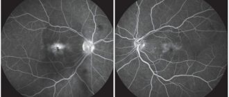
Diagnostics is carried out with the aim of making an accurate diagnosis and includes an initial examination and instrumental techniques. The ophthalmologist performs palpation, examines the patient, collects and analyzes information about him, and also evaluates visual acuity (during visometry using a table), clinical picture, possible causes (for example, recent injuries or past illnesses), and a complete medical history.
Laboratory and instrumental research methods are also used to prescribe correct treatment:
- magnetic resonance and computed tomography (MRI and CT) of the brain, cervical spine and orbital cavities;
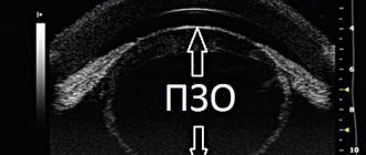
- ultrasound examination performed in B-mode (sonography);
- radiography;
- blood tests: general and clinical, for hormones and for some infections;
- exophthalmometry (measuring the size of the eyeballs with a special instrument).
Only after a comprehensive differentiated diagnosis as prescribed by a doctor can treatment begin.
Surgical treatment of anophthalmia
At an early age, surgical treatment is not performed, since external canthotomy does not cause an increase in the palpebral fissure, but its serious deformation. This is the reason for the impossibility of subsequent prosthetics. In this regard, surgical treatment is carried out only when prosthetics are completed. Most often, this manipulation is performed at 7-8 years of age.
For the purpose of primary correction, plastic surgery of the outer and inner corners, elimination of entropion of the eyelids, delayed plastic surgery of the stump, correction of lagophthalmos, formation of the fold of the upper eyelid and correction of the location of the eyelashes are performed. In case of anophthalmia, operations with transplantation of flaps of the mucous membrane of the lip or skin are not performed.
Treatment
Treatment is prescribed by a doctor and involves eliminating not only the external defect, but also the causes of the condition. Therapy methods are selected individually, taking into account the nature of the pathology, the nature of the course, the patient’s health and concomitant diseases. All treatment methods can be divided into conservative and radical. But in any case, with successful therapy, the prognosis is favorable.
Conservative treatment
Prescription of conservative treatment of enophthalmos is possible only with a slight displacement of the eyeball (no more than two millimeters), as well as in the absence of concomitant complex conditions, for example, interposition of extraocular muscles (their introduction into bone structures after fractures) and serious visual impairments, such as diplopia.
For conservative treatment, the following medications are prescribed:
- Glucocorticosteroids that relieve complex inflammation.
- Hormonal drugs that normalize hormone levels and the functioning of the endocrine system.
- Non-narcotic analgesics and non-steroidal anti-inflammatory drugs (NSAIDs) to eliminate severe pain.
- Saline hypotonic solutions are administered to eliminate tissue swelling.
- Antibiotics are prescribed for infections. Local preparations in drops are indicated for minor bacterial lesions of the tissues of the organs of vision. Broad-spectrum antibacterial drugs are prescribed for inflammation localized in the retrobulbar tissue, located in the muscular funnel between the optic chiasm and the orbital cavity.
- In case of severe general damage to the body, detoxification is sometimes recommended.
Diagnosis of enophthalmos
If the displacement of the eyeball is caused by injury, the doctor determines the degree of injury. He must establish the nature of the damage and the current clinical picture of the pathology. Visual acuity is also checked, and studies are carried out for the presence of inflammatory and infectious diseases. If primary ophthalmological examination methods do not provide a complete picture, methods such as:
- general blood analysis;
- test for human immunodeficiency virus (HIV) and hepatitis;
- Ultrasound of the eyes;
- computed tomography and MRI of the brain.
Based on all the data obtained, a decision is made on the treatment method. It can be conservative or surgical.
Radical treatment methods
Surgical treatment methods include the following:
- Injections of adipocyte suspensions. They are prescribed if enophthalmos manifests itself in the form of a decrease in the volume of fiber filling the retrobulbar space. The patient's own fat tissue, taken from the subcutaneous layer of fat of the abdominal anterior wall, is injected into this area. The risks of rejection and allergies are minimal.
- Surgical introduction of implants made of titanium, polymer compounds or dense silicone into the retrobulbar area.
- Reposition of bone fragments. This operation is performed in the treatment of post-traumatic enophthalmos. Access is provided in different ways: transciliary, transantral or transconjunctival. If the damage is minor, the intervention is carried out using endoscopic equipment in a minimally invasive manner.
Possible complications
In the absence of effective and timely treatment, complications may develop. First of all, there is a cosmetic defect that significantly spoils the appearance, provoking the development of complexes and a decrease in self-esteem.
In addition, if the treatment was incorrect or untimely, then enophthalmos may cause visual impairment. Due to disruptions in the trophism (nutrition) of the tissues of the oculomotor muscles, the progression and deterioration of the condition leads to weakening and complete atrophy of the muscle fibers. The eye simply stops moving and functioning normally, strabismus (strabismus), deformation of the eyelids, decreased visual acuity and miosis are observed.
Important information! Some lesions and pathological changes are irreversible, so treatment must be started as early as possible.
Preventive measures
Prevention includes the following measures:
- Compliance with safety rules while working in potentially hazardous industries (for example, at high altitudes, with constant contact with current or aggressive chemicals).
- Regular scheduled examinations.
- Timely treatment of any emerging diseases.
- Maintaining a healthy lifestyle with a balanced diet, optimal rest, moderate physical activity and no bad habits.
- Strengthening the immune system.
Enophthalmos, which you learned about on our website, is a pathological condition that spoils the appearance and negatively affects visual functions. Competent diagnosis and correct timely treatment will eliminate the problem and avoid consequences. Contact a qualified professional and follow his/her recommendations.