Structure of the eye: functions of the human eye
Human eyes are the most complex sensory organ in the human body. From muscles and tissues to nerves and blood vessels, each part of the human eye is responsible for a specific action. Moreover, contrary to popular belief, the eye is not perfectly spherical; instead, they are two separate segments joined together. It is composed of several muscles and tissues that together form a roughly spherical structure. From an anatomical point of view, the human eye can be broadly divided into external and internal structures.
Human eye
External structure of the eye
The parts of the eye that are visible from the outside include the following parts:
- Sclera: This is the visible white part. It is made up of dense connective tissue and protects the inner parts of the eye.
- Conjunctiva: lines the sclera and consists of stratified squamous epithelium. It keeps our eyes moist and clean and provides lubrication by secreting mucus and tears.
- Cornea: It is the clear front or front part of our eye that covers the pupil and iris. The main function is to refract light along with the lens.
- Iris: This is the pigmented, colored part of the eye visible from the outside. The main function of the aperture is to adjust the diameter of the pupil depending on the light source.
- Pupil: This is a small hole located in the center of the iris. It allows light to enter and focus on the retina.
Internal structure of the eye
Inner parts of the eye:
Lens: The clear, biconvex lens of the eye. The lens is attached to the ciliary body by ligaments. The lens, along with the cornea, refracts light so that it is focused on the retina.
- Retina: This is the innermost layer of the eye. It is light sensitive and acts like camera film. It contains three layers of nerve cells, these are ganglion cells, bipolar cells and photoreceptor cells. It converts images into electrical nerve impulses for visual perception by the brain.
- Optic Nerve: Located at the back of the eye. The optic nerves carry all nerve impulses from the retina to the human brain for perception.
- Aqueous fluid: This is the fluid found between the cornea and the lens. Nourishes the eyes and keeps them puffy. In other words, it is a lens.
- Vitreous: This is a clear, jelly-like substance found between the lens and the retina. It contains water (99%), collagen, proteins, etc. The main function of the vitreous body is to protect the eyes and maintain its spherical shape.
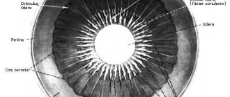
The lens is a thin elastic capsule consisting of anucleate cells and certain fibers. It plays the role of a biconvex lens.
Structure of the eye diagram of the structure of the human eye
The lens and its suspensory ligament divide the eyeball into two chambers. The anterior chamber is filled with liquid, aqueous humor, which is constantly renewed. The posterior chamber is filled with a transparent gelatinous substance, the vitreous or vitreous. The vitreous humor influences intraocular pressure and therefore the shape of the eye.
Middle layer of the eye, pathology, treatment
The iris determines the color of the eyes.
Eye diseases can appear at any time in our lives; the risk of their occurrence increases with age. To determine the extent of damage, eye pathology requires a high-quality, complete diagnosis and periodic preventive examinations.
The examination is used:
- ophthalmoscope;
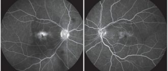
- Angiography determines the condition of the blood vessels and reveals damage to Bruch's membrane.
- ultrasound examination.
Pathology of the middle membrane of the eye
Changes in the middle shell can be congenital or acquired. A congenital pathology is the absence of the choroid in a certain area. Purchased items include:
- Dystrophic lesions of the choroid.
- Inflammation of the choroid can occur together with damage to the retina.
- Detachment of the membrane that appears when intraocular pressure increases, for example, during glaucoma surgery.
- Rupture and hemorrhage of the membrane due to eye injury.
- Nevus (mole or birthmark) of the choroid.
- Neoplasms of benign and malignant nature.
- Iridocyclitis is an inflammatory process in the iris and ciliary body.
Eye like a camera
If you compare the structure of the eyeball with a camera, then it can be defined as the most complex and most successful of the existing ones. In this highly complex organ, each element has its own role and meaning.
The human eye is the best camera in the world
Cornea: lens responsible for illumination
The cornea acts as a lens through which light enters the eye. It plays an important role in focusing light on the retina. In fact, the cornea should always be perfectly clean and transparent. Regular closing of the eyelids (blinking) and secretion of tears protects the surface of the cornea from contamination.
Lens: the role of “zoom”
The main purpose of the lens is to allow for the necessary adjustments to focus objects at all distances. This focusing occurs due to a change in curvature, which occurs either through tension or relaxation of the tendons that attach the lens to the inner wall of the eyeball. The lens bulges to focus near objects and becomes flatter when at rest to allow the eye to see distant objects clearly.
Iris: eye color
The iris is responsible for eye color. This color depends on the thickness of the layer that formed the pigment and the concentration of melanin in it. The thicker the iris and the more melanin, the darker the eyes. In general, the iris is thinner than silk. The iris is nourished by moisture, which washes it and some small arterioles.
Iris and pupil
Watery liquid
The aqueous humor is a clear, continuously filtered and renewed fluid that, together with the vitreous humor, maintains the pressure and shape of the eyeball. Consisting mainly of water, but also vitamin C, glucose, lactic acid, and proteins, it is completely renewed in 2–3 hours.
Pupil: eye diaphragm
The pupil is a structure consisting of the free space in the center of the iris. The latter includes two muscle groups:
- one, consisting of radiant fibers (arranged like the spokes of a wheel), dilates the pupil,
- the other containing round fibers that narrow it.
Their action changes the diameter of the pupil and regulates the amount of light entering the eye, just as the aperture of a camera determines the aperture diameter of a lens.
Retina: photographic film
The retina is a quarter-millimeter membrane located at the back of the eye, on the inner wall. Ultra-sensitive, it allows us to distinguish very weak light at a distance of up to 10 kilometers, even in complete darkness.
Retina of the eye under a microscope
The retina consists of hundreds of millions of photoreceptor nerve cells:
- Cones (about 6-7 million): These cells interpret the colors of an image by breaking it down into three primary colors: red, blue and green.
- rods (about 130 million): these cells analyze light.
The outer layer of the retina contains photoreceptors containing photosensitive pigment that responds to light by chemically modifying it to convert light energy into electrical energy.
This electrical signal is then transmitted to the brain through the ganglion cells found in the innermost layer. The visual information is then restored through a complex process that requires the help of other cells.
Optic nerve
It transmits information received by the eyes to the brain at the level of the visual cortex. Its diameter is 4 mm, length 5 cm. It is the optic nerve that allows the brain to record, interpret and translate images. Efferent nerve fibers exit the eye through the optic nerve. At this exit point, the retina is quite naturally interrupted: it is a blind spot (because it cannot detect light stimuli due to the lack of photoreceptors). Next to this blind spot is the macula (with the fovea), which is the point of the retina with the best visual acuity: here light rays enter directly with the least interference and it is here that the density of photoreceptors is greatest.
Diagram of the human visual cortex
Useful video
If you follow a daily routine, exercise, and a balanced diet, your eyes will acquire natural beauty, in which there is no need for an additional increase in the size characteristics of the eyeball.
Author's rating
Author of the article
Alexandrova O.M.
Articles written
2031
about the author
Was the article helpful?
Rate the material on a five-point scale!
( 1 ratings, average: 5.00 out of 5)
If you have any questions or want to share your opinion or experience, write a comment below.
Vision Explained: How the Human Eye Works
The way we see things is part of a complex process: different things happen in the eye and in the brain before we see anything. This process follows the retinocortical pathway, which begins in the eye and continues to our brain. In short, the way vision works is that the human eye absorbs ambient light and collects it on the cornea. The result is the first visual impression. Each eye then transmits this image to the brain via the optic nerve and converts it into what we call “vision.” Light is the source of everything we see. In complete darkness we are practically blind.
This means that a certain amount of light must illuminate an object for us to have even the slightest chance of seeing it. This light is then reflected by the object and processed by our visual system. If we look at a tree, our eyes absorb the light it reflects: the rays first pass through the conjunctiva and cornea, then through the anterior chamber and pupil. The light then reaches the lens, which absorbs it and transmits it to the retina, the photosensitive organ (= sensitive to light). The retina collects visual information and sorts it: the rods are responsible for night vision, the cones for daytime vision and colors. This information is transmitted to the optic nerve, which transmits it directly to the brain, where it is evaluated again.
Although we already have detailed information about the anatomy of the human eye and its structure, many questions remain regarding how our consciousness works. We know, for example, which areas of our brain are most active when we see something, but we don't actually know what worldview is.
Near and far vision
Healthy eyes adjust automatically, without any assistance, so that we can see near and far and perceive objects clearly, no matter how far away they are. This ability to see objects clearly at different distances is called accommodation. The elasticity of the lens is a key factor in this ability. In the absence of problems, the lens can change shape and adapt to near or distance vision, depending on what we want to see. Typically, the lens is flat and elongated - the ideal shape for distinguishing distant objects. But if we look at an object up close, it bulges out to allow us to see up close and allows us to see nearby objects.
Vision in very bright environments (light vision or day vision) is provided by sensory cells responsible for color vision: cones. The pupil is also involved in daytime vision: the more light there is, the more it contracts. The pupil adapts to different light intensities and regulates the amount of light entering the eye. This is called visual adaptation.
Now you know more about the structure of the eye, in order to remember this topic well - look again at the diagram of the structure of the eye and watch the video.
Alternative medicine
One of the methods of alternative medicine based on the structure of the eye is iridology. The diagram of the iris helps the doctor make a diagnosis for various diseases in the body:
This analysis is based on the assumption that different organs and areas of the human body correspond to certain areas on the iris. If an organ is sick, this is reflected in the corresponding area. These changes can be used to determine the diagnosis.
The importance of vision in our lives cannot be overestimated. In order for it to continue to serve us, we need to help it: wear glasses to correct vision, if required, and sunglasses in bright sunshine. It is important to understand that age-related changes occur over time, which can only be delayed by prevention.
Related articles:
- Structure, functions, diseases, cornea transplantation
- Binocular vision: definition, treatment, research
- Iris of the eye: structure, functions, diseases and features
- Lens of the eye: structure, functions, replacement surgery (price, consequences)
Brief questions on the topic
What are the external structures of the eye?
The external structures of the eye include:
- Sclera
- Conjunctiva
- Cornea
- Iris
- Pupil
What is the function of the conjunctiva?
The conjunctiva lubricates the anterior surface of the eye. It also protects your eyes from debris, dust and germs that cause infection.
What is the function of the iris? How many layers does it have?
The iris regulates the amount of light entering the eyes by controlling the diameter and size of the pupil. The iris consists of two layers:
- Stroma, anterior pigmented fibrovascular layer
- Pigmented epithelial cells.