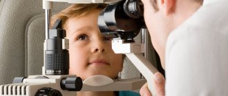
Keratotopography is a non-invasive diagnostic method aimed at determining the refractive power and radius of the cornea, as well as the refraction of the eye. Analysis of the research results is carried out by specialized software, after which the computer produces a printout in the form of a graph. This is a keratotopogram.
Carrying out diagnostics does not require much time and is indispensable before laser corneal correction surgery. Thanks to it, the ophthalmologist can plan the operation as accurately as possible and determine its volume.
You can undergo diagnostics at the Sfera ophthalmological clinic of Professor Eskina. We have a unique set of diagnostic equipment that allows us to conduct comprehensive studies for diagnostic purposes, as well as in preparation for laser correction. With us you are guaranteed to receive everything you need to restore your vision. You can find out the price of a keratotopogram in our clinic in the corresponding section of our official website or by calling us at +7 495 139‑09-81.
What is corneal keratotopography?
Keratotopography is a painless, non-contact diagnostic method. During the examination, eye contact with the equipment is excluded, which helps prevent injury and infection. The result of keratotopographic diagnosis is a color map of the ocular surface, on which healthy and pathologically altered areas of the cornea are marked. The importance of this study can be understood from the anatomy of the cornea.
It is the anterior, most convex part of the eyeball, which performs the important function of a light-refracting medium. Rays of light pass through it and are collected on the retina, forming a clear “picture”. Abnormal thickening of individual areas or the entire cornea, along with its irregular curvature, lead to disturbances in refractive power, which, in turn, cause deterioration in visual acuity.
Keratotopography of the cornea of the eye allows you to identify existing irregularities on the ocular surface, determine the direction of the visual meridians and the degree of their severity. Based on changes in the relief of the cornea, the ophthalmologist can accurately determine the causes of visual acuity disorders. Moreover: keratotopography is the only technique that allows us to identify keratoconus, in which the cornea takes on a cone shape.
Additional techniques
The area of the anterior chamber angle is not visible from the outside, as it is covered by the opaque tissue of the limbus. Therefore, a gonioscope is used to examine it. It is a contact lens, inside of which there is a mirror located at such an angle that it reflects the area of the anterior chamber angle. Before the examination, anesthetic drops are placed in your eyes. You are then seated at the slit lamp and asked to place your chin on the instrument stand. The doctor applies the gonioscope to the eye being examined and, slightly rotating the lens, examines the area of the anterior chamber angle through the eyepieces of the biomicroscope.
The technique is used in glaucoma to clarify the cause of impaired outflow of intraocular fluid, and in case of eye injury to exclude foreign bodies and damage to the structures of the anterior chamber angle.
Tonography
Tonography is a method for studying the hydrodynamics of the eye.
After preliminary instillation of anesthetic drops, a tonograph sensor is installed on the cornea of the eye being examined. Based on the results of an extended (4 minutes) measurement of intraocular pressure, the device calculates indicators of the hydrodynamics of the eye: the level of intraocular pressure, the coefficient of ease of outflow and the rate of production of intraocular fluid, and some others.
These data are used to clarify the cause of increased intraocular pressure in glaucoma, when choosing a treatment method and to monitor its effectiveness.
Keratotopography
Keratotopography is a method of mapping the optical power of the cornea.
During the test, a series of light rings are projected onto the cornea. Their reflections are recorded by a video camera, subjected to computer processing and converted into a color refractive map of the cornea.
The method is used for accurate diagnosis of astigmatism, keratoconus, and when planning laser vision correction.
Wavefront Analysis
Aberrometry (wavefront analysis) examines the propagation of light within the eye and records the distortions that occur. They are called aberrations and are manifested by visual impairment.
During the examination, which takes only a few seconds, you only need to place your chin on the device’s stand and fix your gaze on the luminous mark. A ray of light directed into your eye is reflected from the retina and returned back. By comparing the source and reflected light rays, the device creates a map of your eye's aberrations, sometimes called an optical fingerprint.
This data is used to create a vision correction program (LASIK, implantation of an artificial lens, selection of glasses or contact lenses).
Ultrasonography
Ultrasound examination (ultrasound) allows you to determine the size of the eyeball and its structures, identify pathologically altered areas and their location based on the characteristics of the propagation of ultrasound in the tissues of the eye.
The study is carried out by contact or immersion method. In the first case, after preliminary instillation of anesthetic drops, the doctor touches your eye with an ultrasound probe. In the second case, the examination is carried out through closed eyelids after applying a special gel to them, or a nozzle containing an immersion liquid in which a diagnostic probe “floats” is installed on the eye. Ultrasound aimed at your eye is reflected from certain structures and recorded by the device's sensor. Echoes form an image of a cross section of the eyeball.
The method is irreplaceable for diagnosing foreign bodies, tumors, retinal detachment, and for determining the size of the eyeball and its structures.
Optical coherence tomography
Optical coherence tomography (OCT, OCT) allows you to image the microscopic structure of the eye in a cross section. The method is based on the analysis of a beam of coherent light reflected from various structures of the eye.
During the examination, you only need to fix your gaze on the luminous mark in the camera lens for 2-3 seconds. The visible picture and the scanning direction using a video camera are displayed on the monitor. The computer generates an image of a cross section of the area under study (cornea, anterior chamber angle, retina, optic nerve head), determines the thickness of the tissue and its optical density.
This information is especially valuable in cases of corneal opacities, the formation of strands in the vitreous body, macular holes, age-related macular degeneration, and macular edema.
Indications and contraindications for corneal keratotopography
| Indications | Contraindications |
| The procedure has no contraindications for the eyes and can be performed even if the patient has ophthalmological diseases of various natures. However, it is not carried out if the patient is under the influence of alcohol or drugs, or if he suffers from severe mental disorders. |
Indications for the use of keratotopography of the eye
This examination method is prescribed by an ophthalmologist for the purpose of:
- early diagnosis of diseases of the cornea such as keratoconus, keratoglobus, epithelial dystrophy and other pathologies of the epithelial tissue of the eye;
- quantitative assessment of astigmatism and various corneal deformations when wearing contact lenses;
- obtaining information about the condition of the horny tissue before or after surgery, keratoplasty. Before laser vision correction, the doctor must prescribe this examination to exclude keratoconus;
- choosing lenses and assessing the effectiveness of wearing them.
Keratotopography allows the doctor to obtain an objective picture of the condition of the cornea of the eye. Using this method, it is possible to diagnose various diseases of the visual organs at an early stage and begin treatment on time, thereby increasing the chance of a full recovery.
What does a keratotopogram show?
A diagnostic study is carried out using an ophthalmological apparatus - a keratograph. Its design provides for the presence of recording rings, which are designed to record the results obtained from 8 thousand positions, by which the condition of the cornea is assessed.
Based on these data, a special computer program constructs a topographic map on which you can see:
- areas of red or yellow color, displaying thickening of the cornea while maintaining its integrity and relief above normal;
- areas of purple and blue color, showing thinned areas with relief below normal.
Thus, the keratotopogram provides the ophthalmologist with all the data necessary to:
- make an accurate diagnosis;
- evaluate treatment results;
- determine the scope of the upcoming laser correction.
Based on these data, the curvature of the cornea, the power of refraction, and the degree of distortion of the visual meridians are identified.
What is the keratotopography method?
Keratotopography of the eye
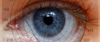
Keratotopography of the eye is an ophthalmological procedure that allows the doctor to study the condition of the patient’s cornea and its sphericity. The name of the method, translated from Greek, literally means “determining the geometry of the horny tissue of the eye.”
With the help of this study, it is possible to determine pathological processes in the cornea of the eyes and, if necessary, to competently plan the extent of the operation to improve vision.
The topographic map obtained during the examination contains information about the irregularities of the cornea and the radius of its curvature. This method began in the 80s of the 20th century by the Portuguese Antonio Placido. The ophthalmologist was able to create a special device for diagnosing pathologies of the surface of the corneal tissue of the eye, which was called the Placido disc.
The device looked like a flat disk in which white and black rings alternated. During diagnostics, a bright lighting element was installed behind the subject's head.
The rays emanating from the light source were reflected from the disk and displayed on the cornea. Using a device in the center of the disk, the specialist could study the image obtained on the cornea.
Currently, keratotopography of the eye is carried out using computer technology. A special laser scans the surface of the eye. The computer processes the received data and produces the finished result in the form of color topographic maps, on which normal and abnormal areas are indicated in different colors.
The video will tell you what keratotopography and keratotopogram are:
How is keratotopography performed?
This diagnostic procedure is simple and does not require special preparation from the patient. The use of anesthetic drops before the procedure is not necessary, since it is not only painless, but also non-contact. Despite the difference in the types of keratotopographic systems used, the scheme for performing a diagnostic study does not differ.
Before starting the study, the patient needs to remove contact lenses or glasses, as well as remove decorative cosmetics from the face. He will be asked to sit in front of the keratograph and fix his head on its stand. During the scan, which lasts several minutes, head and eye movements must be eliminated in order for the study results to be as accurate as possible.
There are various types of keratotopographic systems, each of which has its own advantages and disadvantages. One of the most modern and accurate systems is based on the use of the Hartmann-Shack wavefront sensor. It allows you to analyze light waves at ten different points, which allows you to achieve the highest quality data obtained.
At the Sfera clinic we use just such a keratotopograph: “HRK-7000”, which allows you to obtain data even on the periphery of the eye, which, in turn, allows you to achieve the desired accuracy when selecting contact lenses.
Interpretation of keratotopography results
Displaying data on a keratotopogram is carried out in different forms.
| In what form is the data displayed? | What does the information received say? |
| Numbers | The resulting diagram contains information about the corneal curvature in diopters (D) in ten concentric areas in 1 mm increments. |
| Profile view | The resulting image includes the flattest and steepest meridians of the cornea, and the difference between them in diopters (D). |
| Photokeratoscopy | The appearance of Placido rings on the cornea reveals the location of the apex of keratoconus. |
| Keratometry | Allows you to identify the symmetry of astigmatism and determines the decision on the need for laser correction. |
Capabilities of the PENTACAM device
Let us list the capabilities of this unique device:
- Scheimpflug images allow for excellent clear visualization of the structures of the anterior segment of the eyeball (from the anterior surface of the cornea to the posterior surface of the lens).
- Pachymetry involves analyzing the thickness of the cornea at any point from limbus to limbus, with the data displayed in the form of digital values and color pachymetric maps.
- Corneal topography allows specialists to analyze the anterior and posterior surfaces of the cornea. Data is displayed in the form of sagittal, tangential and elevation maps. The results of corneal wavefront analysis are presented as Zernike polynomials.
- Analysis of the state of the lens and densitometry. PENTACAM allows you to perform densitometry of the lens at any point with a quantitative assessment of optical density parameters on a 100-point scale.
- Index report. The main parameters of the anterior segment of the eye are automatically correlated with data from reference databases for a normal population with typical pathologies.
- Screening method for the presence or absence of keratoconus using the Belin-Ambrosio method. During the study, the analysis of corneal ectasia is carried out on the basis of optimized topography maps of the anterior and posterior surfaces of the cornea, the progression of pachymetric changes, the determination of the thinnest point of the cornea and the value of pachymetry in it are taken into account. The obtained data are correlated with the regulatory framework for patients with refractive pathologies and are displayed in the form of coefficients of deviation from normal values.
The cost of a screening study of the curvature and structure of the anterior and posterior segments of the cornea and wavefront analysis using a Scheimpflug camera on a PENTACAM device (Oculus, Germany) starts from 1850 rubles. You can obtain detailed information about this study from our consultants.
Advantages of keratotopography at the Sfera clinic
Diagnostics in this area in our clinic are carried out using a modern autorefractometer “HRK-7000”, manufactured at the production facilities of the South Korean company. This is a modern device that allows for a full analysis of possible violations of the optical system of the eyes, having a highly sensitive matrix and the “Super Luminescent Diode” system, which guarantee image clarity. Moreover: it is equipped with a special noise reduction system, which allows for the most accurate diagnostic studies even with pathologies such as cataracts, ametropia, and if the patient has an IOL.
Undoubtedly, even the most high-tech equipment will be useless without an experienced specialist who knows how to handle it... But the ophthalmologists of the Sfera clinic have been certified and work in accordance with international standards. If you want to be sure that the diagnosis will be carried out in the best possible way, contact us!
In order to get an appointment with our specialists, use the online form on our website or call us: +7 495 139‑09-81.

