Farsightedness and nearsightedness are the most common visual impairments in humans. The main cause of these diseases is the anatomical features of the visual organs, which differ in myopia and hypermetropia. What is the structure of our eye in these cases and what does it depend on? We'll tell you in more detail in the article.
In this article
- Differences between myopia and farsightedness
- The structure of the eye with farsightedness
- The structure of the eyes in myopia
- Eyes with simultaneous myopia and farsightedness
- Treatment of myopia and hypermetropia
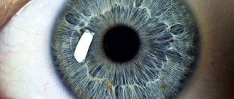
The human eye is a complex optical system. Our eyeball is shaped like a ball with a normal diameter of approximately 23-25 mm. Light reflected from surrounding objects enters the eye, passes through the cornea and lens, and is projected onto the retina. Light-sensitive cells located on it process information and transmit it to certain areas of the brain along the optic nerve.
The lens, a natural biconvex lens, is responsible for accurately focusing light on the retina; with the help of the ciliary muscle, it is able to change its curvature. When looking at distant objects it becomes flattened, and when seen close it becomes more convex and refracts light more strongly. This property of the lens to change the refractive power, as well as the focal point of the eye, is called accommodation.
When seeing at long or near distances, the size of the eyeball itself also changes; special muscles are responsible for this. To view an object close up, the eye stretches a little, and, conversely, it rounds when looking at distant objects. In the presence of pathologies in the organs of vision, light rays can be focused behind the retina, which causes farsightedness, or in front of it, which leads to myopia. Let's take a closer look at the structure of the eye in these two diseases.
Differences between myopia and farsightedness
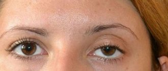
With farsightedness, a person sees objects located nearby blurry, it is difficult for him to read text and work with small details, but he clearly and clearly distinguishes objects at long distances. With myopia, on the contrary, near vision is characterized by high quality, but objects at a distance become blurred.
Myopia and farsightedness also differ in that myopia is most often caused by a genetic predisposition and manifests itself in early childhood, while pathological hyperopia (as opposed to physiological hyperopia, inherent in all people at birth) usually begins to develop after 40-45 years (age-related farsightedness) . This is an inevitable process for all people.
Lens pathologies
Diseases in the area of the human biological lens can be congenital or acquired. The second form is most common, and progresses as a person ages. There are the following types of pathology:
- mechanical damage, capsule rupture;
- disruption of formation during embryogenesis;
- change in location in the eyeball;
- color change;
- a change in the composition of the internal fluid, which becomes cloudy, causing cataracts.
Since this area is not supplied with blood vessels, there is no nervous regulation, the formation of an inflammatory process is impossible.
The most common disease of the lens is cataract. It can be congenital and acquired. If the disease is at an early stage, it can be controlled using conservative treatments. Drops are used that eliminate insoluble proteins within the liquid. If there is excessive clouding, surgery is used.
The structure of the eye with farsightedness
With this pathology, the optical focus is not exactly on the retina, but behind it. There may be several reasons for this:
- shortened size of the eyeball. Normally in humans, this organ has a diameter of 23-25 mm. If its size is too small (19-22 mm), the focus “goes” behind the eye, bypassing the retina;
- the cornea is too flat and has low refractive power;
- displacement of the lens forward, which leads to improper focusing of light rays. He is forced to constantly strain to concentrate on an object nearby;
- abnormalities of the lens: microphakia (too small), aphakia (complete absence of the lens), or the placement of this natural lens in the wrong place (displacement).
Physiological farsightedness is inherent in all people at birth. The baby is born with a slight degree of hypermetropia of approximately 2-4 diopters. This is explained by the fact that the visual organs of a newborn are not yet fully developed, and the size of the eyeball is only 17-18 mm. As a child's body grows, so do the eyes. Normally, by the first year of life, the degree of farsightedness should be no more than 2.5 diopters, gradually decreasing, and, in the absence of pathologies, hyperopia should disappear by the age of 14.
Farsightedness is much more difficult to recognize than myopia, especially with weak and moderate degrees. In fact, our eyes themselves fight hyperopia by constantly straining the ciliary muscle, which allows a person to see objects equally well at different distances. But by the age of 40-45, when the muscle weakens due to age and is not able to work at full strength, presbyopia, also called senile farsightedness, appears. At the same time, people suffering from small degrees of myopia have more advantages - negative diopters are compensated by positive diopters, and near visibility even improves slightly. Those who previously had normal vision begin to wear glasses or lenses with a “plus” sign.
The main changes in the eyes with presbyopia occur in the lens. Its age-related degeneration begins: it becomes inelastic, the core becomes denser, and accommodation decreases. As a result of such transformations, the lens loses the ability to increase the radius of curvature when viewing closely located objects, and they have to be moved further and further from the eyes.
With severe degrees of farsightedness, blurred vision is diagnosed both near and at very far distances, and with this form of hyperopia there is a risk of developing glaucoma.
Too short an axis or forward displacement of the lens can lead to partial blockage of the drainage pathways through which intraocular fluid is drained, which increases pressure in the eyeball and increases the risk of glaucoma.
Features of visual perception
First of all, the optical system of the eye is designed to obtain information about the world around us through vision. This concept has many characteristics and features.
The sense of light allows the human eye to perceive daylight and artificial light, as well as to distinguish the degree of its intensity. And thanks to the natural adaptation of the eyeball, the optical system is able to independently adapt to illumination of varying brightness without outside help. Light sensitivity is determined by the natural threshold of light stimuli. Few people know that a person with good eyesight can see even a small light at a distance of several kilometers.
The sensitivity of the visual apparatus primarily depends on many factors, such as the intensity of the light source, its angular size and wavelength, as well as the time that the light stimulus acts on the eye. Due to the deterioration of the optical characteristics of the sclera with age, the sensitivity of the eyeball can greatly decrease.
The structure of the eyes in myopia
Unlike farsightedness, with myopia, on the contrary, the eyeball has an increased size, and there are two types of myopia. If the ocular axis is elongated - the distance from the edge of the cornea to the retina, then such myopia is called axial. If the cornea has an overly convex shape, then light rays are refracted too much, and this type is called refractive myopia. Usually they are combined with each other.
Myopia poses a greater risk to eye health than farsightedness. This disease begins to develop, as a rule, with the beginning of school, when the child’s visual load increases sharply. At the same time, his body is growing rapidly, all organs, including the eyes, increase in size. Too sharp growth along the anteroposterior axis may be accompanied by disturbances: the retina is stretched due to an enlargement of the eyeball, and this can lead to its detachment or rupture. During this period, it is important for parents to pay attention to the state of the child’s vision and, if there are alarming symptoms, contact an ophthalmologist. Successful correction and treatment of myopia depends on timely diagnosis.
In the presence of this pathology, it was previously forbidden to give birth naturally, since at the time of birth intraocular and blood pressure increases greatly, and the eyes experience great tension, which often leads to rupture or detachment of the retina. Nowadays, pregnant women with high degrees of myopia undergo laser coagulation of the retina, which allows it to be strengthened and firmly connected to the choroid, so there is virtually no risk of damage.
In childhood, the only eye surgery allowed to stop progressive myopia is called scleroplasty. A small strip of biological tissue is attached behind the eyeball, which strengthens the sclera and prevents it from stretching. However, no method provides an absolute guarantee of stopping the development of myopia.
Symptoms associated with myopia
The most common symptoms that may raise suspicion of a myopia condition are:
- Vision deteriorates in relation to distant objects, and close ones become sharp.
- Recurrent headaches caused by tension in the eye muscles trying to correct focus.
- Frequently squinting the eyes in an attempt to improve image clarity.
- Difficulty with night vision , but day vision is normal.
- Injuries to the fundus and retina caused by excessive strain.
Eyes with simultaneous myopia and farsightedness
It also happens that a person has myopia and hypermetropia at the same time. This may occur due to the following factors:
- curved shape of the cornea;
- presbyopia;
- presence of astigmatism;
- disorders in the visual center of the brain and others.
In the case of presbyopia, the elasticity of the eye lens decreases and its ability to accommodate decreases. With the development of age-related farsightedness against the background of slight myopia, this happens unnoticed by a person, but with high degrees of myopia, one has to wear either two pairs of glasses or complex multifocal contact lenses, since vision is blurred at different distances.
Astigmatism can be myopic, hyperopic and mixed, when a person has myopia and farsightedness. Most often, they occur in different eyes, but in case of complications, these defects can be observed simultaneously in one of them.
With astigmatism, the eyes quickly get tired, as they are under constant tension. It is best to get rid of it with the help of microsurgical operations that will restore clarity of vision near and far.
Prevention of myopia
If myopia is an anatomical feature, it cannot be prevented, but it is quite possible to avoid progression or significant deterioration of visual characteristics. Here are some recommendations.
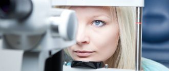
- Visit your ophthalmologist regularly. This is especially important for children, when the risk of developing myopia is high.
- Organize proper workplace lighting.
- Maintain a visual exercise regime, take breaks when working at the computer for long periods of time, reading and other visual work.
- Visual gymnastics and special exercises for the eyes for myopia are good for relieving tension.
- Move more, walk in the fresh air.
- Watch your diet: your diet should contain proteins, vitamins and microelements, such as Zn, Mn, Cu, Cr and others.
Treatment of myopia and hypermetropia
There are several ways to restore good vision by changing the surface of the cornea or replacing the lens in the eye with an artificial one - an intraocular lens. This procedure is called a lensectomy. LASIK and LASEK operations, which are performed using an excimer laser, make it possible to shape the cornea in such a way that light rays passing through it will be focused precisely on the retina. Such operations are performed in clinics around the world using modern ophthalmological equipment and guarantee high clarity of vision for many years.
Thus, modern medicine is able to restore good vision even with the most complex visual impairments; it is only important to diagnose the pathology in time and seek help from a specialist.
Main functions
In a person with normal vision, the lens performs many functions:
- The passage of a ray of light from the cornea to the retina.
- Refraction. The beam of light entering the eyes has a large volume. The cornea and lens refract it so that the projection on the retina appears in the form of a thin beam.
- Change in the shape of the lens (from convex to stretched, in the form of a lens). This action is carried out using the ciliary muscles. When the ciliary muscles are tensed or relaxed, the shape of the lens changes. Accommodation occurs quickly, so a person can move his vision from near and far objects, and vice versa.
- Barrier function. The lens is a kind of barrier to the protrusion of the vitreous body forward. It retains its structure and is located in its anatomical location.
- Protective function. If pathogenic microorganisms have penetrated the internal structures of the eye, they will first stop in the area of the lens. It prevents the passage of bacteria and viruses into the vitreous body and retina. This action is carried out temporarily; in the absence of therapeutic measures, the protective power of the lens may not withstand it.
The biological lens performs many functions that make vision possible. But if clouding of the internal structures has formed, the doctor may advise replacing the formation with an artificial model, which will not be inferior in function to a real lens.