The eyes are friction organs that perceive not only surrounding objects, but also color.
This function is performed by the retina, which contains nerve endings that transmit information to the brain.
If any structures along the path of light transmission are disrupted, a person stops perceiving 1, 2 or completely all colors. The disorder can be congenital or acquired. For diagnosis, consult an ophthalmologist, conduct research, and use treatment if possible.
Causes of color blindness
The most important cause of impaired color perception is a hereditary factor. The pathological gene is inherited from parents to offspring. If the cause of the disease is color blindness, the pathological gene is associated with the X chromosome, which manifests itself only when interacting with the Y chromosome. This means that women can be carriers, but only boys will be affected.
In a healthy person, cones are located on the retina of the eye. They perceive colors: blue, yellow, red. A hereditary disease can cause the absence of one of them, or several at once. In the latter case, the person will have black and white vision with the appearance of some color shades.
A person may be exposed to damaging factors that result in acquired color vision impairments:
- infectious and inflammatory diseases of the brain that affect the center of vision;
- mechanical damage to the brain or skull, which leads to damage to the center of vision;
- the growth of benign and malignant tumors, which with their edges compress the center of vision;
- inflammatory diseases of the optic nerve;
- infectious and inflammatory eye diseases that spread throughout the internal structure and spread to the retina;
- retinal hemorrhage;
- a complication of diabetes mellitus, as a result of which conglomerates of carbohydrates and fats form in the blood, which clog the microcirculation vessels of the eyes;
- narrowing and damage to the blood vessels of the eye microcirculation, resulting in reduced nutrition of the retina;
- mechanical damage to the eyes, as a result of which the retina, lens, and vitreous body suffer;
- increased intraocular pressure, which occurs due to an increased volume of intraocular fluid, as a result of which the chambers of the eyes increase in size, compressing the retina and optic nerve;
- increased exposure to ultraviolet radiation, which damages the retina;
- clouding of the lens, as a result of which the light beam passes insufficiently to the retina, reducing the perception of surrounding objects.
The acquired condition is characterized by the fact that when the damaging factor is eliminated, the color perception function is completely restored. That is why the doctor must identify the root cause in order to tell the patient about the prognosis of the disease and possible methods of therapy.
Study of color perception (colorimetry)
The patient is seated with his back to the window and asked to keep his head straight. The doctor picks up Rabkin’s polychromatic table (ed. VIII or IX) and places it strictly vertically at eye level of the subject, 0.5-1.0 m from him. Each table test is demonstrated for 5-10 s. If errors are detected in reading the tests presented, their numbers are recorded, and then a protocol card is drawn up according to the sample available in the table.
In accordance with the Kris-Nagel-Rabkin classification, the following types of color perception are distinguished: normal and abnormal trichromasia, dichromasia (in the form of protanopia, deuteranopia or tritanopia) and monochromasia (achromasia). These provisions should be used to guide the assessment of the results of the study performed using the methodology described above.
Since 1986, Rabkin’s polychromatic tables, now discontinued, began to be replaced by new spectral colorimetric tables of Yustova-Alekseeva*. They serve to determine the color discrimination thresholds of the subjects in three primary colors (red, green and blue) and consist of 12 cards measuring 130 x 130 mm.
* Their introduction into clinical practice was facilitated by many years of research by V.V. Volkov, V.A. Roslyakov and V.P. Sergeeva.
Each of them is filled with squares (9x9 mm) of two colors to be compared. Moreover, each specific pair of colors always has two equivalent color coordinates (in terms of lightness and saturation) in the physiological RGB* system. They differ thresholdly only in color.
Squares of one color are arranged in such a way that they form a “U”-shaped test figure, and squares of another color create a “camouflage” background around it. The results of the study are expressed by the maximum number of unperceived thresholds, i.e. discretely changing, color differences between the test and its background. For receivers R and G there are 4 of them (5, 10, 20 and 30), for B - three (5, 10 and 15). The corresponding numbers are indicated on the back of each card. They characterize one of the three possible degrees of color weakness of the test subject for each color receptor of the retina (tests with a number of thresholds equal to 30 are used to identify color blindness). Hence the emergence of a new idea about the existence of three types of color deficiency - proto-deficiency, deuterodeficiency and tritodeficiency of three different degrees.
In terms of classification, color weakness occupies an intermediate position between normal trichromasia and dichromasia.
In newborns and children in the first months of life, it is practically impossible to determine the state of color perception. To identify gross violations in this area, children ~1.5 years and older are shown color pictures and try to determine whether they correctly distinguish between primary colors, in particular red and green. However, it must be borne in mind that individuals with serious color pathology usually have pronounced deviations in the acuity of central vision. Abnormal trichromasia cannot be detected using this research method.
* Specified in case of failure to distinguish any one test (for example, number 1, 5 or 9) or simultaneously several of them within the same color receiver * Abbreviated name for the colors - red (R), green (G) and blue (B).
Symptoms of color vision disorder
If a person has a color perception disorder, a type of disease in which only some shades are changed, he may not be aware of it. Sometimes a certain color is replaced by another. For example, colorblind people may see green instead of red.
Parents may not immediately notice the disease in their children. When they are just learning to speak, they express the opinions they hear from their parents. Only when they grow up do they develop their own opinions. Then parents begin to notice that children see red grass instead of green, and the sun turns blue.
If the disease is hereditary, there will be no other symptoms other than color vision disorders. If the cause is a primary disease, it is supplemented by characteristic clinical symptoms. Most often it is neuralgia.
Color vision disorders - symptoms, treatment, tests
Contents
hide
1 Color vision disorders - symptoms, treatment, tests
2 Color vision disorders - color blindness
3 Color vision disorders - features of birth defects
4 Color vision disorders - achromatopsia
5 Color vision disorders - protanopia, deuteranopia and tritanopia
6 Color vision disorders - acquired diseases
7 Color vision disorders - causes
8 Distinctive colors - tests
9 Color vision disorders - difficulties in life
10 Color vision disorders - treatment
11 Color vision disorders - chromosomes in hereditary diseases
12 Risk of having a child with color blindness 12.1 Bibliography 12.1.1 Related entries:
The most common color vision disorder is color blindness, an inherited disorder. In this case, there is a problem with distinguishing between red and green. We can divide defects into congenital and acquired. What is the difference? Learn the causes, symptoms and treatments for color vision problems.
The ability to see colors is due to the presence of photoreceptors in the visual epithelium of the retina, which is one of ten layers of this complex structure. The receptors mentioned are rods and suppositories. The latter are responsible for color vision. They contain visual pigments - incl. rhodopsin and cyanopsin, the chemical activity of which converts light energy into the corresponding nerve impulse. There are essentially 3 types of cones, and each is sensitive to a different wavelength in the visible spectrum of light. Some are sensitive to red, others to green, and the third group of suppositories is responsible for the perception of blue. The colors mentioned are called primary.
By summing them up correctly, you can get the impression of white (equal proportions of individual colors) and about 160 shades of colors in the visible light spectrum (when mixing primary colors in different proportions). The normal human eye is capable of perceiving 3 primary colors. This condition is called trichromatopsia.
Classification of color vision disorders
To make a diagnosis, a doctor must classify the type of color vision disorder in a child or adult. Most often, the patient does not perceive only one color. But there are times when a person sees the world around him in black and white.
There are several types of color blindness:
- Protanopia. When a person has the disease, there are no cones on the retina that perceive red colors. The pathology is quite rare and occurs more often in men. In such patients, the difference between red, orange, and yellow colors is reduced.
- Deuteranopia. If the patient has the disease, there are no cones on the retina that perceive a green tint. The disease belongs to a rare category that most often occurs in men.
- Tritanopia. A disease in which the perception of many colors is impaired. This is a fairly rare disease that occurs in both men and women. When sick, a person does not perceive short light waves. Red and green colors are overly dull for him, almost gray. Patients see the violet shade as red.
- Lack of perception of any colors. Pathology refers to both congenital and acquired forms. A person does not perceive all shades; surrounding objects are seen in black and white colors.
- Retinal damage. Only acquired diseases fall into this category. The reasons may be neurological conditions, retinal nutritional disorders.
The main difference between congenital and acquired defects is in treatment. The first type cannot be treated. The patient will need to learn to live with color vision impairment. Acquired defects are completely eliminated after treatment, if possible.
Types of color vision disorders
The perception of shades by the visual apparatus depends on the length of the spectrum emitted by the color:
- yellow and green shades belong to the mid-wave range;
- red and orange color spectrums emit long wavelengths;
- cyan, indigo and violet colors are characterized by short wavelengths.
Depending on the degree of sensitivity to color, there are:
- Trichromasia. Abnormal shade perception, in which the patient cannot distinguish colors.
- Dichromasia. Partial cone disorder in which one or more shades cannot be distinguished.
- Monochromacy. The world around us is perceived in shades of gray, black or white.
According to the inability to distinguish a certain color, the following types of color perception are divided:
- Protanopia. The male category of the population is affected. It is quite rare. In pathology, the red-green spectrum of colors suffers. Patients with protanopia can learn to distinguish shades according to the brightness of an object.
- Deuteranopia. The condition is based on the complete absence of green spectrum cones in the visual apparatus. The patient cannot distinguish between red, green and yellow shades.
- Tritanopia. The disorder occurs in both men and women. In pathology, short-wave shades are not perceived.
Depending on the reasons for the formation of violations, several types are distinguished:
- Congenital. Passed from parents to children through genetic inheritance. According to statistics, it occurs in 8% of the male population and only about 1% in the female population. Anomalous trichromasia is the most common type of color disorder caused by heredity. The pathological condition always affects both eyes. It is determined in children during the first year of life.
- Acquired. Impaired perception of shades develops against the background of retinal diseases, optic nerve dysfunction, or disorders in the central nervous system. Both or one organ of vision is affected.
Acquired color disorders are divided into types according to immunity to a certain shade:
- Erythropsia. Patients see everything in a red hue.
- Chloropsia. People with a pathological disorder perceive everything as green.
- Cyanopsia. Patients perceive the world around them in shades of blue.
- Xanthopsia. The entire field of view is yellow.
Diagnostics
To diagnose the patient’s condition, the doctor uses several ophthalmological techniques:
- Anamnesis collection. This is data obtained from the words of the patient or his close relatives.
- General inspection. If the disease is hereditary, there will be no additional clinical symptoms. In acquired conditions, changes in the surface structures of the eyeball and neurological symptoms may occur.
- Application of color tables. They depict dots of different shades. In the center, these points form a figure (number, letter). If the patient does not recognize the figure or confuses the shade, the doctor diagnoses color vision impairment.
- The use of specialized equipment that delivers sources of different colors into the patient's eyes, which he must identify.
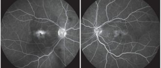
- Fundus examination. First, a solution is instilled onto the mucous membrane, which temporarily disrupts the accommodation of the pupil. The doctor assesses the condition of the chambers of the eye, lens, vitreous body, retina, and microcirculation vessels. Based on the study, it can be assumed that there is an acquired color vision disorder.
- MRI, brain and eyeballs. Organs appear on the monitor simply by chance. Using the technique, the doctor can identify blockage of blood vessels, inflammatory state of nervous tissue, damage, stroke. All these factors can contribute to the development of color vision impairment.
Based on diagnostic methods, the doctor will make a reliable diagnosis. Only after this is treatment prescribed.
Risk of having a child with color blindness
As you can see from the examples above, the risk of having a boy with color blindness is much greater than the risk of having a girl with the disease. In most cases, girls inherit one or two dominant (“healthy”) alleles and therefore do not have color vision impairment.
Bibliography
- Nizhankovskaya M.Kh., Ophthalmology. Clinical base, PZWL Medical Publishing, Warsaw 2007
- Trzcińska-Dąbrowska Z., Practical ophthalmology, PZWL Medical Publishing, Warsaw, 1995.
- Kański JJ, Clinical Ophthalmology – Compendium, Medical Publishing House Urban & Partner, Wroclaw, 2006.
- Orlovsky V.Yu., Modern ophthalmology, Państwowy Zakład Wydawnictw Lekarskich, Warsaw 1986
Vorobyova Marina
Neurologist of the highest qualification category (work experience 14 years), doctor of neurofunctional diagnostics (work experience 12 years); author of scientific publications on vertebroneurology; participant of scientific conferences on neurology and functional diagnostics of all-Russian and international significance.
Treatment
At the moment, no specific treatment has been developed for patients suffering from hereditary color vision impairment. A person needs to learn to live without distinguishing certain shades.
If a patient is diagnosed with an acquired form of the disorder, treatment procedures will depend on the underlying cause. The most common therapeutic agents are:
- anti-inflammatory drugs;
- antibacterial agents;
- drugs that promote blood resorption;
- adjusting insulin dosage;
- chemotherapy;
- surgical methods for the treatment of benign and malignant tumors;
- surgical methods to repair damage to nerve tissue;
- drops that promote the resorption of protein inside the lens;
- if the cataract is at a severe stage, the lens is removed and replaced with an artificial model;
- drops that reduce intraocular pressure due to the outflow of fluid.
If color vision is impaired, patients' standard of living decreases. If the condition is hereditary, there is no cure. If an acquired form is detected, it is recommended to carry out urgent treatment, since at a late stage of the disease it is impossible to restore impaired color perception.