Retinal rods
These photoreceptors are cylindrical in shape, with a length of approximately 0.06 mm and a diameter of approximately 0.002 mm.
Thus, such a cylinder is really quite similar to a stick. The eye of a healthy person contains approximately 115-120 million rods. The human eye rod can be divided into 4 segmental zones:
1 - Outer segmental zone (includes membranous discs containing rhodopsin), 2 - Connecting segmental zone (cilium), 3 - Inner segmental zone (includes mitochondria), 4 - Basal segmental zone (nerve junction).
Rods are highly photosensitive. So, for their reaction, the energy of 1 photon (the smallest, elementary particle of light) is enough. This fact is very important for night vision, which allows you to see in low light.
Rods cannot distinguish colors; this is primarily due to the presence of only one pigment in them - rhodopsin. The pigment rhodopsin, otherwise called visual purple, due to the included protein groups (chromophores and opsins), has 2 light absorption maxima. True, one of the maxima exists beyond the range of light visible to the human eye (278 nm - the region of ultraviolet radiation), therefore, it is probably worth calling it the wave absorption maximum. But the second maximum is visible to the eye - it exists at around 498 nm, located on the border of the green and blue color spectrum.
It is reliably known that rhodopsin, present in rods, reacts to light much more slowly than iodopsin, contained in cones. Therefore, rods are characterized by a weak reaction to the dynamics of light fluxes, and in addition, they poorly distinguish the movements of objects. And visual acuity is not their prerogative.
Cones
Peripheral processes are conditionally conical in shape. This type of cell converts light signals into nerve impulses. The cones contain the pigment iodopsin, consisting of chlorolab, which reacts to the yellow-green part of the spectrum, and erythrolab, which reacts to the yellow-red part of the spectrum.
Cones are smaller in size than rods - their length is ~0 µm, and their diameter is 2-4 µm. Cones perceive light several orders of magnitude weaker than rods, but respond better to rapid movements.
The structure of the cone includes
- outer segment with constantly renewed and emerging membrane half-discs;
- communication department;
- internal segment (includes the nucleus, mitochondria and polyribosomes);
- synaptic region that forms synapses with bipolar cells.
Cones of the retina
These photoreceptors also got their name due to their characteristic shape, similar to the shape of laboratory flasks. The length of the cone is approximately 0.05 mm, its diameter at the narrowest point is approximately 0.001 mm, and at the widest point - 0.004. The retina of a healthy adult contains about 7 million cones.
Cones have less sensitivity to light. That is, to excite their activity, a luminous flux will be required, which is tens of times more intense than to excite the work of rods. But cones process light fluxes much more intensely than rods, so they perceive their changes better (for example, they better distinguish light when objects move, in dynamics relative to the eye). They also define images more clearly.
The cones of the human eye also include 4 segmental zones:
1 - Outer segmental zone (includes membrane disks containing iodopsin), 2 - Connecting segmental zone (constriction), 3 - Inner segmental zone (includes mitochondria), 4 - Synaptic connection zone or basal segment.
The reason for the above-described properties of cones is the content of the specific pigment iodopsin in them. Today, 2 types of this pigment have been isolated and proven: erythrolab (iodopsin, sensitive to the red spectrum and long L-waves), and chlorolab (iodopsin, sensitive to the green spectrum and medium M-waves). A pigment that is sensitive to the blue spectrum and short S-waves has not yet been found, although the name has already been assigned to it - cyanolab.
The division of cones according to the type of dominance of color pigment in them (erythrolab, chlorolab, cyanolab) is due to the three-component vision hypothesis. There is, however, another theory of vision - nonlinear two-component. Its adherents believe that all cones contain erythrolab and chlorolab at the same time, and therefore are able to perceive colors in both the red and green spectrum. The role of cyanolabe, in this case, is played by the faded rhodopsin of the rods. This theory is also confirmed by examples of people suffering from color blindness, namely the inability to distinguish the blue part of the spectrum (tritanopia). They also have difficulty seeing at night (hemeralopia), which is a sign of abnormal rod activity in the retina.
Diseases and symptoms of damage to the rods and cones of the retina
Retinal damage can occur due to:
- Myopia, farsightedness.
- Brain or eye injuries.
- Congenital pathologies of the retina.
- Poisoning, severe stress or surgery.
- The presence of diabetes mellitus, rheumatism, liver pathologies, atherosclerosis, blood diseases and some other diseases that, at first glance, have nothing to do with the organs of vision.
Damage to the retina, which consists of rods and cones, leads to the development of ophthalmological diseases, accompanied by a significant decrease in vision or complete blindness.
Macular degeneration
With macular degeneration, damage occurs to the central part of the retina (macula). The pathology is characterized by dysfunction of the choroid and vessels supplying the retina. As a result of oxygen starvation and nutrient deficiency, the function of the macula is impaired, which leads to loss of central vision and distortion of images. This disease develops in patients over 50 years of age. The most common cause of its occurrence is thinning of the macular tissue caused by age-related changes. Also, the cause of the development of pathology can be the accumulation of pigment in the macula and the formation of foci of detachment.
The danger of the disease lies in its asymptomatic course and the high risk of vision loss. Macular degeneration can be detected by decreased central vision and the appearance of distorted images.
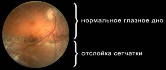
Retinal disinsertion
With this pathology, the inner membrane of the eye is separated from the vascular layer, which leads to a disruption in its nutrition. As a result, the phytoreceptors die off. If untimely or incorrect treatment is used, a person may lose vision forever, so it is very important to detect the disease in time.
Its symptoms are:
- Deterioration of vision.
- Narrowing of visual fields.
- Floaters, dots, a veil before the eyes.
- Deformation of images.
- Loss of lateral vision.
According to statistics, retinal detachment is most often diagnosed in people suffering from myopia, who have suffered a hypertensive crisis or injury.
Diabetic retinopathy
The pathology develops against the background of diabetes mellitus. Lack of insulin in the body leads to metabolic disorders and destruction of cells that maintain small blood vessels in tone. As a result of weakening of the vascular walls in the retina, hemorrhages occur, and due to the pathological formation of new blood vessels, irreversible retinal detachment can occur and complete blindness can develop.
The disease is characterized by slow and asymptomatic development even at advanced stages. As a rule, the development of diabetic retinopathy occurs 5-10 years after the onset of diabetes. Left untreated, it often causes irreversible blindness, which is why people with diabetes need regular eye exams with an ophthalmologist. According to statistics, about 40% of patients suffering from diabetes have diabetic retinopathy.
Our eye
The back wall of our eye shell has photoreceptors. They are light-sensitive cells and come in two types – rods and cones. The perception of black and white images is provided by rods, of which there are about 130 million in the human eye. Cones are responsible for human perception of color; their number in our retina is 7 million. Also, this type of cell perceives rapid movements much better.
When light enters the cones, photochemical reactions occur that transform the light into a signal. The optic nerve sends this signal to the brain. This entire process provides visual perception of the surrounding world.
Cones are light-sensitive cells due to their specific iodopsin pigmentation. This pigment, in turn, has several pigments responsible for vision:
- Chlorolab, which is responsible for the perception of the green and yellow color spectrum;
- Erythrolab, which senses the red-yellow spectrum.
The source of color cells, that is, cones, is the X chromosome. As everyone knows, men have only one X chromosome, while women have two. As a result, the female eye has more cells responsible for color perception. Thus, women's definition of colors is much broader than men's.
There is such a thing as an extra cone in the retina. It can only occur in women, but is very rare. What the extra cone of the retina looks like - photos can be found on the Internet or in special medical journals and literature.
Major retinal diseases
Macular degeneration (central retinal degeneration) is one of the most serious eye diseases, leading to complete loss of central vision. With this disease, a gray transparent spot first appears in front of the eye, which darkens over time and loses transparency, as a result of which the person cannot see the objects he is looking at. One of the reasons for the development of macular degeneration is high myopia.
Peripheral dystrophy is diagnosed during an examination of the fundus with special lenses. According to statistics, this disease in the vast majority of cases (40%) develops in people with myopia. Its main danger lies in the absence of symptoms at the development stage, so it is usually diagnosed by chance. However, if the patient does not regularly visit the ophthalmologist’s office, and pathological processes begin to develop in the periphery of the retina, then due to retinal detachment, visual acuity will occur unexpectedly for the patient. Often people come to the ophthalmologist only when the retinal detachment has reached the central zone and there is an accompanying feeling of a veil before the eyes. This is considered an advanced stage, and in this case only surgery will help, which cannot guarantee complete restoration of visual functions.
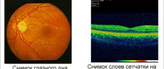
Dystrophic changes in the retina can be detected by an ophthalmologist through a thorough examination of the fundus. As a rule, if there is a suspicion of problems with the retina, the specialist suggests conducting an in-depth examination of the peripheral area. The laser surgeon is responsible for the latter. There are two options for the development of events: either the specialist will recommend dynamic observation, or will carry out restrictive laser coagulation, which allows you to limit the affected areas of the retina. This procedure allows you to avoid complex and dangerous operations and permanent loss of vision.
Video about what the cones and rods of the retina look like
The video demonstrates a conditional semantic retinal image. It consists solely of photoreceptors and several layers of nerve cells. This organ contains about 7 million cones and 130 million rods.
They are unevenly distributed, complex photochemical processes take place in them, and there is also an excitation of the light from the bottom itself, thanks to which a person has an excellent opportunity to see. If you are interested in learning more about the structure, I recommend watching the video to the end.
Cones - their meaning and structure
A distinctive feature of cones is the presence of the pigment iodopsin, which is divided into chlorolab and erythrolab. The first one mainly covers the yellow-green visibility spectrum, while the second one covers the yellow-red spectrum. In general, they are capable of capturing almost the entire spectrum cavity.
In addition, cones have another ability that is responsible for recognizing objects in motion, due to their better adaptability to the dynamics of light particles. They have three main sections:
- Outer. It contains several visual pigments that are located in certain places on the plasma membrane. It also has a very important property - the ability to update.
- An elastic molecular structure consisting of proteins and lipids forms the so-called constriction, formed from cilia and designed to distribute energy.
- High metabolism zone. In this area there is an energy accumulation of cells, the structure of which is made up of mitochondria, which release a large amount of energy for visual operations.
- The last zone consists of two neurons, or a neuron and a cell, which receives signals.
There are also three types of photoreceptor cells - L-type, M-type and S-type. Each of them is responsible for specific colors: L for red and yellow, M for green-yellow, and S controls blue.
Diagnostic technique
To diagnose pathologies associated with the work of cones and rods, a whole range of examinations is prescribed:
- study of the width of visual fields;
- study of the condition of the fundus of the visual organs;
- comprehensive test for the perception of colors and their shades;
- UV and ultrasound of the eyeball;
- FA - an examination that allows you to visualize the state of the vascular system;
- refractometry.
Correct color perception and visual acuity directly depend on the functioning of rods and cones. It is impossible to answer the question of how many cones there are in the retina, since their number is in the millions. With various diseases of the retina of the visual organ, the functioning of these receptors is disrupted, which can lead to partial or complete loss of vision.
General picture of sticks
These photoreceptor cells are distributed in great numbers throughout the retina, their number ranges from 115 to 120 million. These cells have the shape of cylinders, which is why they were conventionally named. Their length is small, about 30 times their diameter.
The most significant difference from other cells is that they contain rhodopsin, a visual pigment belonging to the group of chromoproteins, with the help of which the greatest light sensitivity of the eye is achieved. It stands out with a red tint, which was found out during various analyzes and studies. Rhodopsin is divided into a colorless protein and a yellow pigment.
The main thing is that it responds to light particles by disintegrating and irritating the optic nerve. During the daytime, sensitivity moves to the blue zone, and visual purple transforms into nighttime within half an hour, which is not able to distinguish colors, but perfectly captures small flashes of light with the energy of one photon.
By the time everything is completely rebuilt, the organ adapts to dim light and begins to see more clearly, and this process is considered better for the eye. The structure of the sticks consists of four components:
- Membrane discs.
- Cilia.
- Mitochondria.
- Nervous tissue.
Important! The rods are indeed too light sensitive and only need one photon for the reaction to occur. Thanks to the smallest elementary particles of light, a person is able to see quite well even at dusk!
Functions
The rods and cones of the retina perform different functions, but only the participation of both groups of photographic receptors can ensure the smooth operation of the entire visual apparatus.
If the rods, due to their structure, are capable of perceiving even very small light stimuli, at low levels of illumination, but do not distinguish the shades of the light spectrum at all, then the cones, on the contrary, allow the visual system to appreciate the entire richness of the color palette of the world.
Therefore, both receptor groups are equally important and necessary for our vision to obtain a high-quality, reliable image at any time of the day and regardless of weather conditions.