Different retinal diseases require different treatment methods. Some of them are treated exclusively surgically. But a number of diseases can usually be treated in the initial stages with conservative methods - using medications (eye drops, injections).
The main goal of conservative treatment of retinal pathologies is to relieve inflammation, proliferation of newly formed vessels, as well as eliminate degenerative changes and swelling of the retina. In some cases, prescriptions are made for prophylactic purposes (“to strengthen the retina”). At the same time, the use of drugs in the form of drops is not always effective, because they do not penetrate very well into the back of the eye (in this case it is recommended to use injections or tablet forms).
Anterior and posterior parts of the eye
Retinal angiopathy - treatment with drops
Angiopathy is a pathology of retinal vessels caused by various negative factors (hypertension, diabetes mellitus). In order to eliminate its manifestations, an ophthalmologist in the early stages of the disease or as part of complex therapy prescribes certain eye drops to patients.
The goal of such therapy is to activate the metabolic processes of the organ of vision, stimulate the speed of local blood flow, and improve the process of tissue nutrition. Eye drops are becoming one of the components of therapy for symptomatic treatment. Eye drops are becoming one of the components of therapy for symptomatic treatment. Read below what medications are prescribed.
Eye drops are convenient, but not very effective.
Taufon
The main component of the solution is taurine. Apply the solution 1-2 drops in each eye three times a day - for a long time. The drug is produced in bottles of 5 and 10 ml.
Indications for use of Taufon:
- Moderate retinal angiopathy.
- Corneal injuries.
- Early stage cataract.
- Dystrophic changes in the retina and cornea.
- Open angle glaucoma.
Expected effect:
- Stabilization of cell membranes.
- Acceleration of the regeneration process of traumatic corneal injuries.
- Activation of metabolic and energy processes.
- Normalization of IOP.
Emoxipin
This is a synthetically produced antioxidant, a solution of which is instilled daily, three times a day, 1-2 drops in each eye. Treatment lasts several days or for a long time - as prescribed by a doctor. The drug is not prescribed to pregnant women, as well as to persons with allergies to the components of the solution.
Indications for use:
- Diabetic retinal angiopathy.
- Cerebral circulation disorders.
- Various origins of hemorrhage in the eye.
- Corneal burns.
- Complicated myopathy and glaucoma.
Expected effect:
- Resorption of small hemorrhages on the retina.
- Protects the mesh shell from bright light.
- Strengthening the walls of blood vessels, reducing their permeability.
- Stimulation of local blood flow.
Emoxipine and Taufon (taurine) are the most popular eye drops for retinal diseases
Quinax*
Expected effect:
- Stimulation of metabolic processes in tissues.
- Activation of antioxidants.
- Increased lens transparency.
*At the moment, the drug is no longer available.
Aisotin
Able to increase and restore visual acuity in case of eye diseases. Available in 10 ml bottles. It is instilled three times a day, 1-2 drops, for 2-3 months.
Indications for use:
- Diabetic angiopathy.
- Restoring visual function after eye surgery.
- Conjunctivitis.
- Eye burns.
- Redness of the eyes.
- Glaucoma.
It is better to discuss the use of any medications with your attending ophthalmologist.
Emoxy optic
Substitute for Emoxipine at a lower cost.
- Indications for use:
- Eye burn.
- Bleeding in the eye.
- Progressive myopia.
- Inflammatory processes of the organ of vision.
Expected effect:
- Normalization of the permeability of vascular walls.
- Stimulation of local blood circulation.
- Resorption of hemorrhages in the eye.
- Elimination of the effects of oxidative stress.
- Increasing tissue resistance to hypoxia.
Modern methods of treatment of central and peripheral retinal dystrophies
To date, treatment for retinal dystrophy does not completely eliminate the disease. Using various techniques, doctors can only stop the development of dystrophic processes. In some cases, ophthalmologists are able to achieve a temporary increase in visual acuity.
However, in the absence of supportive therapy, the disease soon begins to worsen again.
Any dystrophy causes irreversible organic damage to the retina of the eye, due to which it ceases to function normally. Since the retina has a complex structure, it is almost impossible to restore its structure and functions.
This means that a person with dystrophy is unlikely to be able to see well again.
Which doctor treats retinal dystrophy?
The diagnosis and treatment of pathology is carried out by a retinologist - an ophthalmologist who specializes in diseases of the retina. If surgical intervention is necessary, a vitreoretinal surgeon comes to his aid. This specialist performs complex operations on the posterior segment of the eyeball.
Methods
Using what methods and how to treat retinal dystrophy? Medical tactics depend on the location of dystrophic foci, their size and type. The choice of treatment method is also influenced by the presence (or absence) of concomitant diseases and complications.
Uncomplicated peripheral dystrophies are usually treated with medications. If a patient is diagnosed with excessive thinning or ruptures of the retina, he undergoes laser treatment. But for macular degeneration, laser is not used, since this is fraught with serious complications.
In the initial stages, medications are used to combat central degenerations. If the wet form of macular degeneration develops or macular edema appears, the patient is administered anti-VEGF drugs.
Due to the abundance of forms and types of retinal dystrophies, the question of treatment tactics for each of them is decided on an individual basis. First, the doctor carefully examines the patient and makes a diagnosis. After this, he tells the patient in detail about his illness and possible methods of combating it.
Based on the information received, the person himself makes the final decision. No one has the right to force a patient to buy expensive medicines or agree to an expensive operation.
Medication
Drug treatment is used for age-related, pigmentary and peripheral retinal dystrophies. Patients are prescribed drugs that improve blood supply to the tissues of the eyeball and saturate the retina with nutrients. Medicines can be taken orally or administered parabulbarly.
Drugs used to combat retinal degeneration
DrugActionFeatures of use
| Eufillin | It has an antispasmodic effect, that is, it relieves spasm from the vessels supplying the retina of the eye. Improves blood circulation in the tissues of the eyeball, thereby slowing down the course of the disease | The drug is taken orally, in the form of tablets or capsules |
| Emoxipin | The medicine strengthens retinal vessels, has an antiplatelet and antihypoxic effect. Thanks to this, it improves blood circulation in the retina, which slows down its destruction | For the treatment of retinal dystrophy, the drug is used in the form of parabulbar or subconjunctival injections |
| Cavinton | Antispasmodic agent with antihypoxic effect. Normalizes microcirculation, thins the blood, slows down platelet aggregation, protects tissues from the effects of free radicals and oxygen starvation | The drug can be prescribed in tablets or administered intravenously. |
| Retinalamine | The manufacturer positions Retinolamine as a drug that improves metabolism and normalizes energy processes in the retina. Restores some damaged cells and prevents the death of others | The medicine is administered intramuscularly or parabulbarly |
| Adruzen Tsinko | It is a dietary supplement containing a lot of active ingredients. Improves metabolism, preventing retinal destruction | Available in capsule form to be taken orally |
| Visiomax | The medicine contains a complex of vitamins, minerals and pigments that actively nourish the retina | Taken orally in tablet form |
Physiotherapy
Along with medications, physiotherapeutic methods are used to treat central and peripheral chorioretinal dystrophies of the retina. They are prescribed in courses lasting 10–14 days. A person must undergo at least two courses of physiotherapy per year.
Methods that may include in treatment:
- magnetic therapy;
- photo- or electrical stimulation of the retina;
- stimulation of the retina with low-energy laser radiation;
- electrophoresis with nicotinic acid, No-Spa or heparin.
If there is no therapeutic effect from conservative therapy, the patient is recommended to use more effective methods.
Intravitreal administration of anti-VEGF factors
In modern ophthalmology, anti-VEGF drugs are used to treat the wet form of macular degeneration and macular edema of the retina. The drugs are administered intravitreally, that is, directly into the eyeball. The procedure is performed under local anesthesia.
https://www.youtube.com/watch?v=nJbs89TFFYQ
The administration of Lucentis or Aylia helps to remove macular edema and even improve the patient’s vision. To get a good, lasting result, you need to perform several injections. Due to the high cost of drugs, not every person can afford this procedure.
Description of anti-VEGF factors
LucentisIlia
| Active substance | Ranibizumab | Aflibercept |
| Mechanism of action | Medicines suppress angiogenesis - the process of growth of new blood vessels. Thus, they help to avoid neovascularization, macular edema, and hemorrhage. They also reduce the permeability of the vascular walls, thereby preventing hemorrhagic leakage of the retina | |
| Indications for use |
| |
| Possible side effects and complications |
| |
| Average cost per package | 42,500 rubles | 38,000 rubles |
| How many injections are needed? | Three injections at 1 month intervals |
Avastin is also an anti-VEGF factor. However, the drug is intended exclusively for the treatment of cancer. It should absolutely not be used in ophthalmology. Intravitreal administration of the drug can lead to unpredictable consequences and serious complications.
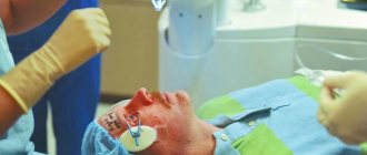
Surgical intervention
Central retinal dystrophy, complicated by a macular hole, is treated surgically. The essence of the surgical intervention is to remove the vitreous. After this, an air bubble or a special liquid is introduced into the cavity of the eyeball, which puts pressure on the retina and prevents its detachment.
Peripheral chorioretinal retinal dystrophy can also be treated surgically. The indication for vitrectomy is the formation of adhesions between the retina and the hyaloid membrane of the vitreous body. Removal of the strands in this case is necessary to prevent retinal detachments and ruptures.
Stages of surgical treatment:
- Exploratory survey . Before the operation, the patient is sent for consultations with specialists and for tests. This is necessary to identify concomitant diseases that can cause intraoperative complications.
- Preoperative preparation . A few days before surgery, the patient begins to put drops prescribed by the doctor (antibiotics, non-steroidal anti-inflammatory drugs) into the eye. Such a preventive measure helps reduce the risk of complications in the postoperative period.
- Pain relief . The operation is performed under local retrobulbar anesthesia. The anesthetic is injected into the postorbital space.
- Operation . During surgery, the patient lies on his back. His head is carefully fixed. The operation usually takes about 30-40 minutes. After this, the patient is transferred to the recovery room.
- Rehabilitation . After the operation, the patient must use the medications prescribed by the doctor for some time. In the future, he will need to be monitored by an ophthalmologist.
Laser
Laser coagulation is used to protect areas of degeneration and suppress pathological vascular growth. This allows you to slow down the development of the disease and reduce the risk of complications. Timely laser coagulation of newly formed vessels makes it possible to avoid hemorrhages, which are fraught with severe deterioration or even loss of vision.
The laser is used to treat lattice degeneration, retinal pigmentary degeneration, cochlear track and cobblestone dystrophies. Laser coagulation is not performed in the macular area due to the high risk of complications. Because the mesh in this area is very thin, it can tear or peel off.
Since it is impossible in principle to cure retinal dystrophy, laser coagulation has only a temporary effect. Don’t think that it will help you forget about the disease forever. Unfortunately, over time, the patient may develop new degenerative lesions. Therefore, after the procedure, it is necessary to continue to be observed by an ophthalmologist.
Traditional methods
How to treat retinal dystrophy with folk remedies and is it worth doing it at all? Unfortunately, traditional medicine is in many ways inferior to traditional medicine. It is powerless against macular edema, ruptures and detachments. The use of various tinctures and decoctions in this case will only harm the patient.
Treatment at home using folk remedies is possible only for slowly progressive peripheral and age-related retinal dystrophies. It should be noted that traditional medicine methods can only be used after consultation with your doctor.
Folk remedies that are used for dystrophies:
- crushed sprouted wheat;
- infusion of nettle leaves;
- a decoction of blueberries, rowan and sea buckthorn;
- a collection of chamomile, lingonberries, calendula, dandelion and dried cucumbers;
- blueberry infusion.
The above remedies are taken orally. But to wash the eyes, use a decoction of nettle and lily of the valley leaves, aloe juice, a mixture of water and fresh goat milk. It is believed that these remedies nourish the eyes and help cope with the disease.
Is eye dystrophy curable? Yes, there are many methods that can successfully combat the disease. However, the second question is, is it possible to completely cure retinal dystrophy and forget about the problem forever? Unfortunately, this is impossible to do these days. However, despite this, sick people should not despair.
Thanks to the achievements of modern medicine, it is possible to slow down the progression of the disease and preserve vision. To combat the disease, medications, physiotherapy, surgery and laser methods are used to treat retinal dystrophy. They can be used separately or combined.
Alina Lopushnyak, ophthalmologist, specially for Okulist.pro
about treatment of the retina
(1 5,00 out of 5)
- retinal dystrophy treatment
Source: https://okulist.pro/bolezni-glaz/setchatka/lechenie-distrofii-setchatki-glaza.html
Intraocular injections
Intravitreal injections are the injection of a drug directly into the eye cavity (into the vitreous body). Such injections are performed for hemorrhages (partial hemophthalmia), wet form of macular degeneration, retinal edema, the appearance of newly formed vessels and other serious eye diseases.
In our clinic, intravitreal injections are performed by ophthalmologists with extensive experience in complex intraocular injections in sterile procedure rooms, in compliance with appropriate medical protocols. Such therapy for retinal pathologies, using effective medications, allows one to preserve visual function and also achieve a significant improvement in visual acuity.
Lucentis
The active component of the drug is ranibizumab, a substance that suppresses excessive angiogenesis (the formation of pathological blood vessels), which is characteristic of age-related macular degeneration. In addition, it helps relieve macular edema, normalizing the thickness of the retina. The drug quickly penetrates completely into the layers of the retina, reducing the size of the lesion, preventing possible hemorrhages and further progression of the growth of newly formed vessels with a pathological wall. More about Lucentis >>>
Treatment regimens for retinal angiopathy
Drug therapy in the case of angiopathy and its vascular complications should be comprehensive. If necessary, treatment can be continued in courses. Before prescribing it, an ophthalmologist develops a specific, most effective regimen for using drugs. As a rule, these are the following appointments:
- Drug therapy aimed at activating blood circulation in the vessels of the eyes is necessary. This is a course treatment with Emoxipin, Vazonit, Mildronate, Arbiflex, Solcoseryl, Trenatal. They help activate local microcirculation processes. Another property of these drugs is improving the plasticity of red blood cells. This allows them to move faster through the small vessels of the eyes.
- Pentoxifylline and Curantil help prevent possible thrombus formation. Xanthiol nicotinate and nicotinic acid improve blood rheology.
- To reduce vascular permeability, ginkgo biloba and calcium dobesilate are prescribed.
- Actovegin injections are designed to provide better nutrition to the eye tissues. To improve metabolic processes in eye tissues, ATP and cocarboxylase are used.
- It is mandatory to take vitamin complexes to maintain eye health - Lutein-intensive, Anthocyanin Forte. It is important to take ascorbic acid, which accelerates microcirculation processes in blood vessels and maintains visual acuity.
- For diabetic retinopathy, it is necessary to adhere to a special diet that can neutralize diabetes mellitus and improve blood flow in the retinal vessels. It is worth remembering that prohibited foods include any food rich in fast carbohydrates (baked sweets, sweet soda, etc.). The same applies to excessively high-calorie foods. Limiting salt is also important, because it helps normalize metabolism and improve recovery processes.
- Vigorous physical exercise, included in the daily routine to give energy to the muscular system, also improves the condition of the eye vessels.
- When treating angiopathy caused by hypertension, it is mandatory to lower blood pressure levels through medications and specially designed dietary adjustments. To do this, you should contact a cardiologist and nutritionist.
- It would not be superfluous to prescribe physiotherapeutic procedures. In the case of angiopathy, magnetic therapy courses and acupuncture can show good results.
- Another physiotherapeutic method is “Sidorenko Glasses”. This device combines the effects of pneumomassage, infrasound, phonophoresis, and color therapy. The powerful effect on the retina of such a complex makes it possible to achieve good results in a short time.
- Treatment also helps with massage courses on the cervical spine.
Drugs to strengthen the retina
The eyes are the most important organ of the human body, thanks to which people are able to see and perceive the world around them.
The retina is an integral part that contributes to the formation of pictures.
That is why the eyes, more than other organs, need protection, proper care and maintenance of normal performance. For this purpose, there are a number of different drugs that have been used in ophthalmology for many years.
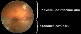
Retinal dystrophy
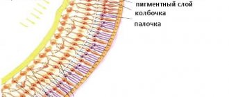
The retina of the eyes has the most complex structural structure, due to which it is able to distinguish between light pulses.
The functioning of the optical apparatus of the eyes and the visual department of the brain, where all the information seen is transmitted, depends on the retina.
A common cause of retinal dystrophy is the presence of problems with blood vessels. The most striking example is older people who lose their vision over time.
Dystrophy is a pathology of the retina in which the photoreceptor cells are damaged, thanks to which a person is able to distinguish colors and see into the distance. In the early stages, the disease can develop without any pronounced symptoms.
The following forms of the disease should be distinguished:
- Central and peripheral dysplasia. The second form is most typical for myopic people, since it develops due to a decrease in normal blood flow in the vessels of the eyes. As a result, there is a deterioration in the supply of oxygen and other necessary elements to the retina.
- Congenital (genetic) dystrophy and acquired.
- Senile form of dystrophy. Most often occurs in people over 60 years of age. It occurs against the background of processes caused by the aging of the body.
- Development of pigmentary dystrophy. It manifests itself as problems with photoreceptors, which are responsible for twilight vision. It is one of the rarest forms of the disease that occurs as a hereditary factor.
- The dotted white form of the disease manifests itself in childhood. In the absence of appropriate treatment, it develops along with the child.
Selection of means for recovery
The retina of the eyes has the most fragile structure, so it constantly needs reliable protection. Every person should remember this. At the same time, there are categories of people who are at risk, for whom proper eye care should be the basis.
These categories include:
- People whose work involves constant eye and vision strain. Most often this is associated with working with magnification devices, computer and office equipment.
- People working with the risk of small parts (dust, shavings, etc.) getting into their eyes.
- Athletes and workers whose work involves lifting heavy weights. In such cases, there is a risk of developing dystrophy due to increased pressure.
- People with bad habits. Everyone knows about the negative effects of tobacco smoke not only on the body, but also on the eyes and retina separately.
Ophthalmologists convince us that the body itself will tell you when to use drugs that maintain the retina in normal condition.
You just need to listen to the smallest changes in the state of the body.
Depending on the symptoms that appear, it is possible to select individual medications that will help restore the normal functioning of the visual organs and prevent the development of an irreversible process - dystrophy.
Drug groups
In ophthalmology, drugs are used that have an auxiliary effect and thereby help reduce the manifestation of unpleasant symptoms of many diseases, including retinal dystrophy. Most often, treatment consists of using complex therapy, which necessarily contains eye drops that help slow the development of the disease.
Vitamins
Treatment of dystrophy must necessarily include the use of vitamin complexes. With their help, the normal functioning of the eyes is restored, tissue nutrition is enhanced and the progression of the disease is stopped. Vitamins A, E and B become necessary.
The most effective vitamin products for the eyes are:
- Vitrum Vision;
- Complivit Oftalmo;
- Lutein-Intensive Evalar;
- Lutein Complex.
Retinoprotectors
Most often used in the treatment of chronic retinal diseases. Treatment with drugs of this group is carried out twice a year as part of complex therapy. The drug composition is selected by a specialist based on the patient’s individual characteristics, as well as focusing on the complexity of the pathology.
Angioprotectors
Angioprotectors consist of drugs that are used to normalize the permeability of vascular walls. After using such drugs, there is a decrease in the severity of tissue edema, an improvement in the metabolic process and metabolism in the retinal tissue.
Vitamin P also has similar properties, as well as drugs that belong to the group of non-steroidal anti-inflammatory drugs.
Angioprotectors include:
- Doxium – helps strengthen the vascular system of the eye;
- Ascorbic acid – helps restore the permeability of capillary cells;
- Rutin – enhances the elasticity of capillaries. Has a positive effect when used in combination with vitamin C;
- Troxevasin - helps reduce swelling and significantly reduces inflammation.
Absorbable drugs
Necessary for thinning the blood, helping to dissolve clots that can clog blood vessels.
Anticoagulants
Helps stimulate the fibrinolytic activity of blood cells. The drugs also help reduce the concentration of cholesterol and b-lipids.
Antiplatelet agents
When using drugs in this group, a positive anti-inflammatory effect is observed. There is a decrease in platelet aggregation in the blood. The drugs are used to reduce the possible risk of blood clots in the lumens of blood vessels.
Drops for retinal dystrophy
One of the most complex eye diseases is retinal dystrophy. As a rule, it develops when the nutritional process of the pigment tissues of the eyes is disrupted. The pathology most often occurs in people suffering from myopia.
In this case, several factors can be identified that can provoke the development of the disease:
- chronic cell diseases;
- presence of diabetes mellitus;
- transmission of viral diseases;
- other complex eye diseases;
- presence of bad habits such as smoking, etc.
The only method of completely curing retinal dystrophy is surgery. In this case, in the early stages of development, treatment using laser and medications is used.
The most effective drops for dystrophy are: