The retina of the eye is responsible for human vision. Due to the development of macular degeneration, this part of the eye is damaged. Therefore, a person sees a blurry picture. In the presence of macular degeneration of the retina, drug or surgical treatment is prescribed depending on the form and severity of the disease.
What is retinal macular degeneration
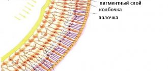
Macular degeneration of the eye is a lesion of the central part of the retina. The main reason for the development of pathology is insufficient blood flow. A yellow pigment accumulates near the macula. Because of this, the light-sensitive cones are damaged. First, vision deteriorates in one eye, and later in the second.
Dystrophic changes rarely appear at 17–18 years of age. The disease most often occurs in people over 50 due to age-related changes. The disease usually affects both eyes.
With macular degeneration, the first symptoms do not appear immediately. In such cases, people turn to an ophthalmologist when they have the last stage of the disease. Therefore, it is impossible to completely restore vision.
In the presence of macular degeneration, disability is given, but the group depends on the severity and form of the disease. If visual acuity decreases to 0.03, the person is assigned disability group 1. With a slight decrease to 0.1, group 3 is given. In other cases, group 2 is assigned.
Macular degeneration does not lead to complete loss of vision. The disease affects a person’s ability to work. It makes it difficult to drive a car, watch TV, recognize others, or count money.
Types of dystrophic changes
Retinal dystrophy can be central or peripheral. It depends on the localization of the process.
If the central part, the macula, is affected, then this is central dystrophy or macular degeneration.
It mainly develops in older people. Causes: age-related changes. Although there are hereditary forms of pathology - pigmented and dotted white.
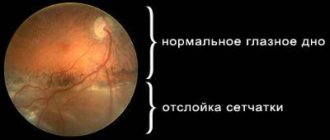
In the case of peripheral dystrophy, this is damage to the lateral parts of the retina. Usually there are no symptoms of pathology, especially at the initial stage. However, peripheral dystrophy is not so harmless. Causes: it can lead to retinal detachment and significant vision loss if not treated promptly.
In most cases, peripheral changes are observed in myopic people. Because with medium and high myopia, the eyeball enlarges and the retina stretches. As a result, some parts of it become thinner and torn. This can lead to vitreous fluid getting under the retina and causing it to detach.
Peripheral dystrophy can also affect people with normal vision, including women after pregnancy.
Classification
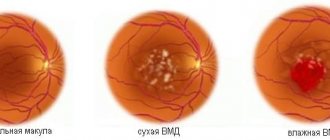
In ophthalmology, pathological changes in the macular zone of the retina come in dry and wet forms. In the first case, the disease is treated successfully. Macular degeneration in the dry form is characterized by slow development. Wet macular degeneration of the retina is dangerous for humans as it progresses quickly. Therefore, vision rapidly deteriorates.
Wet macular degeneration with age-related changes is characterized by the proliferation of blood vessels that are directed towards the macula. Therefore, fluid from the new capillaries flows out and permeates the retinal tissue. As a result, the area swells.
The wet form of macular degeneration is the result of advanced dry macular degeneration. Unlike the dry form, the wet form leads to complete loss of vision. Dry dystrophic changes in the macula are distinguished by stages of development:
- Initial. There are no symptoms. The disease is diagnosed randomly during a medical examination. An ophthalmological examination of the fundus of a person reveals yellow spots. These growths are called drusen. At the beginning of the development of macular degeneration, they are small, almost invisible.
- Intermediate. The examination reveals medium and large drusen. A person with this stage is no longer able to distinguish objects.
- Expressed or last. When light-sensitive cells are destroyed, vision decreases sharply. Dystrophic changes lead to disruption of central vision. A person sees one continuous spot of black color.
Signs of the disease
The pathological process, as a rule, begins asymptomatically. In some cases, patients note a decrease in visual acuity, the occurrence of blurred vision or distortion of some of its areas.
Complications of the disease are accompanied by the appearance of holes in the foci of lattice lesions, which in the future often become the cause of rhegmatogenous retinal detachment. In this case, rapid progression of detachment processes occurs due to the release of fluid into the subretinal space. Most often, this complication is observed in young people with myopia.
People over 50 years of age who suffer from myopia usually experience a complication of a different nature. It is associated with traction from the vitreous, which also leads to retinal detachment along the ethmoidal edge. Such a detachment is usually accompanied by the release of subretinal fluid, which can cause acute detachment of the cervical cavity.
Symptoms
Macular degeneration of the retina varies according to the stages and forms of the disease. Let's look at the general symptoms:
- distortion of vision;
- hallucinations in the form of distorted straight lines;
- difficulty reading books;
- poor visibility in the dark;
- spots, dots before the eyes.
The listed symptoms appear in one or two eyes. In the second case, the person gradually gets used to the new state, and therefore triggers the disease.
How does a person with macular degeneration of the retina see:
You can watch the following video for more information about the symptoms of this disease:
Causes of retinal degeneration
Medicine has never found the exact causes of the degenerative process. Some experts believe that the disease is hereditary. Often it begins to manifest itself only in old age. This phenomenon is associated with the accumulation of metabolic substances in nerve tissues. The main factors are considered to be the following.
- Impaired blood flow in the body. The cause may be increased blood pressure, diseases of the vascular system, elevated cholesterol and blood sugar.
- Poisoning or infection of the body.
- Myopia.
- Diabetes.
- Excess weight.
- Bad habits such as smoking and drinking alcohol.
- Exposure of the visual organ to direct ultraviolet rays.
- Unhealthy diet, where fatty foods predominate.
- Lack of vitamins in the body.
- Constant stressful situations.
Retinal degeneration can also develop at a young age as a result of:
- diseases of the cardiovascular system;
- diseases associated with the endocrine system;
- pregnancy;
- injury to the visual organ.
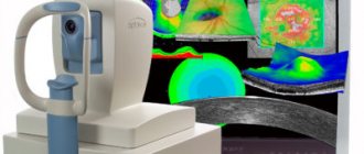
Diagnostics
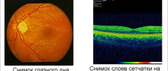
First, the ophthalmologist conducts an examination of the fundus. Special drops are dropped into both eyes of a person to dilate the pupils. Afterwards, the ophthalmologist examines the eye for 15 minutes.
Sometimes a diagnostic method is used at home - the Amsler test. In this case, a special mesh and fluorescein angiography are used. In the presence of dystrophic changes in the central region of the retina, a person does not see a spot in the center of the field.
Diagnosis of retinal degeneration disease
Often, patients do not immediately seek help from a doctor, which has a bad effect on the further functioning of the visual organ. In order to accurately diagnose the form of the disease, the doctor prescribes an examination, which includes the following steps.
- Perimetry.
- Visometry.
- Examination of the fundus.
- Fluorescein angiography.
- Ultrasound diagnostics of the eye.
- Electrophysiological examination of the visual organ.
- Lab tests.
Treatment
Treatment of dry macular degeneration of the retina is carried out using medications. During therapy, a person is prescribed special drops and ointments. In the treatment of wet macular degeneration of the retina, surgical and laser operations are used.
Conservative
Treatment of the initial stage of macular degeneration of the retina involves the use of drops. To cure the disease, visual pigments, antioxidants and drops containing zinc and copper are instilled into the eye.
A person may be prescribed vitamin-mineral complexes, the task of which is to slow down the development of pathological changes in vision. Complete vitamin-mineral complexes against retinal macular degeneration contain the following components:
- zinc;
- vitamins of groups A, C, E;
- copper;
- lycopene;
- lutein;
- omega-3 fatty acids;
- beta carotene.
After diagnosis, the doctor prescribes medications containing vitamins and minerals:
- “Complivit Ofthalmo”, prescribed for periodic visual fatigue;
- “Lutein Complex”, used as a food supplement for vitamin deficiency;
- “Metmorphine” is used as a prophylactic against ophthalmic complications for people with diabetes;
- “Vitrum Vision Forte” is prescribed for changes in the macular area in the central or peripheral part.
In addition to taking these medications, a person should eat a balanced diet. If there are dystrophic changes in the central part of the retina, it is forbidden to eat fatty foods:
- rich soups;
- pork;
- smoked meats;
- fried, salty foods.
When sick, it is recommended to eat green vegetables, fruits and berries. Lenten dishes predominate in the diet.
Surgery and laser
Laser coagulation of the retina is used for the wet form of macular degeneration. During the operation, fragile, leaking vessels that have formed over the course of the disease are removed. The laser is aimed at the vessels and destroys them, preventing vision loss.
But sometimes surgery and laser surgery damage healthy blood vessels. Because of this, vision deteriorates. The surgical treatment method is used only when the newly formed vessels are located away from the central fossa of the macula.
Watch a video about laser coagulation of the retina below:
Modern methods
Today, age-related macular degeneration of the retina is treated with progressive methods. This therapy is effective for severe dry or wet forms. The essence of modern treatment is to prevent the proliferation of small vessels and destruction of the macula. For this purpose, special glasses Argus 2 were invented in the UK.
Folk
It is impossible to completely restore vision in the presence of dystrophic changes in the retina. Treatment of macular degeneration of the retina involves the use of folk remedies. Let's look at recipes for non-standard medicine:
- The washed, sorted wheat is laid out in a thin layer and filled with water. After germination, the wheat is washed. The resulting consistency is passed through a meat grinder. The resulting medicine is stored in a cool place. People with macular degeneration should eat up to 5 tsp of this wheat every morning. Berries and honey are added for taste.
- To restore vision you will need mumiyo infusion and aloe juice. 5 g of mumiyo is mixed with 100 ml of fresh juice. The resulting solution is stored on the top shelves of the refrigerator. It is necessary to instill the prepared drops 2 times a day. The course of treatment is 15 days. Then there is a break for a week. If necessary, therapy is resumed.
- Following a diet prevents further development of the disease. It is important to eat foods that are filled with vitamins and beneficial microelements.
Prevention
Age-related dystrophic changes in the retina now appear at an early age. Therefore, it is necessary to observe measures to prevent macular degeneration:
- Rejection of bad habits. It has been proven that alcohol and cigarettes age the body.
- Eye protection in sunny weather. Ultraviolet exposure negatively affects the structure of the retina. Therefore you need to wear sunglasses.
- Annual examination by an ophthalmologist. Timely consultation with a doctor is a guarantee of health.
- Partial exclusion from the diet of foods high in cholesterol.
- Arterial hypertension cannot be ignored. The disease must be treated.
- Take vitamin complexes if there is a hereditary predisposition to the disease. Many beneficial elements are found in vegetables, berries, fruits and seaweed.
At the first suspicion of retinal macular degeneration, you should consult a doctor. If treated in a timely manner, the disease will not lead to irreversible consequences.
Have you encountered this disease? Tell us about your story of fighting the disease. Share the article with your friends on social networks. Be healthy. Anti-inflammatory eye drops, read our article.