Angiosclerosis of the retina belongs to hypertensive diseases that cause organic changes in blood vessels. The pathology provokes enlargement and tortuosity of the arteries. If the destructive process affects the retina (retina), its blood supply deteriorates and swelling develops. The disease is also dangerous due to other complications, which include ischemia of the optic nerve, hemorrhages in the vitreous region, partial or complete loss of visual function.
Mechanism of development of angiosclerosis
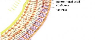
The manifestation of angiosclerosis is preceded by periodic rises in blood pressure, causing narrowing of the vascular lumens and uneven thickening of the walls of blood vessels. The pathological process worsens the condition of the retina, an important structure of the eye, the purpose of which is to conduct and transform light rays, provide color perception, and create a three-dimensional image. The development of angiosclerosis in this part of the eyeball is dangerous due to thrombosis and defective visual perception.
Hypertensive angiosclerosis of the retina is characterized by gradual development. At the initial stage, the disease is characterized by good sensitivity to treatment and correction.
Hypertensive angiosclerosis
Hypertensive angiosclerosis of the retina has dire consequences and leads to the formation of blood clots, hemorrhages and aneurysms. Treatment of pathologies of the eyeball is carried out with the participation of a therapist and an ophthalmologist. The first method aimed at combating the disease may be the prescription of medications that reduce pressure in the blood vessels.
In an advanced stage of the disease, few specialists can make a favorable prognosis. The disease is destructive not only to the visual organs, but also to the entire organism as a whole. Particularly difficult cases are considered when complications are caused by renal retinopathy, and the optic nerve has been affected by the disease. In such cases, the physician is obliged to prescribe a course of antihypertensive and atherosclerotic drugs.
In most cases, treatment of retinal angiosclerosis requires surgery. Slowing down can significantly increase the risk of vision loss. Ophthalmologists recommend annual examinations, and at the first sign of problems with the visual organs, immediately seek help.
Retinal angiosclerosis is the second stage of angiopathy, characterized by uneven thickening and narrowing of the lumen of blood vessels.
Reasons for development
The main causes of pathology include:
- arterial hypertension;
- generalized form of atherosclerosis.
There are also various factors, the presence of which significantly increases the likelihood of pathology in the retinal area. The risk of developing the disease increases if the patient has:
- injuries to the eyeballs, head;
- diabetes mellitus;
- cervical osteochondrosis;
- high intracranial pressure;
- impaired blood clotting;
- autoimmune diseases leading to inflammation of the inner choroid.
Angiosclerosis of the ocular retina tends to affect people with bad habits (alcohol, nicotine addictions) and the elderly.
Changes in retinal vessels in the presence of arterial hypertension
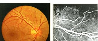
With a tendency to increase blood pressure, damage to the vascular walls in various body systems is observed. The fundus of the eye becomes one of the main areas experiencing the negative effects of hypertension.
Often, ophthalmologists diagnose hypertension after identifying angiosclerosis of the retinal vessels, which provokes thickening and hardening of the arterial walls.
In the presence of pathology, the retinal vessels change their usual characteristics and are distinguished by the presence of a whitish reflection (the “silver wire” symptom). The veins acquire atypical curves and move deeper into the retina. Sometimes they completely disappear from the area of intersection with the arteries. In some cases, the walls of the arteries accumulate lipid deposits, due to which they acquire a peculiar (golden) hue.
In addition to angiosclerosis, hypertension leads to other changes affecting the vessels of the fundus - hypertensive forms of angiopathy, retinopathy, neuroretinopathy.
Retinal changes in generalized atherosclerosis
The generalized form of atherosclerosis is a vascular disease that occurs with the appearance of atherosclerotic plaques. The pathological process is associated with impaired lipid metabolism. Generalized atherosclerosis affects various groups of blood vessels and is often chronic. Affecting the retina, it provokes vascular angiosclerosis, hemorrhages in the vitreous body, and in the conjunctival area.
Ways and methods of diagnosis
In order to diagnose angiosclerosis, there are a number of special procedures. All of them are painless and do not take much time. These include:
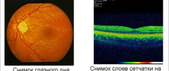
Ophthalmoscopy - This is the study of the bottom of the eyeball. This procedure takes from five to ten minutes. The ophthalmologist illuminates the pupil with an ophthalmoscope if this is direct ophthalmoscopy. In addition, a specialist can install a lens between the eye and the device and redirect light through it into the eyeball. He can also use a special lamp (slit), and place a lens between it and the eye.- Ultrasound - with the help of this study, you can identify violations of the structure and mobility of the eye muscles and detect areas of the retina that do not receive sufficient blood supply.
- Retinal tomography is an expensive procedure that allows you to examine all layers of the retina and detect even barely noticeable swelling.
Angiosclerosis does not have the status of an independent disease, but is a consequence of other pathologies that cause deformation of the retinal vessels. Therefore, the international classification of diseases gives a code only to the disease that caused this pathology (mechanical damage to the eyes and head, diabetes mellitus, etc.).
Stages of development
Angiosclerosis of the retina goes through 2 stages of development:
- First stage. It has a relatively easy flow. At this stage, the pathological condition of the retina is more associated with angiopathy. Changes in blood vessels caused by changes in blood pressure are observed, but their normal structure is still preserved. Examination of the fundus reveals redness, often covering the optic disc. High-quality treatment of this stage of the disease allows you to restore the full state of the visual organs.
- Second stage. The disease becomes more severe. Experts note the presence of angiosclerosis, interlacing of blood vessels piercing the retina, spasms and narrowing of their muscle walls. A typical violation is the lack of adequate blood flow.
In turn, stage 2 of the disease is divided into 3 stages. The first occurs with a narrowing of the venous lumen, increased pressure of the artery on the vein. The second stage is characterized by the formation of a large, deep bend of the vein. At the third stage, the venous bend is clearly defined, part of the vessel becomes invisible and is visualized as a rupture.
Origin of the disease
Angiosclerosis of the retina is a lesion of the walls of blood vessels that envelop the fundus of the eye. The walls of blood vessels become thicker and denser, which leads to the appearance of cholesterol plaques.
This pathology can provoke:
hemorrhage into the membrane of the eye or vitreous body;
- the occurrence of blood clots in an artery or vein;
- retinal ischemia;
- swelling in the membrane of the eye.
Due to the fact that the blood supply to the retina is reduced, swelling occurs. Due to thrombosis and tissue death, vision is impaired. Without treatment, this disease can lead to complete blindness.
Symptoms and diagnosis
The onset of the disease often does not show any obvious signs. This feature is explained by the presence of minor changes in the blood vessels.
At first, the patient feels increased eye fatigue and sees spots, “floaters” or other inclusions in the field of vision. Such a condition is often not given due importance, and all negative phenomena are attributed to ordinary fatigue.
The presence of progressive retinal angiosclerosis is confirmed by the presence of the following symptoms:
- increased tension in the visual organs;
- nagging pain;
- blurry image;
- periodic dizziness, headaches;
- incomplete field of view;
- non-standard reaction of the pupils to lighting (their atypical dilation or narrowing).
Less common signs of pathology include memory impairment, tinnitus, problems falling asleep, and periodic nasal bleeding.
To diagnose retinal angiosclerosis, the patient is prescribed the following:
- Computed tomography.
- Ultrasound.
- Electrophysiological study (EPS).
What are the symptoms of the disease?
The essence of the disease of the blood vessels of the eye is their narrowing, most often due to fatty deposits appearing on the walls, which in turn leads to impaired blood flow.
The patient complains of pain in the eyes, frequent dizziness, and headaches. The eyes get tired much faster. At times, visual disturbances occur. Usually people do not pay attention to such phenomena and attribute them to manifestations of other diseases. Incipient eye problems can only be detected randomly, for example, during an annual medical examination, since all the initial symptoms are characteristic of many diseases.
But the disease is most often identified in later stages, when serious problems with the blood vessels of the eye begin. This ophthalmological pathology can develop to such an extent that it can manifest itself as atrophy of the optic nerve, the appearance of glaucoma, and hemorrhages in the eye tissue.
When a patient with suspected atherosclerosis of the vessels of the eye sees a doctor, first of all a survey is carried out and the presence of chronic diseases is revealed. An ophthalmologist treats this disease. Its task is to assess the condition of the retinal vessels.
The criteria by which the assessment is carried out are as follows:
- The thickness of the vascular wall of the eye is determined.
- The degree of their narrowing is determined.
- The structure and configuration of blood vessels is studied.
The presence of hemorrhages and blood clots is determined.
- The condition of the lumens is assessed.
Diagnosis is carried out as follows:
- As part of the examination, the patient is prescribed visometry, which makes it possible to identify the degree of visual impairment and at the same time determine the condition of the retina.
- Ophthalmoscopy. This type of examination allows you to determine how much damage has affected the arteries and how many of them are damaged.
- Computer perimetry. This test looks for problems in the peripheral retina.
- The traditional examination carried out for all eye pathologies is an examination of the fundus.
- And finally, an MRI of the eye, which is performed to identify abnormalities in all tissues of the eye.
Treatment of retinal angiosclerosis
Pathology can be most successfully treated at an early stage of its development. The drug course prescribed for retinal angiosclerosis consists of the use of the following pharmacological products:
- eye drops that lower IOP (Fotil, Brimonidine);
- angiotensin II receptor antagonists (Candesartan, Losartan);
- beta blockers (Bisoprolol, Carvedilol);
- calcium antagonists (Amlodipine, Felodipine);
- diuretics (Hypothiazide, Indapamide).
For general toning of the visual system, special ophthalmic complexes or instillations with fortified preparations are prescribed.
To treat and prevent retina pathologies associated with generalized atherosclerosis, drugs that lower blood cholesterol are used. These include statins, represented by Atorvastatin, Mevastatin, Rosuvastatin. In case of complications, they resort to laser therapy and injection of corticosteroids. Hyperbaric oxygenation, a modern method based on the use of increased oxygen pressure, is considered an effective therapeutic agent.
A radical therapeutic measure is surgical intervention prescribed to patients with a complicated diagnosis.
Additionally, patients are given recommendations regarding the proper organization of their daily routine and a healthy diet. In case of retinal angiosclerosis, physical inactivity should be avoided, salty foods that cause fluid retention in the body, swelling and an increase in blood pressure should be avoided, smoked foods, canned food, fatty meats, spicy, sweet, and baked goods. Instead of junk food, a variety of vegetables, fruits, cereals, dietary meats and poultry should be present on the patient’s table.
Treatment
Treatment of retinal angiosclerosis without eliminating the cause of the disease - arterial hypertension - is unsuccessful. Therefore, the first measure is the prescription of antihypertensive drugs. The dosage and frequency of taking the drug, the duration of the course of treatment is determined by the therapist.
As the pressure normalizes, therapy is performed aimed at restoring blood circulation in the retina. For this, the ophthalmologist prescribes:
- Blood thinners;
- Eye drops to reduce intraocular pressure;
- Preparations for oral administration to strengthen the walls of blood vessels;
- Eye drops to speed up metabolism in retina;
- Complex vitamins orally and vitamin drops in the eyes.
If drug treatment does not bring the expected result, laser coagulation is performed - strengthening the retina using a laser. The method of hyperbaric oxygenation is also distinguished by its effectiveness - a method of improving visual structures under the influence of increased pressure from oxygen. The last resort treatment measure is surgery.
Prevention of the disease
The main prevention is aimed at achieving normal blood pressure levels. For this purpose, it is recommended to adhere to a healthy lifestyle, avoid severe stress, nervous strain, and give up alcohol and cigarettes. It is important to promptly identify and treat diseases that lead to the development of hypertension. Such pathologies include diseases affecting the nervous, cardiovascular, endocrine, and urinary systems. To prevent sudden surges in pressure, an effective course of physiotherapy is indicated, including galvanization, electrosleep, hydrokinesis, and radon baths.
Patients who experience initial manifestations of angiosclerosis should immediately seek help from an ophthalmologist. Only through timely receipt of medical care can dangerous complications and irreversible deterioration of vision be avoided.
Treatment and its purpose
Medicines
When treating this disease, the ophthalmologist, interacting with the therapist, draws up a treatment plan. The patient is prescribed a course of a number of medications.
These include:
- drugs that keep blood pressure within normal limits;
- vascular tonics;
- vitamins that support metabolic processes;
- agents that lower blood viscosity and reduce the risk of blood clots;
- drops that lower intraocular pressure.
IMPORTANT! Angiosclerosis develops extremely rapidly and, without proper treatment, reaches its final stage in a short time. At this stage, the disease is incurable and its development leads to blindness.
The main goal of treatment is to address the cause that caused the disease. If blood pressure is the cause of angiosclerosis, the goal of treatment is to normalize and stabilize blood pressure. In a situation where blood pressure does not decrease with the use of a single drug, combinatorial treatment with several drugs is introduced.
The problem of normalizing pressure is solved using:
diuretics (Hypothiazide, Indapamide);
- ACE blockers (Ramipril, Lisinopril);
- drugs that weaken hormone receptors, which are aimed at narrowing blood vessels and increasing blood pressure (Losartan, Candesartan);
- drugs that block beta-adrenergic receptors (Nebivolol, Carvedilol);
- calcium channel blockers (Felodipine).
Treatment of generalized atherosclerosis is associated with drugs that lower the concentration of cholesterol in the blood: atorvastatin, rosuvastatin, etc. Before using any drug, consultation with your doctor is necessary. In case of worsening of the disease (swelling of the retina, hemorrhages), laser therapy and the injection of corticosteroids into the vitreous body can be used.
Folk remedies
There are many popularly known recipes aimed at strengthening and cleansing the vascular system. Herbal infusions help get rid of harmful deposits on the walls of blood vessels.
Recipe No. 1. Take a mixture of chamomile and St. John's wort (one hundred grams of each ingredient). One tbsp. l. Pour boiling water over the mixture (five hundred milliliters). After twenty minutes, strain and pour boiling water again. The infusion is drunk every day: half of the infusion is drunk at night, and the other on an empty stomach in the morning. Taken every day, as long as there is a prepared collection of herbs.
Receptor No. 2. Grind one teaspoon of mistletoe to flour and leave, poured with boiling water (1 glass), in a thermos overnight. Take an infusion of two tablespoons twice a day for three to four months.
Receptor No. 3. Drink one tablespoon of parsley juice twice a day.
Receptor No. 4. Infuse dill seeds (one tablespoon) poured with boiling water (two hundred milliliters). Half an hour before each meal, drink one hundred milliliters.
Receptor No. 5. Before brewing tea, add currant leaves and rowan berries to the tea leaves.
Before using folk remedies, you should consult your doctor. And make sure that the ingredients presented in the recipes are well tolerated. We must remember that folk remedies cannot be independent therapy in the treatment of this disease.