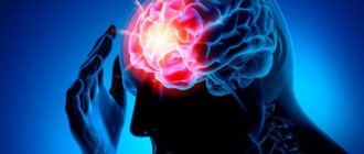
Stroke is a very dangerous disease and is becoming more and more “youthful”: if previously it was mainly older people who suffered from it, now it is not uncommon for young people to be hospitalized with this diagnosis. This happens most often due to overwork.
But older people with chronic diseases still need to take special care - some contribute to stroke, and also make it more difficult to recover.
There are ischemic and hemorrhagic strokes. They differ in many respects and are treated differently, but it is not always easy to distinguish them - many of the symptoms are similar, because in both cases the problem is a violation of blood flow in the head, although its causes are different.
Causes and risk factors
Ischemic stroke can have a variety of causes:
- stress, which provokes the release of catecholamines into the blood - when there is an excess of them, the blood vessels narrow and the blood supply to the brain suffers;
- heart pathology;
- diabetes;
- spasm of the carotid arteries;
- smoking – it damages the endothelium of the arteries;
- other.
Hemorrhagic stroke has its own set of causes, which are very different. Among them:
- amyloid angiopathy;
- inflammation of blood vessels;
- stress;
- constant overwork;
- poor blood clotting;
- incorrect use of certain medications;
- drug use.
Risk factors include:
- overwork leading to chronic fatigue;
- frequent stress;
- obesity;
- bad habits;
- hemophilia;
- a number of other diseases.
Each of these factors increases the likelihood of a stroke, and if several are combined, you should take special care of your health, since otherwise problems are inevitable.
A transient ischemic attack indicates that an ischemic stroke may soon occur. It may cause some part of the body to become numb for a short time and may also cause nausea. a feeling of disorientation, blurred vision and other symptoms.
Usually last no more than 15 minutes (mild) to several hours (moderate), or up to a day (severe).
Other warning signs of stroke:
- excessive excitement or lethargy;
- noise in ears;
- fainting;
- sleep problems;
- decreased performance and inability to concentrate;
- headaches without localization, aggravated by fatigue or changes in weather;
- dizziness, especially severe when walking.
If such signs recur, they cannot be ignored. With timely treatment and initiation of treatment, ischemic stroke can be avoided. Another thing is hemorrhagic. It often develops suddenly, without any warning at all. But sometimes they still exist:
- numbness of half the face;
- sharp pain in the eyes and partial loss of vision;
- difficulty understanding speech;
- inability to maintain balance.
They appear shortly before the onset of a stroke, so measures must be taken immediately.
Precursors of the development of cerebral infarction
Hemorrhagic stroke occurs suddenly. There are clear general cerebral and focal symptoms:
- Loss of sensitivity, paralysis of individual muscle groups;
- Severe nausea with spasmodic vomiting;
- Strong headache;
- Loss of consciousness, convulsions, coma.
Precursors of cerebral ischemia are mild general cerebral symptoms:
- Deterioration of hearing, “congestion in the ears”;
- Violation of the psycho-emotional state (overexcitation or inhibition);
- Darkening of the eyes, fainting.
Headache does not always accompany ischemic infarction.
Symptoms
Following the warning signs, the first symptoms appear. Feeling can deteriorate very quickly after this, so the speed of response is one of the main factors that can ensure survival.
The first symptoms may appear several hours before an ischemic stroke: weakness, headaches and dizziness, fainting. Limbs may be lost on one side or vision may be impaired - in some cases only affecting one eye.
Other signs that can help identify ischemic stroke:
- the patient perceives reality incorrectly;
- his speech becomes unclear;
- pulse quickens;
- the pressure rises and falls;
- auditory perception deteriorates;
- he has difficulty swallowing;
- there is noise in the head;
- movements are not coordinated;
- nausea;
- heat;
- the face is distorted;
- pain appears that a person has not experienced before, for example, the chest or half of the face begins to ache.
The following symptoms indicate a hemorrhagic stroke:
- the head begins to hurt very badly;
- arrhythmia or tachycardia occurs;
- pressure rises;
- breathing becomes loud and hoarse;
- the patient may faint;
- photosensitivity increases;
- the face becomes asymmetrical, turns red or pale;
- convulsions begin;
- the gaze loses its meaning;
- nausea or even vomiting begins;
- limbs or the whole body are paralyzed;
- bowel movements involuntarily.
Differential diagnosis - how doctors detect stroke
Differential diagnosis is extremely important for identifying types of stroke for which treatment is different. Diagnosis is carried out using instrumental studies:
- Computed tomography
. Necessary to determine the amount of hemorrhage and the type of stroke. - Diffusion-weighted tomography
. It is needed to identify areas of the brain with impaired blood circulation and takes 1-2 minutes. - Angiography
. Allows you to assess the condition of blood vessels and identify the presence of blood clots in them.
An MRI is rarely done if a stroke is suspected. This is due to the duration of the study.
Additional studies that are prescribed for illness:
- ECG;
- Ultrasound of the heart;
- ECHO KG.
Differential diagnosis is also performed based on symptoms. Its data is presented in the table
Difference in symptoms:
| Symptoms | Ischemic form | Hemorrhagic form |
| Development speed | Gradual | Swift |
| Change in the patient's appearance | Absent | There is redness of the face and heavy sweating |
| Headache with nausea | Rarely | Often |
| Change in muscle tone | Present | Present |
| Loss of consciousness | Short term | Sequels |
| Increased blood pressure | Yes | Sometimes |
| Problematic speech | Yes | Sometimes |
| Retinal condition | Clean | With hemorrhages |
Data from instrumental differential diagnostics for stroke
| Research | Result in ischemic form | Results for hemorrhagic form |
| ECG | Presence of arrhythmia | Enlargement of the heart |
| Fundus examination | Vascular changes | Hemorrhages |
| ECHO | No offset | With a shift to the healthy hemisphere |
Diagnostics
If warning signs appear, you should consult a doctor as soon as possible. He will conduct a simple test to make a preliminary diagnosis. The patient will need:
- Smile - if the lips on one side do not obey, the smile will turn out crooked.
- Extend your arms - they should be kept at the same level.
- Say something meaningful - for example, your full name.
- Stick out your tongue - it should not deviate to the side or sink.
By how these exercises are performed, it will be possible to determine whether the patient has suffered a stroke or whether the cause should be sought elsewhere. If signs indicate a stroke, the diagnosis is clarified using tomography and other procedures.
Clinical picture
A major stroke occurs with damage to a large area of the brain. The clinical picture is pronounced, and the damage poses a danger to the patient’s life. It is important to seek emergency help if the victim exhibits the following symptoms:
- a sharp decrease in hearing and vision, impaired coordination of movements, inability to concentrate;
- acute headache, dizziness;
- nausea and vomiting;
- redness of the skin and mucous membranes or their pallor;
- decreased skin sensitivity, often on one side of the face or torso, muscle paralysis;
- the appearance of memory lapses;
- fainting.
With a major stroke, there is still a risk of developing coma. This is a condition in which the victim does not respond to any external stimuli, but his breathing and heartbeat continue. The coma can last for a long time, and the chances of further recovery depend on this.
First aid and treatment
An ambulance should be called immediately at the first suspicion. Until she arrives, you will need to provide assistance yourself:
- lay the victim down and raise his head;
- make sure that his clothing does not restrict breathing;
- make sure that your tongue does not sink;
- do not give anything to drink or eat;
- ventilate the room;
- cool your head with compresses;
- when vomiting, make sure that he does not suffocate; his head should be turned to the side;
- If necessary, perform chest compressions or artificial respiration.
Please note: no medications should be used without consulting a doctor; in this condition, taking them can be extremely dangerous.
There are several stages in the treatment of stroke:
- first aid;
- hospital treatment;
- rehabilitation.
It is very important to provide first aid correctly, because it is the first hours of a stroke that are most often decisive: if time is lost, death is very likely. If everything went well, then intensive therapy is carried out in the hospital, with the help of which they try to ensure that the brain is damaged as little as possible, and also to prevent a recurrent stroke.
Gradually, the intensity of therapy decreases, but it continues for several weeks and sometimes months - the patient’s health must be carefully monitored during this time.
First aid for a stroke before the ambulance arrives
It is difficult to provide first aid to yourself for a stroke. Therefore, you need to immediately call an ambulance and call relatives and neighbors.
Basic rules for what to do in case of a stroke before the ambulance arrives, if no one is nearby:
- free your chest from clothing;
- open a window in the room;
- lie on your back so that your head is higher than your body;
- do not move unless necessary.
Algorithm of what to do and how to help a person with a stroke before the ambulance arrives:
- Reassure the patient if possible
. Stress aggravates the condition of the brain and contributes to the rapid progression of the disease. - Call an ambulance at 112 or 103 from your mobile phone
. - Provide resuscitation measures
I. If a person is not breathing, then artificial respiration is given to him (see the picture). If there is no pulse, then first aid is supplemented with artificial heart massage until the ambulance arrives. Massage technique:- the right palm is placed on the left or vice versa;
- The lower palm is placed in the chest area, and the upper palm is pressed on it at intervals of 100 times per minute. Every 30 pressures, perform artificial respiration with 2 breaths into the patient’s mouth.
- Ensure the patient's body is in the correct position
. If he is conscious, then he is placed on his back, so that the chest and head are in an elevated position. If there is no consciousness, the victim is placed on his side. In this position, he will not choke on vomit and saliva.
What to do if you have a stroke at home
It is possible to give medication to a victim for a stroke only if the ambulance is delayed for more than 40 minutes. List of medications that help prevent oxygen starvation of the brain:
- Piracetam;
- Furosemide;
- Lasix.
It is better to administer drugs for a stroke attack intramuscularly. This way they will act faster.
According to WHO statistics, correctly provided first aid helps save 50-60% of patients with a severe form of stroke and 75-90% with a mild one.
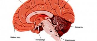
Consequences of ischemic stroke of the right and left side
The consequences of an ischemic stroke will vary depending on the damaged hemisphere. If it is the left one (it is affected much more often), then the right side of the body may be lost. This hemisphere is responsible for logic and speech, hence the other consequences:
- speech is disrupted;
- I have to learn to read and write again;
- loss of speech memory occurs - the patient immediately forgets what he was talking about;
- if help is provided late or incorrectly, he will withdraw into himself and stop communicating.
Restoring memory and speech is difficult in such cases; the patient may not even understand speech. Physical activity is restored much faster.
If the right hemisphere is damaged, then the symptoms clearly appear later, so more time usually passes before seeking help, and the consequences are more serious:
- the patient seems to be completely paralyzed, although this is not the case;
- he forgets what he just did, while he can well remember long-ago events;
- The perception of reality and sense of space are disrupted, depression is observed, and the patient becomes very passive.
Recovery takes a very long time.
Hemorrhagic stroke
Hemorrhagic stroke - hemorrhage in the brain and/or subarachnoid space, occurs four to five times less often than ischemic stroke
- Etiology of hemorrhagic stroke
The main etiological factors of hemorrhagic stroke are hypertension, arterial hypertension, congenital and acquired arterial and arteriovenous aneurysms. Subdural and epidural hematomas are usually of traumatic origin. Less commonly, the cause of hemorrhagic stroke can be hemorrhagic diathesis, the use of anticoagulants, amyloid angiopathies, mycoses, tumors, and encephalitis.
The predominant localization of hematomas is the cerebral hemispheres (about 90% of parenchymal hemorrhages); in 10% of cases, damage to the brain stem or cerebellum is detected. In most cases, there is a rupture of the vessel, much less often - diapedetic hemorrhages.
The clinic of parenchymal hemorrhages has general cerebral and focal symptoms. The clinical picture of subarachnoid hemorrhages includes two main groups of symptoms: cerebral and meningeal. In the presence of these and focal symptoms, we are talking about subarachnoid-parenchymal hemorrhage. Features of the clinical picture of parenchymal hemorrhages depend on the location of the hematoma.
- Hemorrhagic Stroke Clinic
Parenchymal hemorrhages. Hemorrhage into the putamen occurs with severe impairment of consciousness and a neurological defect in the form of contralateral hemiplegia, hemianesthesia, aphasia (with damage to the dominant hemisphere) or spatial hemiagnosia and anosognosia (with damage to the non-dominant hemisphere). The clinical picture is similar to that of middle cerebral artery occlusion.
With hemorrhages in the thalamus, as well as with hemorrhages in the putamen, herniation and coma are possible. Important signs of thalamic damage are greater severity of sensory disorders than motor ones, and unusual oculomotor disorders, often in the form of limited gaze, strabismus.
Table 1. HUNT Scale (Henry JM Barnett, Stroke, 1986) | |
| Degree | Characteristic |
| 0 | Unruptured aneurysm |
| I | Asymptomatic or minimal headache and mild neck stiffness |
| I.A. | Absence of meningeal or cerebral symptoms, but presence of persistent neurological deficit |
| II | Moderate to severe headache, stiff neck; no neurological deficit other than cranial nerve palsy |
| III | Drowsiness, confusion (disorientation in time and space), or mild local deficits |
| IV | Stupor, moderate to profound hemiparesis, possible early decerebrate rigidity and autonomic disturbances |
| V | Deep coma, decerebrate rigidity and signs of agony |
Hemorrhage into the pons is usually characterized by early development of coma, pinpoint pupils that do not respond to light, and bilateral decerebrate rigidity.
Hemorrhage into the cerebellum is characterized by sudden dizziness, vomiting in combination with severe ataxia, abasia, aesthesia and gaze paresis. Consciousness is not impaired, but compression of the trunk can lead to death.
Subarachnoid hemorrhage. Aneurysm rupture. Subarachnoid hemorrhage (SAH) is most often caused by the rupture of a saccular aneurysm, a defect in the internal elastic membrane of the arterial wall, usually occurring at the site of a bifurcation or branch of an artery. In most cases, the gap occurs between the ages of 35-65 years. There may be associated anomalies such as polycystic kidney disease or coarctation of the aorta. Sudden, unexplained headache of any location should raise suspicion of SAH and a computed tomography (CT) scan should be performed. For aneurysms larger than 7 mm, microsurgical obliteration is justified.
Another type of aneurysm is located along the internal carotid, vertebral or basilar artery; Depending on their structure, they are divided into fusiform, spherical and diffuse. Such aneurysms become clinically apparent when they put pressure on adjacent structures or due to thrombosis, but rarely rupture.
Table 2. Classes of social and everyday activity(Schmidt E.V., Makinsky T.A., Research Institute of Neurology, 1979) | |
| I | Return to work and complete independence from others |
| II | Return to work with limitations, independence in activities of daily living, walking without assistance |
| III | Limitation of previous household duties, partial dependence on others in everyday life, walking around the apartment without assistance, walking down the street with assistance |
| IV | The impossibility of returning patients who previously worked and suffered a stroke to work; for those who were engaged in housework, there is a significant limitation in the range of household duties or a complete inability to perform them, significant dependence on others in everyday life. Walking around the apartment with assistance. Sick people don't walk down the street |
| V | Complete loss of any productive activity. Complete dependence on others in everyday life |
A ruptured aneurysm is characterized by a sudden, intense headache. The patient usually says that he has never experienced such a severe headache before. Possible loss of consciousness; sometimes it turns into a coma, but more often consciousness is restored, although stupor remains. In some cases, loss of consciousness occurs suddenly, before the headache appears. SAH often occurs during exercise. When an aneurysm ruptures, the diagnosis is usually simple, but sometimes at an early stage there are no objective symptoms, so if there is a sudden headache, the doctor must think about subarachnoid hemorrhage.
Meningeal symptoms and low-grade fever are often present. Ophthalmoscopy often reveals subhyaloid hemorrhages.
Hemorrhage may be limited to the subarachnoid space or spread to the brain, causing focal symptoms. Sometimes, soon after hemorrhage, an ischemic stroke develops due to impaired blood flow or thrombosis in the arteries affected by the aneurysm.
It is not easy to determine the location of an aneurysm clinically, although it is sometimes possible. Thus, pain in the depths of the orbit and damage to the II-VI cranial nerves indicate an aneurysm of the cavernous part of the carotid artery; hemiplegia, aphasia and a number of other symptoms - for an aneurysm of the middle cerebral artery; damage to the third cranial nerve - an aneurysm at the junction of the posterior communicating and internal carotid arteries; abulia and weakness in the leg - due to an aneurysm of the anterior communicating artery; damage to the lower cranial nerves - an aneurysm of the basilar or vertebral artery.
A transient or persistent focal neurological defect that develops several days after a stroke is usually caused by a spasm of cerebral vessels that occurs in response to blood entering the subarachnoid space. Both an early and late complication of SAH can be hydrocephalus, which sometimes requires ventricular bypass.
Arteriovenous malformations. Arteriovenous malformations usually manifest as epileptic seizures or hemorrhage, but with large lesions, ischemia of adjacent areas of the brain may occur due to large blood flow. Most often this is a combined parenchymal-subarachnoid hemorrhage. People usually suffer from arteriovenous malformations in childhood and adolescence. That is why, for persistent headaches at this age, listening in the area of the orbit, carotid artery, and mastoid process is necessary.
The presence of vascular murmurs in these areas is pathognomonic. In doubtful cases, as well as for the purpose of differential diagnosis of telangiectasia and other angiomas, a CT scan can be done.
- Diagnosis of hemorrhagic stroke
CT is the method of choice. It allows not only to confirm the diagnosis, but also to determine the extent of the lesion in intracerebral parenchymal hemorrhages. CT is the best method for diagnosing SAH, and in most cases reveals blood in the subarachnoid space. This method also makes it possible to diagnose cerebral edema, parenchymal and intraventricular hemorrhage, and hydrocephalus. It is possible to identify the localization of the source in intrathecal hemorrhage.
Magnetic resonance imaging (MRI) compared to CT is more reliable in diagnosing small hematomas localized in the pons and medulla oblongata, as well as hematomas in which the X-ray density of blood clots inside is equal to the density of brain tissue. MRI also makes it possible to identify arteriovenous malformations that are accessible to surgical intervention, which are very difficult to diagnose with CT, especially without contrast enhancement.
Cerebrospinal fluid examination is indicated only in cases where computed tomography is not available. Blood in the cerebrospinal fluid is detected in all cases of SAH, as well as with hemorrhages in the cerebellum and pons; with minor hemorrhages in the putamen and thalamus, red blood cells may appear in the cerebrospinal fluid only after 2-3 days.
X-ray of the skull reveals calcified malformations and aneurysms. As a rule, it is not carried out.
Cerebral angiography is usually performed immediately before surgery to clarify the location and anatomical nature of the aneurysm, as well as to confirm the presence or absence of focal cerebral vagospasm. In severe cases, angiography is best performed only when the diagnosis is unclear and especially when surgical decompression is indicated.
- Differential diagnosis of strokes
Cerebral crises precede cerebral hemorrhage; the disease begins violently, suddenly, often during the day due to physical stress or excitement. Precursors are characteristic (flushing to the face, headache, seeing objects in red); prolonged comatose states develop (sometimes several days); the face is hyperemic; temperature rises; breathing is bubbling, hoarse; pulse intense, rare; accent of the second tone at the top; blood pressure is increased; miosis or mydriasis on the side of the lesion; focal symptoms are identified in the form of rapid development of hemiplegia with a decrease in muscle tone, reflexes, and skin temperature; sometimes epileptiform seizures or early contractures (tonic spasms, protective hyperreflexia) occur; pronounced meningeal phenomena, brainstem disorders (breathing disorders, vomiting, floating movements of the eyeballs); pseudobulbar reflexes are rarely detected, urinary retention or incontinence is observed; retinal hemorrhages are visible in the fundus; cerebrospinal fluid is hemorrhagic, xanthochromic, pressure is increased; leukocytosis in the blood, prothrombin is not increased; in the urine there are red blood cells, sometimes sugar and protein.
Ischemic thrombotic stroke is preceded by transient cerebrovascular accidents. The disease develops gradually, more often at night, in the morning or during sleep; there are warning signs (dizziness, short-term disturbances of consciousness); characterized by incomplete or short-term loss of consciousness; the patient's face is pale, the temperature usually does not rise; slow breathing, weak pulse; heart sounds are muffled; blood pressure is not elevated; the size of the pupils most often does not change; focal symptoms appear in the form of hemiplegia or monoplegia with low muscle tone, one-sided Babinski reflex; hemiplegia develops gradually and is unstable; epileptiform seizures are not typical; there are no meningeal phenomena; stem phenomena are rarely observed (with extensive foci); with repeated strokes, pseudobulbar reflexes occur; sometimes there is urinary incontinence; narrowing and unevenness of blood vessels are visible in the fundus; the cerebrospinal fluid is clear, the pressure is normal; hypercoagulation is detected in the blood; urine specific gravity is low.
Non-thrombotic ischemic stroke is preceded by crises, angina, myocardial infarction, etc.; the disease develops suddenly during the day, more often after physical activity; often without warning; characterized by short-term loss of consciousness, stupor; the face is pale; the temperature is elevated; weakened, slow breathing; pulse is arrhythmic, weakened; muffled heart sounds, sometimes atrial fibrillation; blood pressure is low, pupils are constricted; transient hemiplegia develops with mildly increased muscle tone and a one-sided Babinski reflex; epileptiform seizures are rare; meningeal and stem phenomena are rare; pseudobulbar reflexes are often detected; there is urinary incontinence; sclerosis and narrowing of retinal vessels are visible in the fundus; cerebrospinal fluid is clear, its pressure is sometimes increased; Prothrombin is increased in the blood, traces of protein are detected in the urine.
- Treatment of hemorrhagic stroke
General principles. Along with differentiated therapy for hemorrhagic stroke, basic therapy aimed at maintaining vital body functions plays an important role. The more severe the course of the stroke, the more necessary is multilateral and complex basic therapy, which is carried out individually, under the control of laboratory parameters and the functions of all organs and systems.
In connection with modern pathogenetic concepts, early diagnosis of cerebral stroke, clarification of its nature and organization of emergency medical care at the prehospital and hospital stages are of particular importance. The effectiveness of treatment measures depends on the timeliness of their initiation and on the continuity of therapy in all periods of the disease.
The continuity of treatment measures is determined by the general tactics of patient management and is associated with solving organizational problems: rapid transportation of the patient, clear organization of the work of the emergency department; early clarification of the diagnosis and resolution of the issue of referral to the appropriate department; smooth operation of all levels of assistance.
Neurosurgical intervention. The problem of hemorrhagic stroke, according to most researchers, is largely neurosurgical. If ischemic stroke is a process of development of hemodynamic and metabolic changes, ending mainly a few days after an acute cerebrovascular accident, then hemorrhagic stroke is a fait accompli of hemorrhage, and its pathogenesis involves secondary phenomena of already shed blood.
Removal of a hematoma after an intracerebral hemorrhage, if it is localized in an accessible area of the brain (for example, in the cerebellum, putamen, thalamus, or temporal lobe), can save the patient's life. The operation is indicated as early as possible (24-48 hours) for aneurysm ruptures, if the patient’s condition does not improve and signs of herniation appear. The main operation is clipping the neck of the aneurysm; wrapping the aneurysm with muscle or, less commonly, extracranial occlusion of the internal carotid artery is also performed.
Patients whose condition corresponds to grade 0 - III on the Hunt scale have no contraindications on this scale for hospitalization in the neurosurgical department (Table 1).
Differentiated conservative therapy. Conservative therapeutic interventions for hemorrhagic stroke should be aimed at rapid correction of blood pressure to optimal values for a particular patient; to combat developing cerebral edema and to carry out hemostatic and vascular wall-strengthening therapy.
Correction and control of blood pressure. If possible, blood pressure (BP) should be avoided. They try to keep the blood pressure within normal limits using antihypertensive drugs (beta blockers, calcium antagonists, antispasmodics, ACE inhibitors). To prevent emotional reactions, sedative therapy (diazepam, elenium) is prescribed. Sometimes phenobarbital is prescribed for prophylactic purposes (30 mg orally three times a day), since it also has an anticonvulsant effect.
To avoid straining, laxatives (regulax, glaxena, senade, etc.) are prescribed. It is necessary to create conditions for “protective inhibition”; protect from light and noise.
Hemostatic therapy and therapy aimed at strengthening the vascular wall. Prescribe dicinone (sodium ethmazilate) intravenously or intramuscularly (250 mg four times a day); antiprotease drugs for 5-10 days: gordox (100 thousand units four times a day intravenously) or contrical (30 thousand units immediately, and then 10 thousand units twice a day intravenously).
Calcium preparations (calcium pantothenate, berrocca, calcium gluconate - i.m., calcium chloride - i.v.), rutin, vikasol, ascorbic acid strengthen the vascular wall well.
Antifibrinolytic therapy in the form of gamma-aminocaproic acid up to 30 g per day (100-150 ml of a 5% solution intravenously, then orally) is of great importance. It can be administered with small doses of rheopolyglucin, which improves microcirculation.
Fighting cerebral edema. If lethargy or signs of herniation appear, it is better to prescribe osmotic diuretics mannitol (0.5 - 1.5 g/kg of the patient’s body weight, IV) or glycerin (1 g/kg orally). Less commonly prescribed corticosteroids are dexazone according to the scheme (8+4+4+4 mg IV). More effective are Lasix (20 mg IV twice a day) and/or Reogluman (200 ml IV drip twice a day).
Treatment of spasm of cerebral vessels. Signs of cerebral vasospasm (drowsiness, focal symptoms) appear after two to three days and most often on the seventh day after a hemorrhagic stroke. It is believed to be caused by the release of serotonin, catecholamines, peptides and other vasoactive substances. Prescribed for spasms, and even better for preventive purposes in advance. Calcium antagonists - nimoton (10 mg IV drip) - 10-14 days or nimodipine (30-60 mg orally every four hours). In this case, correction of antihypertensive therapy is necessary, since calcium antagonists affect blood pressure.
Rehabilitation treatment. Rehabilitation therapy is carried out over a long period of time and at all stages of treatment, but it is especially important after the acute period of a stroke. Physiotherapy exercises are combined with physiotherapy, acupressure and classical massage, acupuncture, electrical stimulation, and magnetotherapy.
Occupational therapy is required - training in self-care skills, work on training stands and work simulators. Psychotherapy is effective: individual, group, family; Autogenic, adaptive training, etc. are recommended. Speech therapy classes are required for persons with speech impairments.
The patient needs treatment for a total of at least three to four months. For severe strokes, depending on the patient’s condition, this period can be extended to six months or more. Patients are registered at the dispensary. With a prolonged period of restoration of functions, patients are transferred to disability.
- Work ability examination
Currently, there are five classes of social and everyday activity (Table 2).
The disability group is determined according to the severity of dysfunction and profession. Patients with paralysis of the limbs and aphasia need care from outsiders and are recognized as group I disabled people. In case of deep paresis, when the ability to self-care remains, but the ability to work is lost, disability group II is assigned. Group II patients adapt to working from home: typing, assembling parts, doing dispatch activities on the phone, etc.
Literature
1. Bogolepov N.K. Clinical lectures on neurology, 1971. 2. Internal diseases. Ed. Harrison T. R., vol. 10, 1997. 3. Gusev E. I., Skvortsova V. I., Chekneva N. S. et al. Treatment of acute cerebral stroke (diagnostic and therapeutic algorithms), M., 1997. 4. Allen GS, et al. Cerebral arterial spasm: A controlled trial of nimodipine in patients with subarachnoid hemorrage. //N. Engl. J. Med. 308:619, 1983. 5. Cerebral embolism study group. Brain hemorrhage and management options. Stroke 15:779, 1984. 6. Curling OD et al. An analysis of natural history of cavernosus angiomas. J Neurosurgery 75:702, 1991. 7. Henry JM, Barnett. Stroke: Pathophysiology, Diagnosis, Management. 1986. 8. Manual of Neurologic Therapeutics. Edited by Martin A. Samuels, MD 1995. 9. Nibbelink DW, Torner JC, Henderson WG Interactional aneurysms and subarachnoid hemorrage: A cooperative study. Antifibrinolytic therapy in recent onset subarachnoid hemorrhage. Stroke 6:662, 1975. 10. Origitano TC et al. Sustained increased cerebral blood flow with prophylactic hypertensive hypervolemic hemodilution (“Triple-H Therapy”) after subarachnoid hemorrage. Neurosurgery 27:729, 1990. 11. Wilkins RH Natural History of Intracranial Vascular Information: A review. Neurosurg 16:421, 1985.
Prognosis for patients
With ischemic stroke, complete recovery occurs in only 10–15% of patients. Up to a quarter may return to work. Survival rate is about 50% (one year after stroke). In addition, those who have suffered a stroke are at risk - within 5 years, approximately 55% have a relapse, and the prognosis is even less favorable.
It depends on a number of factors, such as the size of the ischemic lesion and its location, the age of the person and his chronic diseases.
With a hemorrhagic stroke, the prognosis is usually even worse: if a cerebral hemorrhage occurs, in more than 60% of cases it ends in death in the first month. There is a high risk of recurrent cerebral hemorrhage. Of those who managed to survive, about a third become disabled.
In each specific case, the prognosis depends on many factors: the condition of the body as a whole, the location and volume of hematomas, and the promptness of assistance.
With subarachnoid hemorrhage, the prognosis is more favorable, quite often even complete recovery occurs, but the mortality rate is still very high - about 40%.
Rehabilitation
Rehabilitation after a stroke is a very important point. It should start as early as possible. After the specialist assesses neurological disorders, he will prescribe rehabilitation therapy that is appropriate to the situation.
It could be:
- physiotherapy;
- massage;
- paraffin applications;
- electrical stimulation of paralyzed limbs;
- classes with a speech therapist-aphasiologist;
- sessions with a psychologist.
It is advisable that the child’s rehabilitation be carried out in special centers. It has modern medical equipment and qualified specialists. After your condition improves, you can continue recovery at home.
After a stroke, many children retain neurological disorders that negatively affect their quality of life and adaptation in society. Doctors and parents should make every effort to reduce these disturbances to a minimum. Psychological support from loved ones, an optimistic attitude, and complete completion of all rehabilitation courses will help the child recover faster and better after a stroke.
Prevention
To never have a stroke, you need to take care of your health. To prevent it you need:
- to refuse from bad habits;
- eat right, in particular, do not eat fatty foods;
- watch your weight and do exercises.
Nothing complicated, but if you systematically follow these recommendations year after year - preferably from a very young age, the risk of not only a stroke, but also the occurrence of many other diseases will significantly decrease. A healthy lifestyle not only significantly prolongs it, but also allows you to stay in good shape for many years.
It is worth emphasizing several important nuances. The fight against excess weight is very important: every kilogram in excess of the norm leads to stress on the heart and blood vessels, and to the development of related diseases. Eating vegetables and fruits will help make blood vessels more elastic and get rid of high blood pressure.
Among physical activities, aerobic training is especially useful: swimming, cycling, running and walking. They will also help you get rid of excess weight. You need to exercise at least half an hour a day, but if your physical condition is advanced, you should first consult a doctor and monitor your blood pressure.
Interestingly, quitting drinking completely is not necessary. Of course, you should not abuse it, but small portions of alcohol can even be useful in preventing stroke, as they prevent the formation of blood clots. The daily portion is 17 ml of pure alcohol - this is, for example, about 150 ml of wine.
But in case of chronic diseases, drinking alcohol even in such moderate quantities should be discussed with your doctor.
Postoperative management of elderly patients
After the operation is completed, the postoperative period begins. It is divided into near and distant. The immediate period begins immediately after surgery and continues until discharge from the hospital. Remote begins from the moment of discharge until the patient is no longer bothered by general and local disorders.
In elderly patients, the functioning of the respiratory and cardiovascular systems is reduced. In addition, their body cannot, due to age, resist infections. Reparative processes during the healing of postoperative wounds are also deteriorated. These factors make the recovery period more difficult.
Age negatively affects the vital capacity of the lungs, reducing their maximum ventilation and disrupting the drainage function of the bronchial tree. The result is often pneumonia. During rehabilitation, breathing exercises, massage, and bronchodilators are used.
Almost all older people suffer from atherosclerosis and cardiosclerosis. There is a tendency to hypercoagulation - increased blood clotting, which leads to the formation of blood clots
It is important to thin the blood and activate patients. Physical activity reduces the risk of blood clots
Gastric and acid-enzymatic functions of the gastrointestinal tract are reduced, so high-calorie foods that are easily digested are prescribed. In older people, purulent complications develop, so you need to carefully monitor wounds.
From the editor: Symptoms of a microstroke experienced on the legs