Cavernous angioma is a benign neoplasm in the brain, which consists of pathologically developed blood vessels, which determines the nature of the course of the disease. In most cases, it is a congenital defect of the blood flow system. Unlike vascular malformations (improper connection of arteries and veins) of another type, the pathology is usually asymptomatic. Cavernous angioma is often discovered incidentally during an MRI or CT scan ordered for another reason.
What is a cerebral cavernoma
Cavernoma (vascular malformation) is a benign tumor. When pressed, it is elastic and soft, initially disappears, but soon returns to its original shape.
Important! Cavernous angioma may bleed, which can subsequently cause infection.
Cavernoma (ICD code 10 – D33) is a brain tumor in the form of vascular cavities (cavities) containing air or blood breakdown products. These breakdown products consist of blood clots, connective tissue, etc. The size of cavities and their number can be different - several neoplasms can be adjacent to one another or located at some distance from each other. Cavernous angioma has a knobby, bluish-colored surface with a heterogeneous structure; the tumor has clear boundaries that separate it from other tissues; most often it is round in shape. The partitions between the cavities consist of dense fibrous tissue. The size of the cavernoma can vary - from very small to several centimeters in diameter.
Multiple cavities occur in 10-15% of cases. Such cavernomas have a predisposition to frequent hemorrhages, and the medulla that surrounds them becomes yellowish as a result.
Cavernoma is localized in the head and neck area and affects the brain and spinal cord. This pathology occurs due to a genetic mutation during intrauterine development, when the formation of areas of the nervous system is disrupted. The presence of a cavernoma leads to a person’s loss of ability to adapt socially.
Stereotactic radiosurgery of cavernous angiomas of the brain stem
GAMMA KNIFE AND CAVERNOSAL ANGIOMAS OF THE BRAIN
Cavernous angiomas of the brain (cavernomas) are most often congenital vascular anomalies, represented by single, much less often multiple cavities, separated inside by septa (septa) and filled with blood. Despite the supposed congenital nature, they are more often detected in adults than in children, but in children they are more severe. The blood supply to the cavity is carried out from small arterioles and capillaries, and the outflow of blood through venules is of the same order. Due to the small caliber of the feeding vessels, the blood pressure in cavernomas is low, so the draining veins do not hypertrophy and are not visible during SCT/MRI/angiography.
Schematic representation of the structure of cavernous angioma
A special feature of cavernomas is a defective vascular wall, so thin that the cellular elements of the blood - red blood cells - “sweat” through it and settle in the adjacent medulla. This process is called “diapedetic hemorrhage.” The products of the natural breakdown of hemoglobin (hemosiderin), contained in red blood cells, form a zone of chronic, specific and very recognizable changes on MRI around the cavernoma. Cavernomas can be localized in any part of the brain: in the cerebral hemispheres, in the brain stem, cerebellum, subcortical ganglia, in the corpus callosum and in the lateral ventricles. In approximately 30-40% of cases, cavernous angiomas are combined with another type of vascular malformation - venous angiomas. Venous angiomas are an anomaly in the development of venous vessels in the form of a “bundle” of small veins (“Gorgon’s head”) gathering into one large drainage vein.
The incidence of cavernous angiomas is several cases per million population.
WHAT IS THE THREATEN OF THE PRESENCE OF CAVERNOUS ANGIOMA?
Cavernomas can remain asymptomatic throughout life. However, in a number of patients, clinical manifestations can be of two types, both individually and in combination:
Hemorrhage. Sometimes, an increase in blood pressure inside a cavernoma leads to local destruction of the vascular wall and the formation of intracerebral hemorrhage. Unlike AVMs, hemorrhages from cavernomas are never massive and do not pose a threat to the patient’s life, with the exception of extremely rare cases of cavity localization in the lower parts of the medulla oblongata, where the centers of cardiovascular and respiratory regulation are located. However, when the focus of hemorrhage is located in any functional area, even a small volume of hemorrhage can lead to the appearance of neurological symptoms (for example, the development of weakness in the opposite half of the body, when located in the motor cortex or in the deep structures of the cerebral hemispheres).
Epileptic syndrome. In some cases, due to the chronic presence of hemosiderin in the medulla or with the development of acute hemorrhage, a focus of pathological brain bioactivity may form, clinically manifested by epileptic seizures of various structures (convulsive, non-convulsive, absence seizures, vegetative, polymorphic and others). Thus, cavernomas extremely rarely pose a threat to the patient’s life, but can have a significant impact on the quality of life.
HOW TO DIAGNOSE A CAVERNOMA?
- Magnetic resonance imaging (MRI) is the most informative method for diagnosing cavernomas. In T1 and T2 scanning modes, a cavity surrounded by a black rim of hemosiderin may be visible, with signs of acute and subacute hemorrhages. The most sensitive mode is T2* (“T2 with star”), which allows you to diagnose even small (1-2 mm) cavernomas that are not visible in other scanning modes.
- Computed tomography - its diagnostic value is limited, as a rule, to the acute period of hemorrhage (several days), when a fresh focus of shed blood is visible on the image.
- Angiography is not informative in diagnosing cavernomas
HOW TO GET RID OF A CAVERNOMA?
In the case of asymptomatic cases of incidentally detected cavernous angiomas, the generally accepted tactic is dynamic observation. In case of episyndrome formation and/or hemorrhage, two treatment methods are possible:
- Surgical removal of cavernoma is a highly effective method that allows you to once and for all relieve the patient from the risk of repeated hemorrhages. Surgery is also effective if the patient has epileptic seizures, but its effectiveness is reduced in the case of a long history of epileptic syndrome.
- Radiosurgery is performed on the same principle as for arteriovenous malformations - with the goal of causing complete obstruction of blood flow. However, radiation doses in this case may be significantly less than with AVM. Morphological changes in this case are similar to those in arteriovenous malformations - the walls of the cavernoma and the mouths of the feeding vessels undergo hyalinosis, as a result of which the blood flow decreases, pinpoint hemorrhages stop, and the risk of repeated spontaneous intracerebral hemorrhages is reduced by 50-70%. Episodic attacks also become less frequent or disappear completely, but, as in the situation with surgery, the longer the history of the disease, the less likely it is to completely recover from them.
- A meta-analysis of the literature on the effectiveness of the use of Gamma Knife (World Neurosurg. 2021 Mar; 123: 371-377), conducted by researchers from the University Hospital of Sichuan (PRC), proves that the annual risk of hemorrhage is reduced many times and significantly within the first 2 years after radiosurgical treatment, continuing to decrease in subsequent years. Thus, today, radiosurgery is the method of choice in the treatment of patients with cavernous angiomas.
DO YOU NEED TO IRRADIATE MULTIPLE CAVERNOMAS? As in the case of single cavernomas, radiosurgical treatment is carried out only on the lesion that manifests itself clinically - hemorrhage or epileptic seizures.
WHAT TO DO WITH VENOUS ANGIOMA? The question often arises: how to get rid of venous angioma? Firstly, you should know that venous angiomas never manifest themselves as hemorrhages or epileptic seizures. Secondly, although they represent a venous network that is abnormal in structure, they nevertheless take full part in the local venous circulation of the brain.
The only option for the appearance of clinical symptoms associated with venous angioma is the spontaneous development of thrombosis of the draining vein. This situation is more hypothetical than real. In this case, the patient will experience cerebral venous infarction with severe swelling at the site of impaired venous outflow. And only one way can help cope with such a situation - thrombolytic and anticoagulant therapy. Attempts to surgically or radiosurgically influence venous angiomas not only do not make any sense, but will also lead to gross local disturbances of venous blood flow in a manner similar to spontaneous thrombosis. Thus, diagnosing a patient with venous angioma is not a reason for any treatment.
WHAT IS THE BEST TREATMENT METHOD FOR CAVERNOMAS?
Brainstem hemorrhages caused by cavernous angiomas can cause severe neurological deficits due to destruction of neural tissue, ascending and descending brain pathways, and cranial nerve nuclei.
The natural course of brainstem cavernomas differs from similar manifestations in other localizations, because has a higher risk of hemorrhage and permanent neurological deficit. After the first hemorrhage, cavernomas become unstable and the risk of repeated hemorrhages increases.
From the point of view of preventing recurrent hemorrhages, the effectiveness of surgical treatment is higher. However, when cavernomas are localized in functionally important or hard-to-reach areas of the brain (for example, in the subcortical nuclei, brain stem), surgery itself has a high risk of residual neurological deficit, and radiosurgery can largely avoid or reduce the risk of these complications.
Brain stem cavernoma BEFORE Gamma Knife radiosurgery (left) and 2 years AFTER Gamma Knife (right)
Microsurgical resection is the first choice only for accessible brainstem cavernomas located beneath the arachnoid or ependymal surface, and even then carries a significant risk of complications.
Regarding radiosurgery, the results of a meta-analysis of 5 studies (4 using Gamma Knife and 1 using a linear accelerator) including 178 patients with truncal cavernomas revealed a significant reduction in the rate of recurrent hemorrhages after stereotactic irradiation. Thus, radiosurgery should be considered as the treatment of choice for the treatment of brainstem cavernous angiomas in cases with high surgical risk (J Neurosurg. 2014 Apr;120(4):982-7.)
The decision on the optimal method of influence is made individually, taking into account all possible factors, incl. and taking into account the informed choice of the patient himself.
Leading clinics in Israel
Assuta
Israel, Tel Aviv
Ikhilov
Israel, Tel Aviv
Hadassah
Israel, Jerusalem
Causes of cavernoma
Typically, cavernous angioma is considered a congenital neoplasm. During the period of intrauterine gestation, a violation of the structural and functional transformation of tissue cells occurs. The reason may lie in soft tissue trauma, which gives impetus to the formation of a tumor - cavernoma.
Other provoking factors are:
- infectious pathologies during pregnancy;
- difficult or premature birth;
- multiple or late pregnancy;
- birth injuries, placental pathologies;
- bad habits of a pregnant woman (smoking, alcoholism, drug use);
- severe intoxication;
- bad ecology;
- radiation exposure.
Cavernoma symptoms
Cavernoma is often asymptomatic, and the patient does not worry about anything. In such cases, it can be detected during a routine examination.
On a note! The symptoms that appear depend on the location and size of the tumor. The larger the tumor, the more pronounced the symptoms.
Pronounced symptoms of the presence of a cavernoma are observed in patients with a tumor in the brain stem, frontal lobe or temporal lobes (right or left):
- constant headaches;
- convulsions;
- epileptic syndrome;
- vomit;
- sensory disturbance;
- unsteady gait, poor coordination of movements;
- loss of hearing acuity, ringing and noise in the ears;
- disorders of vision, memory, attention;
- decreased mental abilities;
- paralysis.
With severe headaches, there is a high risk (4-23%) of rupture of the cavernoma wall and subsequent hemorrhage. If hemorrhage occurs repeatedly (relapse), then in 30% of cases it leads to disability of the patient.
Symptoms associated with the location of the cavernoma
Some symptoms of the disease are characterized by the localization of the malformation:
| Tumor location | Symptoms |
| Frontal lobe | Difficulty controlling the psycho-emotional state, problems with social adaptation, memory and hand motor skills (handwriting becomes illegible, involuntary twitching of the limbs begins) |
| Left temporal lobe | Speech and hearing disorders. It is difficult for a person to perceive someone else’s speech, the information he receives is not assimilated, and when speaking he often repeats the same phrases |
| Right temporal lobe | Difficulties arise in analyzing noise and sounds. A person has difficulty recognizing the voices of even familiar people |
| Parietal region | Intellectual disorders occur - the patient cannot solve simple mathematical problems, there is a loss of logical, technical thinking |
| Cerebellum | Speech intelligibility is lost, convulsions occur, and a “drunk” gait appears. The patient holds his head incorrectly, takes inappropriate poses |
| Right frontal lobe | Hyperactivity occurs. The person is overly emotional, talks a lot and creates the impression of being inadequate. If infectious diseases spread from the nasal cavities to the brain area, a thrombotic angioma occurs (symptoms: increased temperature, increased sweating, fever) |
Features of the clinical picture
The signs of this disease depend on the location of the vascular formation. The very first sign may be an epileptic seizure with a neurological disorder.
Symptoms are manifested by the presence of:
- headaches, which at the initial stage of development are mild, although periodic, but as the disease progresses, the pain becomes stronger, and medications do not help;
- seizures that have an epileptic form;
- tinnitus or ringing in the head;
- unsteady gait, dizziness, as well as impaired coordination and motor activity;
- dyspeptic disorders in the form of vomiting and nausea;
- the appearance of weakness in the limbs, as well as numbness and paralysis;
- dysfunctions of vision, speech and hearing, as well as deterioration of memory, attention and the appearance of confused thoughts.
How to recognize the disease?
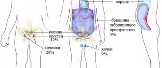
The appearance of symptoms depends on the location of the tumor. Vascular tumor appears in the upper parts of the brain in 80% of patients, in the cerebellum in 8%, and the rest is observed in the spinal cord.
Article on the topic: Good sedatives for depression
Symptoms depending on the location of the tumor:
- Temporal lobe – hearing and speech dysfunction. The patient does not recognize the voices of friends.
- Frontal lobe - memory deteriorates, the patient is unable to perform small movements, and handwriting also changes. Periodic apathy and depression are possible, or, conversely, inadequacy and euphoria. Speech deteriorates (poor vocabulary or excessive talkativeness may occur).
- Parietal region – intellectual disorder occurs.
- Cerebellum – unstable gait, speech disorders and strange movements occur (head turns, strange postures and bends).
Consequences of the disease
The presence of a cavernoma causes consequences associated primarily with neurological disorders and focal brain lesions. The tumor grows, squeezing the substance of the brain, and the symptoms described above begin to appear. After a hemorrhage occurs, the brain substance is saturated with hemosiderin and other metabolic products, the result is the shutdown of some functions. If the frontal lobe is damaged, the patient loses practical skills and cannot critically evaluate himself and others. If the right or left temporal lobes are damaged, loss of visual fields (hemianopia), impaired functioning of the hearing organs, and impaired ability to pronounce words (aphasia) may occur. If the lesion does not occur in the dominant area of the temporal lobe, the development of mental disorders is typical.
If the diagnosis of cavernoma occurred at a late stage of development, when the inflammatory process or dystrophic transformations began, then the following complications may occur:
- hemorrhages;
- cerebrovascular accident;
- rupture of blood vessels and disruption of local blood flow;
- increase in vascular congestions and cavities;
- death.
Treatment
Drug therapy for cavernomas is not necessary. After all, this tumor is not malignant. In addition, this neoplasm does not respond in any way to radiation therapy or treatment with anticancer drugs.
The only treatment method is surgery. Before deciding whether to perform surgery, the doctor, together with the patient, weighs the pros and cons. If cavernomas are small and do not cause any symptoms, then there is no need for intervention, there is only a risk of consequences.
There are strictly defined indications for any operation, because this method of treatment is far from harmless, and sometimes very dangerous for life and prognosis.
- Hemorrhage localized outside the functional areas associated with a cavernoma.
- Epileptic seizures caused by cavernous hemangioma.
- Brain stem tumors.
- Cavernomas affecting functionally significant areas (according to MRI).
For different localizations of the neoplastic process, operations with different approaches are used as a treatment method. Decompensation of concomitant diseases is a relatively contraindication for surgical treatment. The possibility of performing the operation is assessed by a commission (by several doctors).
Diagnosis of cavernoma
This pathology is diagnosed using the following studies:
- CT;
- MRI helps to determine the presence of cavities in the spinal and cerebral structures;
- EEG;
- functional magnetic resonance imaging;
- angiography – to examine blood vessels, identify their condition and detect pathology;
- detailed blood test - to detect the inflammatory process;
- examination of the cerebrospinal fluid to detect the presence of hemorrhage.
Do you want to know the cost of cancer treatment abroad?
* Having received data about the patient’s disease, the clinic representative will be able to calculate the exact price for treatment.
Cavernoma treatment
Cavernous angioma cannot be treated with medication. Surgery is required - it is advisable to remove the tumor. But sometimes due to its location this is not possible. Also, the operation may not be performed if the patient is against the operation, since it does not cause him any inconvenience. The operation is not performed, but the risks remain, and the patient is under the supervision of a neurologist.
In the presence of epileptic seizures, treatment with anticonvulsants is carried out, but surgery to remove the tumor is recommended.
Surgery to remove a tumor is required if:
- the tumor provokes convulsions and epileptic seizures;
- is close to vital areas;
- provokes repeated hemorrhages and neurological disorders;
- located in a functionally significant area.
This takes into account such indicators as: the age and general health of the patient, the presence of concomitant diseases, the size and shape of the tumor, and others.
If surgical removal of the tumor is not possible, there are other removal methods besides the traditional one:
- radiosurgery - gamma knife (cyber knife). Used in case of difficult-to-reach location of the tumor. There is no risk of hemorrhage, the tumor is completely eliminated;
- laser therapy . Used to remove superficial cavities. The risk of bleeding and scarring is minimal;
- Diathermocoagulation is indicated if the tumor is small and there is a high risk of bleeding. Cauterization is carried out with alternating current;
- cryotherapy . Liquid nitrogen ablation is used to remove superficial tumors.
In addition to these methods, the following can be used:
- hormonal therapy . It is used for fast-growing tumors; this method is used to stop its growth or regression;
- sclerotherapy is the introduction of sclerosing drugs into the cavity, which causes its cells to stick together and decrease in size.
In each case, the decision to use one or another method to remove the tumor is made individually.
After surgery to remove a cavernoma, sessions of restorative procedures are necessarily carried out in order to introduce the patient to normal life as early as possible and avoid disability. An individual rehabilitation program is drawn up for each patient in accordance with his needs.
Treatment methods
A brain cavernoma can be:
- superficial in nature, when when the disease occurs, convulsions are frequent,
- the tumor can be located in a dangerous zone and reach large sizes,
- the neoplasm can cause hemorrhages, which may subsequently require treatment for infection of the body.
When pathology is identified and an accurate diagnosis is made, conservative methods of its treatment are ineffective. During treatment, the main method is surgical removal of the brain cavernoma.
Surgical intervention
Surgery permanently rids the patient of the tumor, but the operation can be complicated by the deep location of the tumor in the brain tissue. During surgical treatment, neoplasms are completely excised, thereby eliminating compression of the brain structures. Symptoms completely disappear, and patients with epilepsy experience significant improvements. Manipulations are carried out during surgical treatment in close proximity to the membranes and tissues of the brain, therefore, such treatment is not recommended for elderly people with multiple foci of malformations.
Radiosurgical method
The operation using a cyber knife is a little similar to a regular operation and lasts for five hours. The cavernous angioma is exposed to a beam of ionizing waves that do not affect healthy brain tissue, resulting in no side effects. This treatment method is chosen when the tumor is in hard-to-reach areas. The likelihood of hemorrhage during such manipulations is completely excluded.
Disease prognosis
Removal of a cavernoma when it progresses over 6 months has a better prognosis - in 70% of patients, hemorrhages stop, in 55% of patients with epileptic syndrome, seizures decrease or disappear completely.
If the disease is detected before complications arise, the prognosis is positive, and after surgery and a course of rehabilitation, he returns to his normal life.
Since the disease is mainly congenital, preventive measures are impossible. You can only avoid consequences after surgery by starting a rehabilitation course on time.
Contraindications
When diagnosing cavernomas, as well as other pathologies of the vascular system of the brain, patients must adhere to certain restrictions. Thus, for brain cavernomas the following are contraindicated:
- massages,
- physiotherapeutic procedures,
- warming up,
- attempts at self-treatment.
Unskilled treatment can significantly aggravate the situation and also cause vascular rupture with further hemorrhage.
It is recommended to constantly monitor the dynamics of disease progression in order to promptly prevent complications.