How is conjunctival nevus treated?
Conjunctival nevus consists of so-called birthmark cells of ectodermal origin. It manifests itself in the form of pigment deposits (dark spots) or local irritations. The degree of pigmentation may vary, and the spots may have brown shades (from reddish brown to dark chocolate color). Sometimes there are nevi that are very poor in pigment. Such spots have a yellowish color and often degenerate into cysts. In some cases, nevi are flat, sometimes they rise slightly above the surface of the conjunctiva. Basically, the size of nevi does not change throughout life, but in some cases malignant degeneration may occur. Treatment of conjunctival nevus is prescribed mainly for cosmetic purposes. Other indications are irritation, suspicion of malignant degeneration of the spot. Removal of a nevus is performed through surgery.
gray whites of eyes
It’s worth looking around and you will see what colors are typical for summer in nature. The air trembling from the heat and the blinding sunlight seem to cast a smoky veil over the explosive splendor of summer colors. The yellowish-beige colors of the foliage seem somewhat faded in the sun. The mood of this time of year is determined not by bright, but by muted tones: the cool blue of sea water, the haze that envelops the city on a warm morning, the faded brown and gray colors of dry stones and cracked earth.
Representatives of the summer color type of appearance are also characterized by cool restraint in soft, calm tones, as if muted by a light grayish coating. It is this noble ash shade of hair and eyebrows that usually causes the greatest dissatisfaction among representatives of the summer type and pushes them to the most unimaginable experiments when coloring their hair.
The summer color type is the most common among Europeans, but in terms of dissimilarity of options it surpasses all other types. Suffice it to say that Summer can be Azure, Crystal, Garnet, Opal, Quartz... We will not discuss in detail all the possible options and their differences, we will focus only on the general color concept.
Representatives of the summer type can be either dark or light brown-haired or platinum blonde. Common to this type: there is always an ashy tint in the hair.
The skin is delicate, it can be creamy milky, light pink, pale pink with a beige tint. This type's freckles are always grayish-brown. Tanning in people with the summer color type is usually olive in color.
Eyes: from transparent blue and steel gray to olive and brown. Very often there are eyes of “mixed” colors, for example, blue with a dark border, gray-green, greenish-blue with the typical light cream color of the whites of the eyes. The color of the white usually does not contrast with the iris, but, as with any rule, there are exceptions to this.
Typical representatives of the summer type of appearance are Uma Thurman, Cate Blanchett, Sienna Miller, Cameron Diaz, Marilyn Monroe.
The color principle of the “summer wardrobe”: exquisitely elegant restraint of muted cool tones.
Gray is perfect as the basis of a wardrobe: blue-gray, gray-blue or taupe; soft blues: smoky blue, indigo (denim blue) or plum blue; beige-brown with a grayish or pinkish tint: from light coffee to the color of a withered rose.
Numerous shades of lilac look great in a typical Summer: from pink-lilac to lavender, from muted purple to eggplant; red: from the color of watermelon pulp to raspberry, from burgundy to wine red.
Summer should avoid the cold but bright shades of Winter. Especially undesirable are black, which the summer type can replace with anthracite, and pure white, which can be replaced with vanilla. The summer type often makes the mistake of mistaking the warm brown from the Autumn palette for “theirs.” Summer should remember that its brown always has a grayish or pinkish undertone.
Anti-colors: Eliminate warm, vibrant Spring colors from your wardrobe, such as orange or golden yellow.
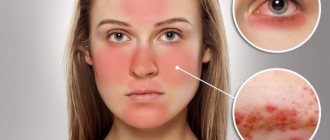
Treatment of melanosis
Dark spots on the whites of the eyes may indicate melanosis.
The disease can be congenital or acquired; accumulation of melanin pigment is observed in the sclera and in the choroid. The spots may have a pale purple or gray color. Most often, this disease is caused by metabolic disorders. For treatment, the patient may be prescribed hormone therapy and vitamin C. Sometimes eye melanosis appears as a result of an inflammatory process, which is successfully treated with anti-inflammatory drugs. For this disease, it is recommended to adhere to dietary standards. In your diet, you should limit the amount of sweets, refined cereals, white bread, and starch-containing foods. It is recommended to exclude fatty, salty foods, spices, coffee, and strong tea. The diet should include fish, seafood, leafy vegetables, eggs, whole grain cereals, citrus fruits, nuts, and honey. Daily eye massage, which should be performed for 2 minutes a day, will be useful. To do this, it will be enough to lightly tap your closed eyes and the area around them with your fingertips. This procedure will help regulate blood circulation. If melanosis is caused by inflammatory processes, traditional medicine recipes can be used to eliminate it. You need to pour two tablespoons of cornflower inflorescences (without baskets) with a glass of boiling water, leave to infuse for two hours, then strain and apply as a lotion to the eye area. They should be done daily for five days. You can wash your eyes with a decoction of oak bark. To prepare it you will need two tablespoons of raw materials and 500 ml of hot water. The raw materials are poured with water and boiled over low heat for half an hour. The resulting decoction is filtered and used to wash the eyes. Spots before the eyes are a symptom of a certain pathological process or severe fatigue, which is characterized by floating dark or colored spots. Treatment can only be prescribed by a doctor, after conducting an appropriate examination and identifying the root cause. Self-medication is unacceptable, as this can lead to the development of serious complications.
Etiology
The cause of this symptom may be the following etiological factors:
- macular edema;
- posterior vitreous detachment;
- macular degeneration;
- violation of the integrity of blood vessels inside the organs of vision;
- age-related macular degeneration;
- Stargardt's disease;
- retinal burn.
Etiological factors of a non-ophthalmological nature include:
- seizures;
- head injury;
- a sharp effect of a light irritant on the eye;
- eye injury;
- increased susceptibility to ultraviolet rays;
- exhaustion of the body;
- severe emotional shock.
Only a doctor can determine the exact cause of this symptom after conducting the necessary examination methods.
Symptoms
In this case, there are no general clinical manifestations. The nature of the symptoms depends on the provoking factor.
If the cause of this symptom is a pathological effect on the central nervous system or brain, then the following symptoms may be added:
- , possibly with seizures;
- sudden mood changes;
With migraines, yellow spots may appear before the eyes and blurred vision.
The appearance of red spots before the eyes may be due to a violation of the blood supply to the brain, or a violation of the integrity of the blood vessels of the eye, which manifests itself in the following clinical picture:
- increased blood pressure;
- headache that may radiate to the back of the head;
- spots before the eyes may appear from time to time or appear only during physical activity;
- , even with minimal physical activity;
- nausea, often with bouts of vomiting, which does not bring relief.
Green spots before the eyes can appear during physical exhaustion, at the stage of pre-fainting, as evidenced by the following symptoms:
- blurred vision, a feeling that everything around is blurry;
- dizziness;
- clouding of consciousness;
- nausea, which is rarely accompanied by vomiting;
- low blood pressure.
If the cause of such a symptom is an ophthalmological disease, then the following symptoms may appear:
- sharp reaction to light stimuli;
- increased;
Regardless of what clinical picture is noted, you need to promptly seek medical help and not self-medicate with medications or traditional medicine.
“Spots before the eyes” is observed in diseases:
Barotrauma is tissue damage that is caused by a change in the volume of gas in the body cavity due to changes in pressure. This pathological process can be observed in the ears, lungs, teeth, gastrointestinal tract, eyes and paranasal sinuses. The clinical picture of such a disorder is quite pronounced, which is why problems with making a diagnosis, as a rule, do not arise. Treatment is prescribed only by a qualified medical specialist.
Hematometra is a pathological condition in which blood accumulates in the uterine cavity, which occurs as a result of a violation of the physiological processes of its evacuation. The pathology occurs infrequently, and in fact, represents a uterine cavity filled with blood, from where the blood cannot be evacuated on its own. Therefore, medical intervention is required to prevent the development of complications.
Hemophthalmos is a pathological condition characterized by hemorrhage into the vitreous body of the eye. It can occur in patients from different age categories. There are no restrictions regarding gender. It is worth noting that hemophthalmos is usually a symptom of some other ailments, especially those that affect the blood vessels.
Destruction of the vitreous body of the eye is a dangerous disease in which the physiological structure of the vitreous body, under the influence of exogenous and endogenous factors, is disrupted. As a result, its fibrils thicken or liquefy. In particularly severe clinical situations, the vitreous body may shrink.
Retinal dystrophy is a dangerous disease that affects the retina of the eye. Whatever the cause of this disease in humans, with untimely and unqualified treatment, the outcome of dystrophy is the same - atrophy or complete death of the tissues that make up the retina. Because of this, the patient will experience irreversible visual impairment, including blindness. It is worth noting that the timing of vision loss directly depends on the type of disease. Retinal dystrophy occurs rather slowly, but as it progresses, the patient’s condition only worsens.
Night blindness is a popular designation for vision pathology, which in medicine is called hemeralopia or nyctalopia. The disease manifests itself in a significant deterioration in visual perception in low ambient light. At the same time, a person’s coordination is impaired, visual fields are narrowed, and there is an incorrect perception of things in blue and yellow shades.
Myopia is a pathological condition characterized by refractive error in one eye or both. In this case, the main optical focus is localized between the retina and the lens of the visual apparatus. Due to such pathological changes, the sick person begins to have difficulty distinguishing between objects that are located at a certain distance from him.
Acoustic neuritis is a disease of the nervous system characterized by the manifestation of an inflammatory process in the nerve that provides auditory function. In the medical literature, this disease is also called “cochlear neuritis.” Typically, this pathology is diagnosed in older people over 50 years of age (more often in the stronger sex). Such people rarely seek help from a qualified specialist, considering the decline in hearing function to be a normal process that accompanies the aging of the body.
When any disease affects the organ of vision, it is of particular concern. After all, any dysfunction of the eye or the integrity of the retina can ultimately lead to loss of vision, partial or complete. Therefore, you need to react adequately and quickly. The surest way, of course, is to see a doctor, without expecting the condition to worsen.
Each of us periodically may notice complete or partial. This especially often happens to those who work at the computer for a long time or are engaged in intense mental activity. When there are no other symptoms: discomfort or pain, a short rest, doing a little eye exercises or preventative drops is enough, and this condition goes away.
Clinical picture
The main accompanying symptoms of blue sclera syndrome are: bilateral blue (occasionally blue) color of the sclera, hearing loss and high bone fragility.
The blue-blue color of the sclera is the constant, most pronounced symptom of this syndrome, observed in 100% of patients. The unusual color is explained by the fact that the pigment of the choroid shines through the particularly transparent, thinned sclera.
Clinical examinations of patients with Lobstein-Van der Heeve syndrome reveal a number of characteristic signs of the disease - thinning of the sclera, a decrease in the number of collagen and elastic fibers, a metachromatic color of the main substance, which indicates a high content of mucopolysaccharides, which indicates the immaturity of fibrous tissue, persistence of embryonic sclera.
There is also an opinion that the blue color of the sclera is not a consequence of its thinning, but of an increase in transparency, which is explained by changes in the colloid-chemical qualities of the tissue. On this basis, a more correct term for denoting such a pathological condition is proposed, which sounds like “transparent sclera”.
The blue color of the sclera in this syndrome can be detected immediately after the birth of the child, since it is more intense than in healthy babies. In addition, the color does not disappear at 5-6 months of life, as it should normally happen. In this case, the size of the eyes is usually not changed, although in addition to blue sclera, other anomalies are often observed. These include: anterior embryotoxon, iris hypoplasia, zonular or cortical cataract, glaucoma, complete inability to distinguish colors, corneal opacities, etc.
The second main symptom of “blue sclera” syndrome is high fragility of bones combined with special weakness of the ligamentous apparatus and joints. These signs are detected in almost 65% of patients with this syndrome at different stages of its course. This became the reason for dividing the disease into three types.
- The first type is the most severe lesion, accompanied by intrauterine injuries, fractures during childbirth or immediately after birth. Children with this type of disease die in utero or in early childhood.
- The second type of blue sclera syndrome is accompanied by fractures that occur in infancy. In this situation, the prognosis for life is more favorable, although due to numerous fractures that occur unexpectedly with little effort, as well as subluxations and dislocations, disfiguring deformation of the skeleton remains.
- With the third type of disease, fractures appear in children 2-3 years old. By adolescence, their number and danger of occurrence are significantly reduced.
The root cause of bone fragility is considered to be extreme bone porosity and its embryonic nature, a serious lack of calcareous compounds, as well as other manifestations of hypoplasia.
The third sign of blue sclera syndrome is progressive hearing loss, which occurs in almost half of patients. It is explained by otosclerosis and underdevelopment of the labyrinth of the patient’s inner ear.
In some cases, the triad of symptoms described above, typical of Lobstein-Van der Heeve syndrome, is combined with other pathologies of mesodermal tissue. At the same time, the most common congenital heart defects are syndactyly, cleft palate, etc.
Treatment prescribed for blue sclera syndrome is symptomatic.
Spot
Occasionally, a small spot may appear on the white of the eyeball. Depending on other manifestations, one can judge what it is, and most importantly, what to do in such a case.
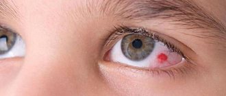
red spot
Actually, the color of the spot is an important indicator of the seriousness of what is happening. A small red spot usually indicates the following:
1. Blood pressure is constantly elevated or there has been a sharp drop. In this case, one or more microvessels may have burst, which ultimately formed a small hematoma. Such a manifestation cannot be treated, but the reason should be an important signal that the body needs help. It would be a good idea to measure your blood pressure daily. 2. High physical activity. For example, during childbirth, a woman’s body experiences enormous stress, including pressure. This can cause capillaries to break. This condition is temporary, it does not require special treatment and will certainly go away on its own. If this happens in the gym or under other circumstances, you should think about reducing physical activity. 3. Eye pressure. This issue is within the competence of an ophthalmologist, therefore, if a red spot appears or it does not go away for a very long time, and there are no other signs, you need to contact a specialist.
At the same time, there are also congenital spots on the white of the eyes, similar to birthmarks. This is a kind of pigment that appears in the eyeball. Such spots are harmless and do not interfere with normal visual function. If they still bother you aesthetically, you should consult an ophthalmologist to see if they can be eliminated.
More serious is a floater on the eyeball. As a rule, such a spot is not detected constantly, but only when looking in one direction, that is, when moving the eyeball. This spot may be associated with. Usually, it is colorless, but when it gets into the pupil area it behaves like a hindrance, the eye feels obvious discomfort, and vision becomes blurred.
Only a doctor can determine the presence of such a floating spot. If it is a particle, it is necessary to carry out laser correction to strengthen it. This is usually a micro-operation that is performed without hospitalization. However, much depends on the severity of the disease. That is why only a specialist can adequately assess the situation. After all, partial retinal detachment can worsen vision and well-being. While complete detachment will lead to an inevitable complete loss of the ability to see. In this regard, it is very dangerous to hesitate or postpone a visit to an ophthalmologist.
Prevention
Strengthening the retina prevents it from detaching. Therefore, it is very important to take vitamin complexes for the eyes, of which there are quite a lot and contain a variety of components.
Complexes with vitamin A (retinol), lutein, and blueberry extract are considered ideal for the organ of vision. These components can inhibit age-related retinal dystrophy, which can also become one of the causes of retinal detachment.
Systematic intake of vitamins, at the same time, can improve vision and relieve discomfort. Vitamins for the eyes are especially important for people who experience daily severe eye strain or are engaged in mental work.
In addition, gymnastics for the eyes is extremely useful. The simplest and most effective exercise is palming. To perform it, it is enough to close your eyes with your palms several times every day and completely relax for 5-10 minutes. This exercise perfectly relieves tension, which can lead to serious pathologies of the organ of vision.
Yellow spot
The yellow spot on the eyeball is called. It is not a disease, but rather a pathological manifestation that is inherent in a person’s age. A similar manifestation occurs due to gradual aging. It may appear on the inside of the eye, closer to the bridge of the nose. Such spots become noticeable in a certain position of the eyes.
A red spot in the eye can appear for many reasons, although the most common is hemorrhage. Then a red spot on the eye may appear due to expansion and damage to one of the many capillaries that are located on the white of the eye. In this case, initially, a red dot in the eye is visible against the background of the white eyeball, although subsequently the size of the spot may increase slightly. A single rupture of a vessel poses virtually no threat to human health, but if this situation occurs constantly, it is recommended to seek help from a doctor.
Reproduction
The mating season for squirrels begins in January and continues until August. During this time, the female is able to give birth to 1-3 offspring, depending on how favorable the conditions are.
For each female individual, 5-6 males simultaneously claim. When they meet, they begin to behave aggressively, making hostile sounds and knocking on trees with their paws. The female evaluates them and chooses the most active one. After this, the couple leaves in search of a favorable place to set up a joint den, where the squirrels will appear.
Squirrel with little squirrel
Pregnancy lasts 35-38 days, with 3 to 9 babies born at a time. Newborn squirrels have no hair and cannot see anything.
Interesting fact : the body of baby squirrels begins to grow hair in the second week of life, and vision appears only after a month.
For the first six weeks, the female feeds the cubs with milk. Baby squirrels adapt to independent life in the third month of life, after which they leave their parental shelter.
Interesting: Animals of the Red Book of Russia - names, descriptions, characteristics, rare species, photos and videos
Factors influencing the development of the disease
If a red spot appears on the white of the eye, the causes of the phenomenon may be the following:
- Mechanical injury or capillary rupture.
- Infection.
- A red spot on the eyeball in an adult appears due to hemorrhage, as blood seeps through the conjunctiva. Usually this situation occurs due to the rupture of several capillaries. It is caused by hypertension or other diseases of the circulatory system.
- The reasons why red dots may appear on the whites of the eyes are due to the accumulation of physical fatigue. This usually happens due to constant lack of sleep.
- If a person is sick, red dots appear on the eyes due to a strong cough.
- In some cases, the reason for the appearance of this symptom in the eyes is excessive secretion of tears, which can occur due to the development.
- There are other reasons for the appearance of this phenomenon - excessive consumption of alcoholic beverages or irritation of the retina by tobacco smoke.
- A red spot appears on the eyes due to severe stress, nervous disorder, the influence of a sharp temperature change, strong wind.
- Often the causes of the development of the disease are long-term work in front of a computer monitor or severe physical stress, for example, heavy lifting, childbirth in women, straining during constipation, etc.
- Red spots may occur due to the effects of certain medications.
But sometimes patients complain to doctors about red spots before their eyes. In this case, the cause of the disease may be the development of cerebral circulatory disorders, minor hemorrhage into the vitreous body, etc. Most often, such diseases affect people over 50 years of age.
Typically, such spots appear in front of the eye with the development of diabetes mellitus or the presence of an injury to the eyeball.
But sometimes bad heredity plays a leading role in the appearance of such spots.
For such patients, consultation with a cardiologist, ophthalmologist, neurologist, and allergist is required.
Sometimes such symptoms indicate damage to the skin around the eye, liver disease, or an allergic phenomenon. Therefore, the diagnosis is made based on a comprehensive examination of the patient.
Kinds
The main types of keratitis that occur in childhood are: bacterial, herpetic, allergic, metabolic (vitamin deficiency) and post-traumatic.
Bacterial
Bacterial keratitis is characterized by the appearance of ulcers in a child's eye . It is rare in childhood. The causative agent is usually a coccal infection, but sometimes the disease occurs due to an injury to the eye. The disease is accompanied by pain, burning, and after eliminating inflammation, a thorn appears on the white part of the eyeball.
Toxic-allergic
Toxic-allergic keratitis has a complex course. Typically occurs in children from birth to 14 years of age and adolescents. The causes of the toxic-allergic form are previously untreated diseases (acute respiratory infections, acute respiratory viral infections), hypothermia, and helminthic infestations.
Toxic-allergic keratitis can be distinguished by characteristic tubercles with vessels crossing the eyeball. Vasodilation causes clouding of the transparent membrane of the eye (cornea) and permanent deterioration of vision .
Metabolic (vitamin-deficient)
Avitaminous keratitis occurs due to a deficiency of vitamins in the child’s body:
- vitamin A. The initial stage of the disease is accompanied by dry eyes, a grayish cloudiness forms on the cornea (the eye becomes cloudy), white plaques affect the conjunctiva;
- vitamin B deficiency and gastrointestinal dysfunction. A pinpoint clouding of the cornea forms, spreading in different areas of the eye. Over time, the cloudiness turns into ulcers that seem to tear the eyeball;
- Deficiency of vitamins PP and E causes eye inflammation.
Herpetic
This form of the disease is most common among young children. Occurs due to the activity of the herpes virus in the body. The causative agent of the disease is most often activated against the background of: hypothermia, psychological disorders (stress), eye injury, febrile manifestations (high temperature, chills).
The course of the disease is possible in mild and severe forms, with the manifestation of complications: the development of secondary glaucoma, cataracts, the appearance of lens transparency .
Primary
Primary herpetic keratitis occurs among children under 5 years of age. The main reason is that children’s immunity is not fully formed. It is characterized by a complex course, accompanied by herpetic blisters on the eyeball and throughout the body (arms, legs, torso, face).
Post-primary
Post-primary herpetic keratitis occurs, like primary, against a background of weakened immunity. Manifested by the following symptoms: pain and pain in the eyes, convulsive spasmodic contractions of the eyelids, no rash on the body.
Discoid
Disc herpetic keratitis involves deep areas of the cornea , in the center of which inflammation occurs in the form of a disc. The eyes turn red, swelling of the cornea, eyelids, and conjunctiva appears, and increased intraocular pressure occurs.
- You may have been looking for: how to cure red eyes in a child
Post-traumatic
Post-traumatic keratitis in children develops due to a careless scratch, entry and removal of a foreign body from the cornea, ultraviolet, thermal or chemical burns. The disease is characterized by direct ingrowth of vascular branches into the cornea , convulsive spasm of the eyelids, and yellow purulent discharge from the eyes.
What diseases can cause red dots on the organs of vision?
The symptom in question may occur with an infection that has entered the eye. For example, most often the appearance of a red dot on the white occurs with conjunctivitis. During illness, microbes penetrate into the organ of vision, affect the mucous membrane, and cause inflammation.
The above symptom may warn of the onset of a disease such as glaucoma. The red spot appears due to a sharp increase in pressure inside the eyes. Glaucoma is diagnosed by their appearance, which turns greenish. The patient complains of severe pain in the head. In this case, it is necessary to urgently take the patient to the hospital for examination by an ophthalmologist, otherwise the person may lose his vision.
Hypertension can cause damage to blood vessels in the whites of the eye. A similar symptom is characteristic of diabetes mellitus. Diseased vessels have low strength and are destroyed even with minimal effort.
If a patient wears contact lenses for a long time, or the process of tear formation is disrupted, then hemorrhages appear in the area of the sclera.
Patients with poor blood clotting and hemorrhoidal diathesis may complain to the doctor about the appearance of red dots in the eye area.
Many of them are interested in what to do if the indicated red lesions are found in the eye? In such cases, you should immediately contact a doctor for help and conduct a full examination.
Types of squirrels
The squirrel family includes 54 species, which have a unique appearance and live in certain places. Among them there are several main ones.
Common squirrel
Common squirrel
The species lives in Russia, Europe and Asia. It has an elongated body and a rounded head on which long ears are placed. The hind limbs are longer than the forelimbs. The color changes over the course of the year from light brown to brown.
Aberta
Aberta
Lives in the southwestern United States and Mexico. The animal has an elongated, rounded body. The belly and underside of the tail are white, and gray-brown fur grows on the back and face. Aberta's ears are long and have fluffy tufts at the tips.
Flying squirrel
Flying squirrel
Individuals of this species are small in size: their body length rarely exceeds 20 cm. Between the squirrel’s limbs there is a fold of skin covered with fur. It acts as a parachute and allows you to fly long distances due to oncoming air flows. In one jump, the flying squirrel is capable of covering 55-60 m. The species lives in the European part of Russia.
Interesting: Why do carrier pigeons fly to the right place? Description, photo and video
Ratuf
Ratuf
This species also has another name - “Indian squirrel”. Individuals can boast of impressive dimensions: the body length reaches 50 cm, and at the tail this parameter can be equal to 65 cm. Ratufs live on the territory of the island of Hindustan.
Red-tailed squirrel
The red-tailed squirrel
lives in South and Central America. The animal has a red belly and an orange-brown back. There is a black tassel at the tip of the tail. The squirrel's head is round in shape, with small ears without tassels at the top.
Western gray squirrel
Western gray squirrel
The animal lives in the forests of America. Its color is gray with a brown tint, its belly is white. In terms of lifestyle and appearance, it is almost no different from an ordinary squirrel.
Yellow-throated squirrel
Yellow-throated squirrel
The species got its name because of the yellow fur growing on its belly and neck. The back, muzzle and tail are red-brown. Individuals are small in size: body length rarely exceeds 17 cm. The animal lives in Venezuela and Brazil.
Symptoms of the disease in children
A red dot that appears on a child’s eye most often develops due to the fragility of the blood vessels. Infants and toddlers may develop these symptoms due to loud screaming or mechanical trauma during play.
OUR READERS RECOMMEND!
To quickly and safely restore vision, our readers recommend the drug Sokolit. For 2 years now, this miracle drug has been available on the European market; its effectiveness is several times greater than laser vision correction and drugs such as Blueberry Forte. It is aimed at eliminating the causes, not the symptoms, of poor vision, its effectiveness and safety have been proven in clinical studies.
Older children suffer from prolonged use of gadgets: addiction to a desktop game console or computer. In such patients, the capillaries cannot withstand overstrain, and cracks appear in them through which blood escapes. To eliminate the disease, eye drops are usually enough, which the doctor will prescribe after examining the child.
It is forbidden to give your child tea, which increases blood pressure in the arteries. If these measures are not taken, the hemorrhage will expand and microbes may enter the affected area. Their activity will lead to the development of inflammation, and then the appearance of an abscess on the eye. In the worst case, a tumor may develop that will have to be removed surgically.
Course of treatment and prevention of the disease
If an infection of the organ of vision occurs, and a dot appears on the eye, then after the examination the doctor will prescribe antibiotics. Self-treatment with lotions or herbs can only worsen the situation.
Along with taking the prescribed drug, the patient must also maintain personal hygiene. He must have his own toiletries. He should wear clothes made of quality materials and wash his hands 3-4 times a day.
During the treatment course, you should not rub your eyes or touch them with your fingers.
As a preventive measure, you can use vitamin C, which will strengthen the arteries, veins and capillaries. The doctor may prescribe other medications that strengthen blood vessels and their walls.
If it is small, then usually a piece of ice is enough, which is wrapped in a thin cloth and then applied to the closed eye. But this can only be done if there is no pain or other symptoms, such as tears.
Such a compress will prevent hemorrhage from occupying a large area. If the size of the spot does not increase after using ice, then no treatment will be required.
After 4–10 days, there will be no trace left of the speck.
In some cases, to strengthen the walls of blood vessels, the doctor may offer the patient special eye exercises. They relax the muscles of the organ. In this case, vitamins and microelements may be required, and for some patients, special sedatives. The ophthalmologist will prescribe all this after a complete examination of the patient.
The patient must be provided with adequate sleep. At this time, the eyes relax and rest. If the sclera is affected, then you need to limit the time you work on the computer. It is recommended to use special moisturizing drops.
If a patient develops diabetes mellitus, symptoms of hypertension appear, or kidney disease is detected, after examination by an ophthalmologist, you should visit a specialized doctor.
Vision is our guide to the world, the source of human activity, pleasure and quality of life. That is why any disease of the visual system causes anxiety in a person. We also worry when spots appear on squirrels. Why do they occur, and what should be the correct action when they are detected?
About red spots
Every person has noticed complete or partial redness of the eyes at least once in their life. Most often, such troubles happen to those who spend a lot of time behind monitor screens, are engaged in intellectual activities and simply do not take care of their eyes. In this case, you just need to have a good rest, make compresses with black tea bags on your eyes, rinse them with sage infusion - and the redness will go away. If a person continues to not maintain visual hygiene during work, then reddening of the whites will become a permanent phenomenon.
When we are talking about one small red spot, the reasons for its appearance may be as follows:
- Constantly high blood pressure.
In this case, blood vessels and capillaries often burst, including in the eyes. As a result, a small hematoma forms. In most cases, such a red spot resolves on its own. And it is necessary to treat not the redness, but the cause of its appearance, that is, control the blood pressure, lose weight, and follow a diet. - Excessive physical activity.
This happens in women during childbirth, during pushing, in men - when carrying heavy objects. Physical effort causes capillaries to rupture. And in this case, no special treatment is required. You just need to draw conclusions and take care of your health, because excessive stress affects not only the eyes, but also the spine and the general tone of the body. - Eye pressure.
We are talking about glaucoma - a condition of increased intraocular pressure. If the red spot does not go away for a long time, headaches bother you, or vision decreases, then rush to see an ophthalmologist.
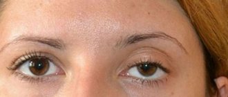
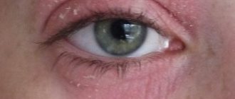
Darkening of the protein is normal
There are physiological reasons why a child may develop gray white in his eyes:
- restructuring of the child’s body, which is typical for him normally; in babies, the whites may become gray-blue, but the condition goes away within 6 months (at the same age the shade of the iris also normalizes);
- close arrangement of vessels that are visible through the sclera, giving a blue color.
Over time, the unusual coloring goes away, and the state of the visual organs normalizes. To make sure the child’s health is normal, you still need to see an ophthalmologist. If it confirms the physiological process, there is no need to worry.
About congenital and floating spots
Many people have small spots of brown or yellowish color on their whites. They are like birthmarks, but are located on the mucous membranes in the form of pigment. Such inclusions do not interfere with visual function at all; they are harmless. Some women may be bothered by these kinds of spots from an aesthetic point of view. Then it is recommended to consult an eye doctor about their removal.
As for floating spots, this is a serious problem. They are visible only from a certain angle (when looking to the side) and can be a signal of retinal detachment. Such formations are usually colorless. When they enter the pupil area, they create interference. The visual apparatus feels severe discomfort. A person complains of blurred vision.
This problem should only be dealt with by an eye doctor. If the formation is a part of the retina, then in most cases laser correction is performed to strengthen it. This micro-operation is now performed without hospitalization. But in therapy, much depends on the severity of the pathology. That is why only a specialist can adequately assess the problem of a floating spot. Partial retinal detachment impairs not only vision, but also a person’s general well-being. But complete is a loss of the ability to see. That is why you need to promptly contact an eye doctor at the slightest deterioration in vision. Strengthening the retina with vitamin complexes helps prevent its detachment. Typically, eye doctors prescribe special vitamins with lutein and blueberry extract to patients with this problem. They inhibit age-related retinal dystrophy. It is this phenomenon that in most cases causes its detachment.
It is especially important for prevention to take the above complexes for those people whose work is constantly associated with serious visual loads. They should not forget about special gymnastics, the need to get enough sleep and eat well.