General information about pathology
Disease:
- occurs infrequently;
- proceeds slowly;
- rarely becomes malignant;
- occurs in people who are at least 35-40 years old;
- more often affects females.
The neoplasm externally resembles a rounded dense nodule, which is located in a capsule formed from connective tissue. Thanks to this, it is relatively easy to get rid of the tumor using surgery.
Neuroma - causes of occurrence
Types of neuroma
Externally, the tumor resembles a dense node of a round or irregular shape with a bumpy surface; its color depends on the blood supply to the nerve area, for example, if there is venous congestion in the area, the color will be bluish. Usually the tumor is enclosed in a capsule of connective tissue, and removal of the neuroma is very successful.
According to the type of structure, neuromas are divided into:
- Epileptoid. They are characterized by a dense arrangement of tumor cells with a small number of fibers.
- Angiomatous. Tumors have many cavernous cavities, which are formed due to pathological dilation of blood vessels.
- Xanthomatous. Tumors of this type exhibit many xanthochrome cells with a high pigment content.
Neuroma is caused by:
- genetic factors.
The likelihood of a tumor increases sharply if relatives suffered from this problem;
- difficult environmental situation
– exposure to harmful chemicals, radiation;
- cigarette addiction
.
Also, a neoplasm appears in people already suffering from another neoplasm.
Types of pathology
There are the following main types of disease:
Acoustic neuroma (aka vestibular schwannoma):
- occurs most often;
- the tumor affects the auditory nerve (usually the vestibular part, rarely the cochlear part). However, the neoplasm gradually grows and begins to penetrate the cerebelloptine angle;
- vestibular schwannoma can harm the facial nerve and even “reach” the trigeminal nerve. The optic and jaw nerves may also be affected;
- The tumor progresses very slowly (by a few millimeters per year). In rare cases, rapid, dangerous growth is observed (up to 30 mm in 12 months), due to which important parts of the brain (primarily the cerebellum) begin to be compressed;
- much more often it develops in only one ear (in two 5% of cases). Bilateral acoustic neuromas are usually caused by neurofibromatosis.
- appears at 40-50 years of age.
Vertebral neuroma:
- usually develops in the thoracic and cervical areas of the back;
- sometimes appears in the lumbar region;
- It is less common than acoustic, but also quite common.
Morton's schwannoma:
- this type of pathology appears on the foot (more precisely, its sole) of one leg, in the area of the 3rd and 4th toes;
- in rare cases, pathology develops on both feet;
- Morton's schwannoma is rare.
What methods of treating neuroma in Israel have proven effective?
Treatment of brain cancer in Israel, including neuromas, directly depends on the area in which it is located, its size and symptoms. In the initial stages and with small sizes of the neuroma, the patient can receive conservative treatment, including taking anti-inflammatory drugs and physical therapy sessions. For example, with Morton's neuroma, cortisone injections are indicated to reduce the size of the atypical nerve, or injections of a drug that destroys the affected nerve. If the proposed therapy does not demonstrate an effect, surgical removal may be required.
Surgery is the only treatment method for neuroma that can provide maximum results. This type of operation is performed by a neurosurgeon. Depending on the location of the tumor, the patient’s health condition and his age, the doctor may use one of the surgical technologies:
- Excision
This requires careful release of nerve fibers from cancerous tissue. This manipulation should be performed by an experienced surgeon, since there is a fairly high risk of injury, as well as the likelihood of preserving individual cancer segments, which can subsequently lead to relapse. In foreign clinics, possible complications are minimized through the use of microsurgical techniques. If the tumor is located in the skull, trepanation is first performed; if the spine is affected, laminectomy is performed. Microsurgery is less invasive than open surgery. To remove the tumor, she uses modern techniques: ultrasound and laser radiation. After surgical removal, radiation is recommended.
- Radiosurgery (gamma knife and cyber knife)
Tumor removal is performed on an outpatient basis (without surgery) for intracranial and spinal locations. Directed ionizing radiation or proton beams, which have fewer side effects, specifically affect cancer cells, leading to the death of most of them, while the remaining ones lose the ability to further grow. Radiosurgical treatment is indicated for neoplasia sizes up to 30 mm. If the tumor is large and the patient has contraindications to open surgery, then this method is used for palliative therapy with a reduction in the size of the tumor.
During radiosurgery at Meir Hospital in Israel, there are no complications typical of open surgery; in addition, there is no need for long-term hospitalization, or subsequent treatment or rehabilitation.
How does pathology manifest itself?
Often the disease is completely asymptomatic and is discovered by chance. This makes it difficult to treat neuroma. Signs of pathology depend on the location of the tumor:
For acoustic neuroma:
- tinnitus (tinnitus) develops;
- hearing deteriorates;
- coordination is impaired;
- severe dizziness appears.
If the facial nerve is damaged:
- tears flow;
- dry mouth;
- There is partial paralysis of the eyelids.
With neuroma of the dorsal vertebrae the following are observed:
- excruciating pain in the back;
- deterioration of sensitivity at the site of the lesion.
4. Morton's schwannoma causes various sensory problems:
- numbness is felt in the area of the feet;
- paresis occurs.
Modern diagnosis of neuroma
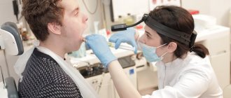
Diagnosis of neuroma always begins with a physical examination and familiarization of the specialist with the patient’s complaints. Depending on the symptoms, the patient’s doctor may be not only a neurologist or otolaryngologist, but also another doctor whom the patient contacted for an initial consultation based on the manifestations of the disease. This is due to the fact that the development of neuroma is largely similar to ordinary damage to the nerve trunk as a result of inflammation, compression or dysmetabolic failure. Additional instrumental influence is indicated to clarify the morphology, which is the root cause of the malfunction of the nerve trunk.
The list of examination procedures depends on the location of the tumor and includes the following types of diagnostics that may be recommended before cancer treatment in Israel or in other countries:
MRI and CT
Carried out in several ways:
- Contrast CT - recommended for cerebral neuroimaging for a tumor in the skull to detect tumors measuring 10 mm or more;
- Cerebral MRI is an informative study that allows you to visualize in detail the state of the brain tissue surrounding the neuroma;
- Isolated MRI is necessary for examining the affected part of the spine; it can show the resulting neuroma of the spinal roots and determine the degree of compression;
- CT or MRI of the neck area - recommended following pharyngoscopy to determine the size of the tumor when localized in the larynx and pharynx;
- MRI - to assess the condition of the soft tissues of the affected limb;
Audiometry
Recommended for complaints of hearing loss - to assess the degree of its deterioration and exclude other causes (for example, age-related) that have led to aggravation of the problem. The procedure is performed by an otolaryngologist and audiologist.
Electroneuromyography (ENMG)
Recommended for assessing the functional state of the nerve trunk affected by the tumor. This procedure is also used after surgery to monitor the recovery process.
Sonography (ultrasound)
This type of examination allows us to detect the presence of local thickening of the nerve canals when the tumor affects the peripheral nerve trunks of the extremities.
Histology of neuroma
Histology helps determine the nature of the tumor (benign or malignant). A biopsy (removal of a sample for histology) is not prescribed as a separate procedure. Histological examination is carried out on surgical material after surgical removal of the tumor.
How is pathology diagnosed?
Treatment of neuroma is preceded by diagnosis. It is produced using:
- computer or magnetic resonance imaging examination. Magnetic resonance imaging is more effective. It is able to detect microscopic tumors, while computed tomography diagnostics detects only tumors whose diameter is at least 1 cm;
- electroneurography;
- audiograms (hearing test).
Treatment of the disease – surgical, radiation, conservative
Usually the neuroma must be removed; conservative treatment is not always effective. True, in the early stages, the tumor can actually be removed using radiation therapy (cyberknife).
Operation
Surgical treatment of neuroma involves a local opening of the skull (trepanation) so that the tumor can be reached.
It will not be possible to do without surgical intervention if:
- the tumor quickly increases in size (for example, after the patient has undergone radiotherapy);
- the disease began to lead to serious complications.
Surgical tumor removal is contraindicated if:
- the patient has problems with the heart or blood vessels;
- The patient is already elderly and has heart problems.
The prognosis is 99% favorable. Relapse of the pathology is a rare phenomenon. After the operation, the patient recovers within 6-10 months.
Possible complications are as follows:
- bleeding may begin;
- there is a small risk of infection;
- the patient's facial nerve may be affected by paresis (especially if the tumor exceeds several centimeters);
- hearing will worsen. If the tumor is larger than 2 cm, there is a high risk that its surgical removal will lead to partial hearing loss.
Radiation (non-operative) therapy
In addition to traditional surgery, there is a safer way to get rid of neuroma. We are talking about stereotactic radiosurgery (cyberknife) - irradiation of the tumor:
- The advantage of this operation is that the tumor is destroyed very precisely. The nerve tissue that surrounds it remains completely unharmed;
- disadvantage - this therapy is effective only if the neuroma is detected in the early stages and does not exceed 30 mm.
Radiation therapy is mandatory if:
- the tumor is located in an “inoperable place”;
- the patient is categorically against surgical intervention;
- the person has already reached old age and has heart problems.
Radiation therapy has side effects. After the procedure, the patient can:
· to feel sick;
· pain appears in the neck (where the stereotaxic frame was located).
What is spinal neuroma and methods of its treatment
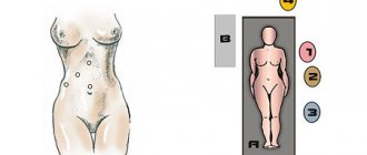
The spine consists of 34 vertebrae, between which discs are located. The majority of all loads on the spinal column fall on its lower sections: lumbar and sacral. At the same time, it was noted that neuroma is found more often in the cervical and thoracic regions. It follows from this that stress on the spine is not the cause of this disease.
What causes a tumor to appear?
There is still no reliable information about the true cause that contributes to the appearance of neuroma. The tumor develops from Schwann cells that line the spinal cord. There is an opinion that the predisposition to the formation of neuroma is most likely genetically determined.
Symptoms of the disease
In the early stages, neuroma does not manifest itself in any way, so it is difficult to identify. Most patients discovered their tumor by chance. Obviously, the sooner this disease is noticed, the easier it will be to monitor its development and prevent the transition to a malignant tumor.
At later stages of tumor development, you can find:
- decreased sensitivity, numbness of the limbs;
- backache;
- deterioration of motor functions of the arms and legs.
Neuroma can also affect other organs, so sometimes the symptoms of the disease include disturbances in bowel movements and sexual function.
Visual signs include curvature of the spine, which can also be a manifestation of the presence of a tumor.
How is diagnosis carried out?
There are five main methods for diagnosing neuroma:
- MRI;
- administration of contrast;
- biopsy;
- ultrasound;
- spondylography.
The first method is used most often because it can give an accurate result, which makes it possible to detect benign tumors in the early stages. But sometimes it is not enough, so a second CT method is used to make sure that the diagnosis was made correctly. In addition, it provides information about the size of the tumor and its location. If it is in soft tissue, then a biopsy is necessary. The fourth and fifth methods are alternative.
Treatment options
The main factors influencing treatment are the nature of the neoplasm itself. However, the stage at which the neuroma was diagnosed can also play a determining role.
In the early stages of tumor development, conservative treatment is most often chosen. It includes diuretics prescribed along with glucocorticoids. These medications are designed to improve blood circulation in the spinal column. In addition, it was noted that these drugs prevent the tumor from developing and increasing in size.
However, not all types of tumors are amenable to conservative treatment. Sometimes doctors are forced to use drastic methods to rid the patient of the disease. They include two types of operations:
- open surgery;
- radiosurgery.
The first method is prescribed to those patients in whom the tumor has reached a significant size.
Neuromas that weigh less than 2 kilograms are removed along with the tumor capsule. This treatment is called endoscopic. In cases where it reaches 2-3 kilograms, this procedure for surgery is unacceptable. First of all, the capsule is opened, the tumor is removed from there, and only then the capsule itself is excised. Complications can arise if the surgeon accidentally touches the nerve endings, and partial paralysis is possible.
Radiosurgery is an alternative method that is used by surgeons when it is difficult to reach the tumor with a scalpel. It is also prescribed to those patients who refuse surgery. The method consists in the fact that the rays affect the tumor from different positions, without causing harm to other organs.
Unfortunately, without treatment, this benign formation turns into malignant.
Author: K.M.N., Academician of the Russian Academy of Medical Sciences M.A. Bobyr