Why does crystallization occur?
What is crystalluria? This is an increase in the concentration of salts in the urine.
Urine contains various salts, and this is considered normal. But in a healthy body, obstacles are created for the precipitation of crystals, even at a time when the concentration of salts in the urine is increased. When an increase in salts occurs, they crystallize, and this entails further stone formation.
And all because of the penetration of an increased amount of proteins into the body.
Why does crystalluria occur in children?
Several groups of its causes can be distinguished. One of them is increased precipitation of calcium oxalate in the urine. Urine is always a saturated solution of calcium oxalate, since at normal urine pH values close to 7 (5.5-7.2), the solubility of calcium oxalate is negligible - 0.56 mg per 100 ml of water. Calcium oxalate reaches its maximum solubility at a pH below 3.0.
The degree of precipitation depends on:
- on the calcium to oxalate ratio (individuals with hypercalciuria excrete more calcium oxalate),
- from the presence of magnesium salts (with magnesium deficiency, precipitation increases),
- from excess or deficiency of substances that support the colloidal properties of urine (citrates, celiac, pyrophosphates),
- from excessive excretion of oxalates.
Causes of the disease
External or internal factors can affect salt metabolism in the body.
External causes of salt accumulation in the body include:
- Living in an unfavorable region with an arid climate.
- Systematic consumption of hard water.
- Eating foods high in protein.
- Excess of vitamins in the body.
- Frequent drinking of alcoholic beverages.
- Taking certain medications: cytostatics, diuretics, sulfonamides.
A person cannot influence or change external causes.
Internal reasons:
- Genetic predisposition.
- Congenital pathologies of the urinary system.
- The presence of infection in the body.
- Hormonal disorders.
- Prolonged stay of a person in an immobilized position.
This set of reasons can be subject to correction by a person. And it should be carried out with the intervention of a specialist.
Causes
There are many factors for the occurrence of crystalluria, which can be divided into two main groups: exogenous and endogenous. Internal factors that provoke the appearance of crystalluria include:
- infectious and inflammatory processes of the urinary system,
- congenital anomalies of kidney development,
- hormonal disorders,
- metabolic disorders,
- hypovitaminosis.
External causes of the development of crystalluria are:
- climatic conditions;
- physical inactivity;
- errors in nutrition - a lot of spicy, salty, sweet, sour foods, which cause thirst and at the same time provoke fluid retention in the body;
- taking certain medications;
- alcohol abuse.
Crystalluria is also caused by the consumption of hard or distilled drinking water. Hard water, like deeply purified water, is quite harmful to the body: if the first contains an increased concentration of salts and minerals, the second is purified not only of harmful, but also of useful elements, including enzymes necessary for the breakdown of mineral salts.
It is also worth noting that crystalluria can develop both with a combination of provoking factors, and against the background of each individual of them.
What types of sediments occur in the kidneys?
Crystalluria of the kidneys and bladder may result from the precipitation of various types of salts. In this case, the patient experiences:
- Oxalate-calcium formations. They often develop in childhood; oxalates are detected for the first time when a schoolchild or teenager is tested. They are formed due to the intake of appropriate food products into the body. And if there is still inflammation in the digestive system, salts are absorbed faster into their mucous membrane.
- Phosphaturia. It is formed against the background of an infection in the human genitourinary system. Due to the breakdown of urine by harmful microorganisms, it becomes alkaline. This leads to the formation of crystals of calcium phosphate salts.
- Uricorosia. Uric acid is formed due to the breakdown of purines, after which crystallization of excess sediment occurs in it. Foods high in purines contribute to the development of the disease, such as alcohol, asparagus, nuts, beans, cauliflower and broccoli.
- Cystinuria. This condition is extremely rare and is caused by a congenital abnormality of the kidney structure. Cystine is an amino acid that is difficult to dissolve, and since it is also poorly absorbed by the kidney tubules, crystalline formations appear.
Main symptoms
Regardless of the form and structure of salt formation, the signs of pathology are mostly similar. At the initial stages of crystalluria formation, the disease practically does not manifest itself. As the pathology develops, the following symptoms occur:
- The need for fluid is reduced, urine is excreted in small quantities.
- Frequent headaches of unknown etiology appear.
- There is pain or discomfort in the lower back and abdomen.
- There is a disturbance in urination; a person may experience false or frequent urges.
- Discomfort occurs when passing urine.
You should immediately consult a doctor if there are blood impurities in the urine or the urine has acquired a cloudy color with an unpleasant odor. All of these symptoms are characteristic of most diseases of the urinary system.
Reasons for development
Doctors call one of the main reasons for the development of pathology hyperparathyroidism - a disturbance in the metabolism of phosphorus and calcium substances and an increase in the amount of serum calcium. Other reasons are divided into:
- external;
- internal.
External include negative climatic conditions (for example, dry climate), which adversely affect the human body. Regular consumption of hard water and foods containing a lot of protein, abuse of diuretic drugs, excessive alcohol consumption - all this inevitably leads to crystalluria.
Internal causes include: impaired metabolic processes at the cellular level, genetic pathologies of the urinary system, prolonged lack of body mobility for various reasons, previous infections with complications. Hormonal imbalances can also lead to metabolic disorders and provoke the development of crystalluria.
Diagnostics
Initially, the patient submits a urine test for crystalluria. If an acidic environment is detected, the presence of oxalates or urates in the kidneys is possible. If an alkaline environment is detected, this indicates the presence of phosphates, which can threaten the development of urolithiasis.
It is worth remembering that an excess of acidic foods in the diet leads to oxalate formations. These could be too sour apples, sorrel, oranges, etc.
Therefore, after the results, the doctor must find out what the patient ate before the analysis, since acidic food will provoke an indicator of increased acidity, which can last for 4-5 days.
If abnormalities in the urine are detected, additional examination is performed. It includes:
- X-ray of the urinary tract.
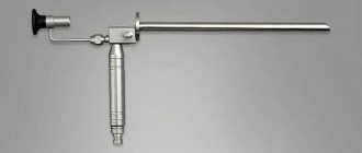
- Cystoscopy.
- Ultrasound examination of the bladder.
Only after all diagnostic measures will the doctor prescribe treatment for crystalluria.
Symptoms and diagnosis
With crystalluria, the patient may experience pain in the lower back and lower abdomen, and pain when urinating. Urine has a rich color, sometimes there is a cloudy sediment in it. Weakness and a feeling of exhaustion appear.
To confirm or reject the diagnosis, the following studies are carried out:
- General urine analysis. Shows exactly which salts are deposited in the patient’s kidneys and what their concentration is. It is more correct to examine daily urine, which allows you to take into account an important criterion - its quantity, which will show whether there is a diuresis disorder. Normal urine should be straw-yellow, transparent, without foreign inclusions (mineral sediments do not exceed the norm), in an amount corresponding to the volume of liquid drunk per day. Any deviations are a reason for additional research.
Ultrasound of the kidneys. Small and large stones are detected during ultrasound examination in the form of echo-positive elements.- A blood test will confirm the presence of inflammation in the body, if any (for example, with chronic pyelonephritis).
Based on the data from all studies, a treatment regimen is developed.
Treatment measures
Crystalluria in children and adults, if detected in a timely manner, is quite easy to treat. Treatment should be selected by a doctor based on test data and the physiological condition of the patient.
The basis of therapeutic measures includes:
- diet;
- taking medications;
- compliance with the drinking regime.
You need to drink up to 2.5 liters of fluid per day, and half of the norm should be drunk shortly before bedtime. This promotes the rapid removal of salts from the body.
Dietary nutrition involves increasing foods containing potassium and reducing foods containing oxalates.
Drug treatment involves taking vitamins from groups A, B and E, as well as magnesium.
Many people are interested in crystalluria - what is it in women and does treatment differ for patients of different genders?
In general, treatment for the disease itself does not depend on gender. But concomitant pathologies, including female gynecological disorders, can provoke the disease. In this case, therapy will be aimed at this disease. It is advisable to also start taking medications that improve intestinal microflora while taking medications. This is "Linex" or "Bifidumbacterin".
Treatment of crystalluria
Therapy for patients with crystalluria consists of three stages:
- Eliminate provoking factors if the disease occurs secondary. Balance your diet. A nutritionist will select the most suitable diet and prescribe a menu, excluding alcohol or heavy protein dishes. Normalization of water balance. The doctor will indicate a specific daily water requirement (from 1.5 to 2.5 liters). Supplementing the diet with missing vitamins and nutrients.
- Eliminate stagnation of urine in the bladder. One hour before bedtime, the patient drinks one liter of clean water to reduce the likelihood of bacterial inflammation due to stagnation of urine. Prescription of natural diuretics to relax the urinary muscles - decoctions of parsley, rose hips, lingonberries and others (only as prescribed by a doctor).
- Prescription of drug treatment. Anti-inflammatory drugs (uroseptics or antibiotics, depending on the course of the disease). Painkillers to eliminate pain and spasms and simplify the process of relieving minor needs. Probiotics (if nephropathy occurs due to intestinal diseases). Enzymes for better digestion of food and acceleration of metabolic processes.
If the patient consults a doctor on time and follows his recommendations, his prospects for recovery are definitely good.
Otherwise, he is at risk of urolithiasis and longer treatment, including surgery.
In addition, following recommendations for nutrition, fluid intake, and physical activity can reduce the intensity of symptoms and speed up the recovery process after an exacerbation of crystalluria.
Buckwheat and rice for crystalluria
Cereals – buckwheat and rice – are good for removing salts from the body. Before going to bed, prepare the following composition: rinse 2 tablespoons of buckwheat and pour a glass of kefir. The next morning the resulting porridge is eaten, and so on for 5 days. During this period, metabolism will improve and the body will be cleansed.
Rice medicine will take longer to prepare and the taste will be less pleasant. But such porridge will serve as an excellent sorbent for removing salts. To prepare, you will need three tablespoons of rice cereal and 1 liter of water, which is used to pour the rice itself. Leave the infusion until the morning, the next morning the liquid is drained and new water is added, after which the product is put on fire and boiled for 5 minutes. Then the water is drained again, new water is added, and boiling is repeated. And so 4 times. For the fifth time, you can eat rice, after which you cannot eat other food for 3 hours.
In general, crystalluria is treated according to the same standard regimen as other urinary tract diseases. But, despite this, you cannot do without medical help, as well as without diagnosis. After the examination, the doctor will be able to select medications in such a way that they will help as much as possible with the illness. Now you know what it is - crystalluria, which means you are already armed against this disease.
Crystalluria in uric acid and calcium oxalate urolithiasis
Journal "Experimental and Clinical Urology" Issue No. 2 for 2015
Konstantinova O.V., Shaderkina V.A.
Crystalluria is one of the important and most informative indicators of the metabolic state of patients with urolithiasis and one of the pathogenetic links in stone formation [1, 2, 3, 4]. When prescribing conservative treatment aimed at correcting metabolic disorders, not only the levels of serum concentration and renal excretion of stone-forming substances are taken into account, but also the presence of crystals in the urine [5, 6, 7]. Often, the appearance of a salt deposit of a different chemical composition than the calculus is a prognostic sign of the course of the disease [8, 9, 10]. The existence of general and specific metabolic disorders in uric acid, calcium oxalate and calcium phosphate forms of urolithiasis suggests the presence of features of crystalluria associated with the form of the disease [11, 12, 13]. For a timely and correct choice of drug treatment tactics, it is necessary to know not only the current condition of the patient, but also to anticipate the possibility of precipitation of lithogenic substances.
In connection with the above, the following goal of the study was set - to determine the biochemical criteria for the occurrence of crystalluria of uric acid or its salts in uric acid urolithiasis and oxalate crystalluria in the calcium-oxalate form of the disease.
MATERIALS AND METHODS
We examined 2 groups of patients, a total of 137 people aged 2174 years. Among them there were 76 women and 61 men. In 49 patients, uric acid stones were diagnosed, in 88 patients - calcium oxalate stones or stones of mixed composition with a predominance of calcium oxalate. 135 patients had recurrent urolithiasis. 93 people were under long-term (2-15 years) outpatient observation. The examination included the collection of anamnestic data, a general urine test, a biochemical study of blood serum and urine for 14 indicators, and an analysis of the chemical composition of the stone. General urine analysis was carried out using the generally accepted standard method. Biochemical research was carried out using an automatic analyzer "Labsistem", the osmolarity of blood and urine was determined using the apparatus "Osmete". To analyze the chemical composition of urinary stones, a Jasco infrared spectrophotometer was used. Statistical analysis of data was carried out using Student's T-test, analysis of variance and correlation [14].
RESULTS AND DISCUSSION
As a result of a survey of 49 patients with uric acid urolithiasis, it was found that the incidence of uraturia (uric acid and sodium urate) in uric acid urolithiasis is 69%. In 24% of patients, crystals of uric acid, sodium urate and calcium oxalate were detected in the urine. Precipitation of ammonium urate salt was observed in isolated cases. Oxalate crystalluria was noted in 7% of patients. With calcium oxalate stones, uraturia was only in combination with calcium oxalate crystals and was recorded in 23% of patients.
When studying 14 metabolic parameters in two groups of patients with uric acid urolithiasis (the presence or absence of crystals of uric acid and its salts), significant differences were found only in urine osmolarity (Table 1). This indicator was higher in the presence of urate crystals than in their absence: 716 ± 29 mOsm/L compared to 548 ± 23 mOsm/L, respectively (p = 0.029). To clarify the obtained data, correlation and variance analyzes were carried out, the results of which confirmed the above (F statistics = 5.121; p = 0.0291; correlation coefficient r = 0.39).
Table 1. Metabolic state in patients with uric acid urolithiasis with and without uraturia
| Biochemical indicator: blood (mmol/l) | Average value of the indicator | T-test | P (significance level) | Significance of differences | |
| no uraturia | there is uraturia | ||||
| Blood creatinine | 0,115±0,003 | 0,117±0,006 | -0,021 | 0,961 | not ven. |
| Total, blood protein | 72,9±1,4 | 73,5±2,1 | -0,531 | 0,674 | not ven. |
| Blood albumins | 46,5±3,3 | 45,4±4,4 | 0,674 | 0,583 | not ven. |
| Blood globulins | 27,9±1,6 | 28,0±3,2 | -0,018 | 0,981 | not ven. |
| Urinary blood supply | 0,382±0,032 | 0,405±0,041 | -1,522 | 0,134 | not ven. |
| Blood oomolarity (mosm/l) | 295±14 | 297±18 | -0,139 | 0,891 | not ven. |
| Urine potassium | 51,1±6,4 | 54,9±7,1 | -0,796 | 0,428 | not ven. |
| Urine sodium | 205,7±12,0 | 206,3±18,4 | -0,027 | 0,915 | not ven. |
| Urine calcium | 5,37±0,31 | 5,56±0,62 | -0,245 | 0,222 | not ven. |
| Urine phosphorus | 25,0±2,8 | 25,6±4,1 | -0,773 | 0,517 | not ven. |
| Urinary acid | 4,46±0,59 | 4,98±0,61 | -0,816 | 0,181 | not ven. |
| Urine oxalates | 0,44±0,12 | 0,55±0,11 | -0,294 | 0,187 | not ven. |
| Urine pH | 5,52±0,41 | 5,68±0,33 | -0,211 | 0,263 | not ven. |
| Urine osmolarity (mosm/l) | 548±23 | 716±29 | -2,262 | 0,029 | reliably |
Table 2. Metabolic state in patients with calcium oxalate urolithiasis with and without oxaluria
| Biochemical indicator: blood (mmol/l) | Average value of the indicator | T-test | P (significance level) | Significance of differences | |
| no uraturia | there is uraturia | ||||
| Blood creatinine | 0,122±0,010 | 128±0,009 | -1,298 | 0,27 7 | not ven. |
| Total, blood protein | 74,2±3,8 | 72,7±5,2 | -0,519 | 0,626 | not ven. |
| Blood albumins | 45,9±6,4 | 45,7±4,1 | 0,021 | 0,979 | not ven. |
| Blood globulins | 28,0±2,9 | 27,1±3,1 | -0,228 | 0,865 | not ven. |
| Urinary blood supply | 0,365±0,017 | 0,411±0,008 | -3,101 | 0,003 | reliably |
| Blood oomolarity (mosm/l) | 296±22 | 291±13 | -0,749 | 0,403 | not ven. |
| Urine potassium | 51,2±4,6 | 53,8±3,7 | -0,381 | 0,712 | not ven. |
| Urine sodium | 196±54 | 233±66 | -1,514 | 0,124 | not ven. |
| Urine calcium | 7,37±0,98 | 8,56±1,12 | -1,499 | 0,162 | not ven. |
| Urine phosphorus | 23,4±2,5 | 24,2±1,9 | -0,427 | 0,651 | not ven. |
| Urinary acid | 4,27±0,79 | 4,54±0,88 | -1,339 | 0,192 | not ven. |
| Urine oxalates | 0,52±0,08 | 0,49±0,06 | -0,629 | 0,538 | v |
| Urine pH | 5,74±0,31 | 5,60±0,24 | 1,315 | 0,243 | not ven. |
| Urine osmolarity (mosm/l) | 675±89 | 748±81 | -1,855 | 0,094 | not ven. |
It should be noted that, despite the absence of significant differences between the levels of uric acid in the blood serum and urine in the two groups of patients, when crystals appear in the urine, a tendency towards an increase in hyperuricemia is revealed - their levels are 0.405 ± 0.041 mmol/l and 0.382 ± 0.032 mmol/l in the presence and absence of uraturia, respectively (p=0.13) and hyperuricuria, and 4.98±0.61 mmol/day and 4.46±0.59 mmol/day, respectively (p=0.18).
In the calcium-oxalate form of the disease, oxaluria was detected in 74% of patients, in combination with uraturia or uric acid crystalluria - in 23% of patients. Analysis of the results of a biochemical examination of patients with calcium oxalate lithiasis in the presence and absence of oxalate crystalluria showed that the metabolic state of patients changes when calcium oxalate sediment appears in the urine (Table 2).
With oxalate crystalluria, the serum concentration of uric acid increases; it is 0.411±0.008 mmol/l compared to 0.365±0.017 mmol/l in the absence of calcium oxalate crystals. The significance of the change in the level of uric acid in the blood serum was confirmed by analysis of variance: F-statistics = 4.495 at p = 0.0375. A tendency to increase the daily renal excretion of uric acid, total calcium, sodium and urine osmolarity was found, which is quite natural: an increase in the serum concentration of uric acid can lead to an increase in its renal excretion, and an increase in the content of sodium in the daily urine can be accompanied by an increase in the daily excretion of total calcium.
Crystalluria is an important link in the pathogenesis of urolithiasis, since it is the initial stage of stone formation. Citrate mixtures have been successfully used for several decades to prevent and treat crystalluria of uric acid and calcium oxalate.
The main mechanism of action of drugs in this group is to alkalize urine. This group of drugs includes blemarene (manufactured by the German company Esparma). The drug is based on three substances: citric acid, potassium bicarbonate and sodium citrate. Research Institute of Urology and Interventional Radiology named after. N.A. Lopatkina has extensive positive experience in prescribing blemarene to patients with uric acid and calcium oxalate urolithiasis for therapeutic and prophylactic purposes. The dosage of the drug was selected strictly individually and varied from 6 to 18 g per day in 2-3 doses. The criterion for the correct dose was the urine pH, which was determined using indicator strips and a color scale supplied with the drug, and should be in the range of 6.2–6.8. When using blemarene for the treatment and prevention of crystalluria of uric acid and its salts or crystalluria of calcium oxalate in 48 patients with uric acid urolithiasis for 1 month, the effectiveness of the drug was 100%. When blemarene was prescribed for a similar purpose to 51 patients with calcium oxalate stones for 1 month, the absence of crystalluria was detected in 49 patients and its effectiveness was 96%. It should be noted that it is possible and advisable to combine the drug with allopurinol, a xanthine oxidase inhibitor that reduces the endogenous formation of uric acid.
CONCLUSION
In uric acid urolithiasis, urine osmolarity can serve as a prognostic criterion for the occurrence of uric acid or sodium urate crystalluria. An approaching value of the indicator to 700 mOsm/l (716±16 mOsm/l) with hyperuricemia and hyperuricuria indicates a high risk of sediment formation. In the calcium oxalate form of urolithiasis, hyperuricemia contributes to the appearance of calcium oxalate crystals in the urine. The level of uric acid in the blood serum, reaching 0.411±0.038 mmol/l in the presence of hyperuricuria and hypercalciuria, can be used as a prognostic sign of oxalate crystalluria. The drugs of choice for the treatment and prevention of uric acid and oxalate crystalluria are citrate mixtures. These include the highly effective drug blemarene.
LITERATURE
1. Asplin J, Parks J, Lingeman J, Kahnoski R, Mardis H, Lacey S, Goldfarb D, Grasso M, Coe F. Supersaturation and stone composition in a network of dispersed treatment sites. // J Urol. 1988. V. 159, N 6. P. 1821-1825.
2. Ryall RL. Urinary inhibitors of calcium oxalate crystallization and their potential role in stone formation. // World J Urol. 1997. Vol. 15, N 3. P. 155-164.
3. Curhan GC, Taylor EN. 24-h uric acid excretion and the risk of kidney stones. // Kidney Int. 2008. Vol. 73, N 4. P. 489-496.
4. Pak CY, Poindexter JR, Adams-Huet B, Pearle MS. Predictive value of kidney stone composition in the detection of metabolic abnormalities. // Am J Med. 2004. Vol. 115, N 1. P. 26-32.
5. Kourambas J, Aslan P, Teh CL, Mathias BJ, Preminger GM. Role of stone analysis in metabolic evaluation and medical treatment of nephrolithiasis. // J Endourol. 2001.Vol. 15, N 2. P. 181-186.
6. Ortiz-Alvarado O, Miyaoka R, Kriedberg C, Moeding A, Stessman M, Monga M. Pyridoxine and dietary counseling for the management of idiopathic hyperoxaluria in stone-forming patients. // Urology. 2011. Vol. 77, N 5. P.1054-1058.
7. Fink HA, Wilt TJ, Eidman KE, Garimella PS, MacDonald R, Rutks IR, Brasure M, Kane RL, Monga M. Recurrent nephrolithiasis in adults: comparative effectiveness of preventive medical strategies. // Comparative Effectiveness Review No. 61. AHRQ Publication No. 12-EHC049EF. Agency for Healthcare Research and Quality 2012.
8. Parks JH, Coward M, Coe FL. Correspondence between stone composition and urine supersaturation in nephrolithiasis. // Kidney Int. 1997. Vol. 51, N 3. P. 894-900.
9. Dolin DJ, Asplin JR, Flagel L, Grasso M, Goldfarb DS. Effect of cystine-binding thiol drugs on urinary cystine capacity in patients with cystinuria. // J Endourol. 2005. Vol. 19, N 3. P. 429-432.
10. Türk C, Knoll T, Petrik A, Sarica K, Skolarikos A, Straub M, Seitz C. Guidelines on Urolithiasis. European Association of Urology 2015.// URL: https://uroweb.org/wp-content/uploads/22Urolithiasis_LR_full.pdf
11. Tiktinsky O.L., Aleksandrov V.P. Urolithiasis disease. St. Petersburg: Peter, 2000. 384 p.
12. Hussain M, Rizvi SA, Askari H, Sultan G, Lal M, Ali B, Naqvi SA. Management of stone disease: 17 years experience of a stone clinic in a developing country.// J Pak Med Assoc. 2009. Vol. 59, N 12. P. 843-846.
13. Eisner BH, Goldfarb DS, Pareek G. Pharmacologic treatment of kidney stone disease. // Urol Clin North Am. 2013. Vol. 40, N 1. P. 21-30
14. SPSS statistical software package
Magazine
Journal "Experimental and Clinical Urology" Issue No. 2 for 2015
Comments
To post comments you must log in or register


