Oxalates in a child's stool
Human health largely depends on the state of the gastrointestinal tract.
Diagnosis of diseases of the digestive system is a difficult task for any doctor. It is almost impossible to obtain a complete understanding of the functioning of the small and large intestines without the use of invasive examination methods, but their use is not always justified.
If any gastrointestinal pathology is suspected, the patient is first recommended to have a stool coprogram.
When is a study prescribed, and what is its essence?
Stool analysis is considered one of the most important tools for assessing the functionality of the intestines, liver, pancreas and gall bladder, allowing a preliminary diagnosis to be made for the patient.
Like a clinical urine test, coproscopy provides a detailed physical characteristic (by appearance), and also allows you to determine the microscopic and chemical composition of the biomaterial under study using special reagents.
Scatology is a way to detect bacteria and occult blood that are not visible to the human eye.
A general stool analysis is rarely prescribed to a patient as a separate study and often acts as an additional, but extremely informative diagnostic tool. He is appointed:
- during preventive examinations of children and adults in the clinic (dispensary examination);
- if you suspect dysbiosis and irritable bowel syndrome (IBS);
- in case of malabsorption (celiac disease, ulcerative colitis, malabsorption);
- in the complex diagnosis of secretory insufficiency and inflammation of the upper gastrointestinal tract (duodenitis, GERD, gastritis);
- for acute and chronic hemorrhoids, rectal hernia;
- in the diagnosis of genetic pathologies, oncology, HIV infection.
A coprogram is also a way to detect the antigen of rotavirus infection in the event of infection with it. In addition to the above, the study allows you to learn about the effectiveness of the prescribed treatment for gastrointestinal diseases.
Coprogram indicators
Scatological analysis includes macroscopic indicators of the resulting sample: volume, consistency, smell, shade, nature of possible impurities. Biochemical examination reveals pigments, protein structures, fats and hemoglobin. Stool microscopy determines the presence of leukocytes, red blood cells, fibers and crystals.
An extended coproscopic examination includes an immunochemical analysis of feces, which allows you to confirm or refute the presence of occult blood (Gregersen test), transferrin and trypsin.
The presence of trypsin indicates pathology of the pancreas, and transferrin and red blood cells can be signs of the development of cancer.
A rapid test for pancreatic elastase is also performed, and a coprocytogram is prescribed to identify parasites.
Coprogram: norms and decoding
To decipher the data received from the laboratory, you can use the summary table below. Typically, a coprogram includes a number of indicators and looks like this:
Standards for adults
| Consistency | Dense |
| Form | Decorated |
| Color | Brown |
| Smell | Fecal, unsharp |
| pH (acidity) | 6.0–8.0 (neutral, slightly acidic, slightly alkaline) |
| Slime | A small amount of |
| Blood | ( – ) |
| Undigested foods | ( – ) |
| Reaction to stercobilin | Positive |
| Bilirubin, ammonia | ( – ) |
| Protein | ( – ) |
| Reaction to occult blood | Negative |
| Protein fibers with striations | ( – ) |
| Protein fibers without striations | ( – ) |
| Connective fibers | ( – ) |
| Neutral fats, fatty acids and their salts | ( – ) |
| Digestible fiber | Single |
| Starch (intra- and extracellular) | ( – ) |
| Pathological impurities | ( – ) |
| Soap | In small quantities |
| Eosinophils | ( – ) |
| Leukocytes | Single |
| Red blood cells | Single |
| Epithelial cells | Single |
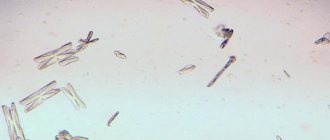
| Crystals (tripelphosphates, cholesterol, uric acid, hematoidin, Charcot-Leyden) | ( – ) |
| Hemoglobin, haptoglobin | ( – ) |
| Detritus | Available in varying amounts |
| Salts (oxalates) | None |
Reasons for deviation from the norm
Feces are formed from the remains of the bolus of food in the large intestine. It consists of detritus, dietary fiber, epithelium, water, bile and enzymes. Their number and type depend on nutrition and other factors. By analyzing all the research data obtained, the doctor will determine in which part of the intestine the pathological changes occurred and whether there is an enzyme deficiency.
Macroscopic indicators
Thick, dense-textured stool is considered normal. This indicates a sufficient amount of liquid in it. When plant foods with a high fiber content predominate in the diet, intestinal motility increases, and feces take on a mushy form. If an increase in the frequency of bowel movements is noted with liquid stool, then they speak of diarrhea (diarrhea).
Hard stools are a consequence of slow peristalsis and dehydration of feces. This condition occurs with constipation or hemorrhoids. These disorders appear due to poor nutrition, as well as in men with prostatitis, in women during pregnancy and after childbirth.
The smell of feces changes with the abuse of protein foods. Fetid occurs during putrefactive processes in the lower intestines, with cholecystitis or pancreatitis. The sour smell is the result of fermentation.
The color of the stool is determined by a pigment called stercobilin, and changes depending on the diet and the use of iron-containing drugs (for example, Maltofer).
Colorless stool indicates a violation of the outflow of bile due to stones in the biliary tract (often with cholelithiasis), occurs with cirrhosis of the liver, jaundice. With hepatitis A and C, the level of pigment also decreases.
A light color is possible when taking antibiotics or consuming fatty foods, as well as in pregnant women due to increased stress on the digestive organs.
Too dark stool is due to excess stercobilin. Dark brown occurs with pleiochromia (high content of bile pigments in bile). Black feces can appear after taking certain medications, in particular De-Nol.
Biochemical indicators
A pH level below normal with an acidic reaction occurs due to problems with the metabolism of fatty acids, as well as an increase in fermentation microflora. The increase in pH (alkaline reaction) is due to the large amount of meat protein in the diet. In this case, rotting of the protein elements occurs. A sharply alkaline reaction is characteristic of putrefactive dyspepsia.
Fatty acids should not be observed in the stool. In a healthy person, they are absorbed by the body. If the number of fats is several times higher than the reference values, then steatorrhea is diagnosed. As a rule, it is observed in cases of problems with the flow of bile, such as cystic fibrosis. Soaps in the coprogram speak of pancreatic dysfunction.
The detected native protein means the development of inflammation in the upper part of the gastrointestinal tract (colitis, enteritis, gastroduodenitis, duodenal ulcer, gastric ulcer, cancer). Be sure to take into account the presence of symptoms. If a person experiences pain in the hypochondrium and bloating, then increased protein indicates acute pancreatitis.
Normally, only altered muscle fibers are detected. If a scatological examination shows unchanged muscle fibers, the gastroenterologist will prescribe an additional examination of the pancreas, since this indicator indicates problems with the breakdown of proteins.
Microscopic indicators
Red blood cells in the stool indicate inflammatory processes in the digestive tract or damage to it by tumors.
Moving through the intestines, fecal lumps injure the inflamed areas, causing bleeding of varying intensity. Another reason why stool contains blood cells may be helminthic infestation.
If occult blood is detected, its quantity will be determined as + for a weakly positive reaction, ++ for a positive reaction.
Mucus is produced by the intestinal epithelium as a protective reaction to irritating factors. In the event of inflammation of an infectious or non-infectious nature, the amount of mucus increases.
This occurs, for example, with intestinal tuberculosis, cholera, ulcerative colitis, IBS or Crohn's disease. Bacteria that can cause fermentation in the intestines (dyspepsia) form iodophilic flora.
Their determination is made using an iodine test: pathogenic organisms differ in the resulting color.
Extracellular starch indicates a decrease in the function of specific enzymes (amylase) that are responsible for its breakdown. Leukocytes and macrophages indicate the presence of any inflammation in the gastrointestinal tract, including those found in large numbers during acute intestinal infection (AIE).
How to prepare for the analysis?
For those who want to undergo a clinical stool test, a referral is issued by a therapist at a clinic or hospital. A few days before the sample is taken, the patient is prescribed a special diet that cleanses the intestines, which affects the accuracy of the result. Two nutrition options have been developed: according to Pevzner and according to Schmidt.
In the first case, they eat meat, bread, sauerkraut and potatoes. In the second, they eat five times a day, consuming a lot of dairy products, eggs, potatoes and meat.
Considering that both diet options include meat products during preparation, vegetarians need to warn their doctor about their dietary needs, since due to the exclusion of animal proteins from the diet, some deviations from generally accepted values will be acceptable.
When preparing for the analysis, you should avoid drinking alcohol, as it affects the properties of feces. If a person takes any medications, then it is necessary to stop them for a while to avoid erroneous interpretation of the indicators. Two days before the test, all coloring foods (carrots, beets, asparagus, prunes) are excluded from the diet.
Unlike most blood tests, in this case there is no need to donate biomaterial on an empty stomach. Women should not undergo coproscopic analysis during menstruation. Following these simple recommendations will allow you to obtain an objective result of the study, and therefore a correct idea of the state of health of the gastrointestinal tract.
How to collect biomaterial?
The morning portion of feces is considered optimal for the study. Evening is also allowed, but the sample must be stored in the refrigerator for no more than 10 hours after collection. There is a certain algorithm for collecting feces for coprogram.
First of all, hygiene procedures are carried out - external toilet of the genital organs. To defecate, you must use a clean container - a pot or a vessel. If defecation is difficult, you can do a Microlax microenema or take Duphalac.
They do not affect the results of the study. You should not use food cans, boxes or use a European toilet as dishes for feces. Feces are collected in a sterile disposable jar or a specialized container with a spoon.
The surname and initials are indicated on it, and the laboratory assistant signs a unique number, date and time of reception of the biomaterial.
The feces are placed in a test tube. For research in laboratories they use a special diachem. kit (test strips) and microscope. When the results are ready, the patient is given a form with a table indicating all the indicators and normal limits for testing.
In case of strange results, which may suggest an error in the study or poor preparation, the analysis must be retaken. The execution time is 1–2 days depending on the selected laboratory. If the analysis result is needed urgently, the study takes less time. In preparation for surgery, tests are valid for 14 days.
If it takes a long time to get to the laboratory, you should ask about the possibility of receiving results online.
Source: https://detki.shukshin-net.ru/oksalaty-v-kale-u-rebenka/
Notes from a laboratory assistant. Causes of oxalate formation
Authors : Kuznyak Irina
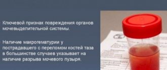
Excess salts in OAM most often appear in “thick”, concentrated urine. If a person drinks a sufficient amount of fluid, then, most likely, he is not at risk of urolithiasis. Oxalates (oxalic acid salts) can be formed in urine with an acidic pH, and with an alkaline environment, and with a neutral one. This is probably the insidiousness of oxalate stones, and the reason that of all kidney stones, oxalate stones occur in the majority of cases. And if for the formation of other stones (microlites) a clear connection to alkaline or acidic urine is needed, which means that, knowing this, you can influence the environment, pH, then, alas, this trick will not work with oxalates.
The only thing that can be advised is to limit the consumption of foods rich in oxalic acid. Oxalic acid interferes with the absorption of calcium, forming insoluble salts. There is also an opinion that excess vitamin C can lead to the formation of oxalate stones, but this is now being questioned, since the reactions that take place in vitro during research are not necessarily repeated in the human body. But personally, I observe 99% of cases of the appearance of oxalates in microscopy precisely after uncontrolled and prolonged use of vitamin preparations. Of these, 90% of cases are after massive intake of vitamin C and 10% after taking vitamin D in the form of an aqueous solution. Firstly, vitamin D contains citric acid, which easily combines with calcium, and secondly, water drops of this vitamin are more difficult for mothers to dose than oil drops. If oxalates suddenly appear in the TAM, then you need to either reduce the dose of the vitamin or discontinue the drug. To be honest, it’s scary to see all the microscopy fields of view in salt crystals, reading the child’s age on the form: 1 month, 2 months, 7 months.
Oxalates have the ability to form insoluble salts with Ca, Mg, Fe ions. Calcium (and magnesium, by the way, too) is a macroelement in the body; oxalic acid salts most often combine with it.
Since iron is a trace element in the human body, the formation of salts with it (and then theoretically) is only possible in the form of sand, which will not be retained in the kidneys. Another thing is the combination of oxalates with calcium. Calcium in the body is a macroelement (concentration in the body is 0.04–2%), that is, with excess intake of oxalic acid from food and a tendency (possibly even hereditary) to disrupt oxalate metabolism, insoluble compounds of CALCIUM OXALATE (CaC2O4) may appear.
Products containing large quantities of oxalic acid, from which oxalates are formed, can easily be found in the literature. I would recommend reading this article about a diet with a tendency to form oxalates.
published 29/11/2018 17:15 updated 30/11/2018 — Nutrition, vitamins, Tests and examinations, Kidney and urinary tract diseases, Tests and examinations, Nephrology and urology
Oxalate crystals in a child's stool
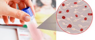
In order for the baby to grow up healthy, it is important to prevent diseases or recognize them in time. For such purposes, mothers are asked to collect the baby’s stool and submit it for analysis.
It is the feces that provide a more complete picture of the state of the internal organs, metabolic processes, the functioning of the intestines, and stomach, and allow one to observe the entire etiology, as well as prescribe proper treatment.
The coprogram will also show crystals of salts, in particular oxalates. Why, why and how to treat oxalates in feces - this is worth talking about in more detail.
Coprogram - why do it?
The coprogram will also show crystals of salts, in particular oxalates
Diagnosis of gastrointestinal diseases, pathologies of the kidneys and ureter in a small child is a problem.
Determining diseases is often impossible using techniques used for adults, so laboratory diagnostics remains one of the most productive methods. When collecting stool for analysis, mothers themselves do not always expect the results that are revealed.
For example, the presence of oxalate crystals is frightening, alarming and forces you to immediately seek the best treatment.
What does a stool test provide:
- identification of disorders of acid-forming and enzymatic activity of the stomach, intestines, and pancreas;
- liver function failure;
- instability of juice evacuation from the stomach/intestines;
- the presence of inflammatory processes;
- disturbances in the state of intestinal and stomach microflora;
- the presence of inflammatory processes in internal organs.
With this analysis, it is easy to identify whether there are crystals, what kind they are, what they are associated with, and choose the desired treatment option. During normal life processes, there should be no oxalate crystals in the feces.
Oxalates in feces: why do they appear?
The cryptogram reveals the presence of several groups of formations, which are “fragments” of cells subjected to destruction during the digestion process
The cryptogram reveals the presence of several groups of formations, which are “fragments” of cells subjected to destruction during the digestion process. Thus, there are crystals:
- Epithelial. These are the remains of epithelial cells that are destroyed under the influence of gastrointestinal enzymes. A small accumulation does not cause concern; an increased level means the presence of an inflammatory process in the colon mucosa.
- Scharko-Leiden. Formed from cells responsible for allergic reactions, they indicate the presence of helminthic infestation.
- Tripelphosphates. They occur during an accelerated reaction of evacuation of intestinal contents and can mean massive bleeding of the gastrointestinal tract.
- Oxalates. Crystals have minimal diagnostic value and appear due to low acidity of gastric juice, as well as from eating vegetarian foods.
Important! The absence of free hydrochloric acid converts calcium oxalate to calcium chloride and is detected by the formation of crystalline formations that precipitate in the feces.