Urolithiasis: symptoms and methods of treatment at home
Urolithiasis (urolithiasis) is a disease that occurs as a result of a metabolic disorder in which an insoluble sediment in the form of sand (up to 1 mm in diameter) or stones (from 1 mm to 25 mm or more) is formed in the urine. Stones settle in the urinary tract, which disrupts the normal flow of urine and causes renal colic and inflammation.
According to medical statistics, urolithiasis is the second most common among all urological diseases, and the third among urological diseases leading to death.
Causes
Urolithiasis is caused by various reasons:
- sedentary lifestyle leading to metabolic disorders;
- infectious and inflammatory diseases of the genitourinary system, which were caused by streptococcus, staphylococcus, E. coli, Proteus vulgaris;
- other diseases of the kidneys and genitourinary system;
- unbalanced diet, disrupted diet, too spicy, sour, salty foods in the diet;
- lack of vitamins A and B;
- drinking low-quality water containing harmful chemical elements;
- some drugs can increase the acidity of urine and affect kidney function;
- unfavorable working conditions, accompanied by physically difficult work or work in the cold;
- tumors in the bladder;
- chronic gastrointestinal diseases (pancreatitis, gastritis and others);
- chronic and protracted pathologies of the kidneys and urinary tract;
- osteoporosis and other bone-related diseases;
- genetic predisposition.
As for women, pregnancy also affects the development of urolithiasis. In women carrying a child, the outflow of urine is often disrupted in the later stages. The uterus enlarges, putting pressure on the kidneys. For this reason, urine can stagnate, causing the development of infectious diseases.
What are the causes of urolithiasis?
The causes of urolithiasis are often associated with heredity, namely metabolic disorders. As a result, changes occur in the chemical and water-salt structure of urine, in particular, excess content of:
- uric acid;
- oxalate salt;
- calcium salts;
- phosphate salt.
Internal causes include digestive diseases, dehydration, infections, vitamin D deficiency and poor diet.
There are also external factors:
- the water we use, or rather its chemical composition;
- climate, amount of ultraviolet rays;
- conditions at work and at home (sedentary lifestyle or, conversely, heavy physical activity).
Classification
Basically, the pathogenesis of urolithiasis develops against the background of metabolic disorders in humans. This leads to the fact that some foods and substances are poorly processed and cannot be completely eliminated from the body. They accumulate as insoluble particles and result in sand or stones in the urine. Stones are classified according to their chemical composition. They come in several types:
- Based on calcium (phosphates, carbonates). They are the most common (more than 60% of all stones).
- Containing uric acid salts (urates). They can be dissolved and are found mainly in elderly patients.
- Based on magnesium salts. Such stones provoke inflammation in the places where they are located.
- Protein stones (cystine, cholesterol). These protein stones occur very rarely.
Studying stones for their chemical composition is of great importance in treating the disease and prescribing a diet.
Magnetic resonance imaging (MRI) in St. Petersburg
Urolithiasis occurs in 12% of men and 5% of women. Most stones form in the distal nephron or at the papilla, that is, the area at the junction of the medulla and the minor calyces, where the collecting ducts open into the common papillary duct. The chemical composition of cameos can be different, most often based on calcium, uric acid and phosphates. Chemical composition plays a role in the choice of treatment, but only dual-energy CT can help determine the composition of the stones.
The purpose of radiation examination for urolithiasis is to identify stones, complications, establish the possibility of stone passage and confirm stone passage. Plain radiography is always performed for abdominal or lower back pain. The role of radiography in the study of the kidneys remains, despite the widespread use of CT.
Survey photographs of the kidneys with nephrolithiasis may reveal enlarged shadows of the kidneys, changes in their shape and contours. However, these symptoms are not constant and can occur with other diseases. A shadow suspicious for a calculus in the projection of the urinary tract may be masked by bone structures. The absence or presence of a shadow in survey photographs does not indicate the absence of a stone or in its favor. Gallbladder and pancreatic stones, fecal stones, foreign bodies and many other solid formations can be mistaken for kidney stones. The diagnosis of staghorn stone can be made with sufficient accuracy even only from plain films. However, survey urography is necessary only to outline tactics for further examination of the patient.
Survey radiograph. Coral stone of the left kidney.
The use of excretory urography makes it possible to establish the presence of a stone in the urinary tract, identify the number of urinary stones and their size, localize stones in the urinary tract and determine the functional state of the diseased and contralateral kidney, as well as observe the disease over time.
Ascending (retrograde) urethropyelography is an invasive research method. The technique allows one to accurately determine stones in the urinary tract. Antegrade pyelography allows you to evaluate
patency of the ureteropelvic segment and the ureter itself.
CT is the method of choice for suspected kidney stones. Sensitivity and specificity approach 100%. Non-obstructive stones are often missed by intravenous urography but are clearly visible on CT. MSCT allows you to study thin sections in one breath-hold from the kidneys to the bladder. Contrast is used only for the differential diagnosis of phleboliths and stones. 50 ml of low-osmolar contrast agent is injected and MSCT is performed with a delay of 3-5 minutes. Phleboliths have a soft tissue component ("comet tail"), but do not have a low-density center.
CT. Stone in the right kidney. There is no hydronephrosis.
With MRI, stones are not visible as such, however, defects in the filling of the collecting system or urethra can be detected.
On ultrasound, stones are visible as echogenic formations with an acoustic shadow behind them. The accuracy of the method is quite high, even stones of 2 mm are already detected by ultrasound. However, the size of stones is more accurately determined by CT. In the presence of stones in the urethra, ultrasound is not reliable enough.
Ultrasound.
Kidney stone. MRI in St. Petersburg USA
Symptoms of urolithiasis
Symptoms of urolithiasis in men occur only when a formed stone moves through the urethra. The pathological condition is characterized by a triad of clinical manifestations:
- pain of varying severity;
- changes in urinary sediment (appearance of blood, pus and other components);
- disruption of the process of urine excretion, up to complete anuria (obstructive genesis).
The pain syndrome can be constant or intermittent, the degree of its severity varies from aching pain to unbearable renal colic, which requires emergency hospitalization of the patient in a hospital.
Painful symptoms are accompanied by complaints of a dysuric nature: frequent and painful urination, disruption of bladder emptying processes. Patients complain of general weakness, decreased performance, a feeling of nausea and vomiting at the peak of pain (it does not bring any relief).
The severity of symptoms of urolithiasis, depending on the location of the stone, is as follows:
- The presence of a calculus in the lumen of the bladder is accompanied by pain in the lower abdomen, and the pain radiates to the genitals, perineum or rectum. There are typical dysuric disorders: frequent and painful urination, which can suddenly be interrupted (symptom of “stream interruption”).
- When the calculus is localized at different levels of the ureter, the pain shifts to the groin area, and is characterized by irradiation to the surface of the thigh and genitals. There are complaints of frequent and painful urination. When a stone completely blocks the lumen of one of the ureters, the pain syndrome becomes unbearable (renal colic).
- If the stone is localized in the pyelocaliceal apparatus of the kidney, then the patient has aching pain in the lumbar region of the corresponding side. Pain is associated with changes in body position and movement of the patient. Traces of blood in the urine often appear.
Often, patients go to the doctor with a stone that has already passed, which is an indisputable sign of urolithiasis.
Features of the course of urolithiasis in women and men
People aged 20 to 55 years are at risk. The highest incidence rate is observed in the countries of Asia Minor. Urolithiasis is diagnosed three times more often in men than in women. However, it is women who tend to form coral-shaped studded stones, which cause many painful symptoms.
Symptoms are almost the same in both sexes. The main difference: in men, pain during renal colic radiates to the penis, and in women, to the labia. Quite rarely (in only 17% of cases) diseases of a bilateral nature occur, i.e. the formation of stones in both kidneys.
Complications
The most common adverse outcomes of the disease are the following pathological processes:
- calculous pyonephrosis (most often, purulent cavities in the kidney tissues occur with a recurrent form of urolithiasis);
- inflammation of the affected kidney due to blockage of the urinary duct (obstructive form of pyelonephritis);
- rupture of the wall of the ureter, bladder or urethra with the development of a septic condition in the patient;
- acute renal failure (observed in patients with urolithiasis of a single kidney);
- cicatricial deformations of the lumen of the ureter and others.
Typical symptoms of urolithiasis in men and women
Signs of urolithiasis are similar in men and women. Only the location of the pain differs. Men often complain of pain in the penis, and in women the pain radiates to the labia.
The most characteristic symptom of ICD is renal colic, that is, acute pain in the lumbar region. However, changing body position does not affect the intensity of pain.
Pasternatsky’s symptom is also typical for urolithiasis, when when tapping the kidney area in the projection of the liver, pain occurs, and the number of red blood cells in the urine increases.
Here are other symptoms that may indicate the presence of ICD:
- pain and stinging when urinating;
- the desire to empty the bladder occurs frequently, while urine is released in limited quantities;
- change in urine color, blood;
- general intoxication, which results from inflammation and insufficient urine outflow - vomiting, nausea, headache, weakness, increased body temperature.
Diagnostics
In order to exclude complications from urolithiasis, urologists recommend not delaying visiting a doctor and seeking medical help at the first signs of illness. A comprehensive differential diagnosis of urolithiasis, which consists of prescribing laboratory and instrumental examination methods, will help to recognize the disease, determine the location of stones, their size, and evaluate the functioning of the organs of the genitourinary system.
Instrumental diagnostics:
- Intravenous excretory diagnostics.
- X-ray – evaluates the kidneys, ureters and bladder, identifies stones.
- CT or MRI of the kidneys is an informative diagnostic method that allows you to evaluate the functioning of the entire genitourinary system and identify the slightest disturbances in its functioning.
- Ultrasound of the kidneys - visualizes all structures of the organ, determines the number of stones and other visible disturbances in the functioning of the urinary system.
Laboratory diagnostics:
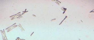
- Urinalysis - determines the pH of urine and the number of leukocytes. Urine tests for urolithiasis are carried out quite often, since they help identify salt crystals and recognize their composition.
- Blood test - allows you to determine the presence of an inflammatory process, as evidenced by an increased ESR and the number of leukocytes.
- Daily urine analysis - allows you to assess the content of various salts in urine.
The essence of pathology
From the name you can guess that the main manifestation of urolithiasis (UCD) is the formation and presence of stones in the kidneys, ureter, bladder and even the urethra.
The more scientific name for these formations is calculi. They consist of insoluble salts and have different compositions, often mixed. When removing a stone, it is important to determine its type and composition in order to select the optimal treatment and avoid relapses.
Stones interfere with the normal functioning of the urinary system and can occupy a large space in the renal pelvis, block the ureter, or cause severe pain when passing through the urethra during urination. At the same time, people often have no idea that they have urolithiasis in the early stages unless they do an ultrasound of the kidneys and bladder or don’t take a urine test. Symptoms of urolithiasis and the general condition of a person significantly depend on the position and size of the stones.
Treatment of urolithiasis
ICD is a group of serious diseases that, if not properly treated, can lead to death. Self-medication for this disease is unacceptable, so at the first signs of the disease you should seek medical help. Any form of urolithiasis is treated comprehensively using:
- medicines;
- diet;
- herbal medicine;
- physiotherapy;
- correct lifestyle;
- ultrasonic crushing of stones;
- removal of stones.
A conservative method of treating urolithiasis in men is carried out taking into account an integrated and systematic approach and involves taking certain medications.
Medicines are prescribed depending on the composition of the stones:
- Citrate suppositories, diuretics and vitamins (if stones are of oxalate etiology);
- Diuretics, anti-inflammatory and diphosphonates (if the detected stones are of phosphate etiology). With this course of ICD, many doctors recommend home treatment with herbs as an auxiliary therapy;
- Medicines that slow down the process of urea synthesis. Drugs are also prescribed that change the degree of acidity of urine, which leads to the dissolution of stones (in the presence of stones of urate etiology).
Drugs for the treatment of urolithiasis are divided into the following groups:
- Painkiller medicine. Medicines relieve pain during an attack of renal colic (Tempalgin, Baralgin and others).
- Antibiotics. Mandatory point of therapy. The antibiotic is selected individually by the urologist.
- Medicines to help pass the stone. The purpose depends on the size, composition, location (Furosemide).
- Antispasmodics. They remove the cause of the spasm, relax the walls of the ureter, facilitating the passage of the stone (Papaverine, No-shpa, Diprofen).
- Drugs that dissolve stone. Selection of products based on the composition of the stone (“Fitolysin”, “Solimok”, “Urodan” and others, as well as dietary supplements - “Prolit”, “Litovit”).
The goal of drug therapy is to prevent exacerbation of urolithiasis, alleviate the general condition of a person, relax the muscles and walls of the ureter (kidney), dissolve possible stones and painless removal.
Urolithiasis disease
Category: Prevention.
Urolithiasis is a disease characterized by the appearance of hard stone-like formations in the urinary organs (kidneys, ureters, bladder). At their core, urinary stones are crystals formed from salts dissolved in urine.
The appearance of foreign bodies in the urinary tract leads to damage to the mucous membrane and inflammation, causing a typical clinical picture of the disease.
CAUSES OF URINOLOGICAL DISEASE
This disease is polyetiological, that is, several factors lead to its development. Most often, urolithiasis develops in people aged 20–45 years, and men suffer from it 2.5–3 times more often than women.
Factors that contribute to kidney stone formation include:
- genetic predisposition;
- drinking water rich in certain mineral salts;
- insufficient water regime - consumption of a small amount of liquid;
- sedentary lifestyle;
- eating foods rich in purine compounds (meat, vegetables - spinach, beans).
A special place among the causes of urolithiasis is occupied by diseases of various organs:
- Infectious and inflammatory diseases of the urinary tract: pyelonephritis, cystitis, urethritis.
- Diseases of the stomach and other organs of the digestive tract: hepatitis, gastritis, pancreatitis and others.
- Congenital and acquired anomalies of the kidneys and ureters.
- Metabolic diseases: gout, hyperparathyroidism.
All of the above conditions lead to changes in the acid-base balance in the body, which leads to the formation of kidney stones.
URINOLOGICAL DISEASE: SYMPTOMS OF THE DISEASE
Signs of urolithiasis vary in their diversity - from the complete absence of clinical symptoms to such serious phenomena as renal colic and kidney block.
The leading symptoms of urolithiasis, or what patients complain about:
- burning and pain above the pubis and in the urethra during urination - are explained by the spontaneous release of small pebbles, the so-called “sand”;
- lower back pain associated with a sudden change in body position, sudden shaking, heavy drinking (especially after drinking liquids such as beer and pickles). Pain occurs due to slight displacement of stones;
- hyperthermia (high temperature) - indicates a pronounced inflammatory reaction to the stone at the site of its contact with the mucous membranes, as well as the addition of infectious complications;
- renal colic is the most unpleasant complication of urolithiasis, manifested by sharp pain in the lower back with irradiation (spread) along the ureter, irradiation of pain may be noted in the leg, in the abdomen;
- patients often note cloudiness of the urine, as well as the appearance of blood in it (typical of renal colic).
The nature of the pain and its location can give the doctor information about the location of the stones: in the kidney itself, ureter or bladder. X-rays and ultrasound can help confirm this assumption.
In the earliest stages, the disease may not manifest itself in any way - kidney stones are often discovered by chance during a medical examination. Sometimes even large stones do not show up until the patient experiences an attack of renal colic.
TREATMENT OF URINOLOGICAL DISEASE
We recommend reading:
- Treatment of kidney stones: features of surgical and conservative treatment
- In the treatment of urolithiasis, both conservative methods are used - using tablets and injections, and surgical methods - operations are performed to remove stones.
CONSERVATIVE TREATMENT OF URILOSTICAL DISEASE
Analgesics are used to eliminate pain; even narcotic drugs can be used in the ambulance and in the hospital. Antispasmodic drugs also relieve pain - with good antispasmodic therapy, the stone can come out on its own.
To dissolve stones, drugs are used that change the acid-base balance of the blood and change the acidity of the urine. The drug is selected taking into account the type of stones, of which there are several types: cystine, oxalate, phosphate.
Thiapramine, Uralit can be used to dissolve cystine stones; oxalate - Prolit, renal collections No. 7 and 8; phosphate - Marilin.
Important: the drug is selected by a urologist or nephrologist based on urine and blood tests of the patient!
Among the conservative methods of treatment, physiotherapy is also used: patients are prescribed magnetic therapy, amplipulse therapy, inductothermy and other methods.
URILOSTICAL DISEASE: TREATMENT AT HOME
At home, in the absence of pain, and also to prevent relapses, you can use traditional methods. For phosphate stones, the effect is observed with regular drinking of rosehip or barberry decoctions. Combined herbal preparations are also used, consisting of several herbs that have moderate diuretic, antispasmodic and uroseptic effects.
Important: only the attending physician can give accurate recommendations!
For urate stones, you can use oatmeal decoction. For cystine and struvite stones, traditional methods in the treatment of urolithiasis are ineffective, as well as conservative treatment, since these stones almost do not dissolve.
SURGICAL TECHNIQUES
Large urinary stones that cannot be dissolved are broken into small fragments, which either come out on their own or are removed surgically. Stones are destroyed by lithotripsy, affecting them with a shock wave. There are several types of lithotripsy:
- ESWL - external shock wave lithotripsy - is a non-invasive method in which the impact on kidney stones is carried out without any skin incisions or other invasive techniques.
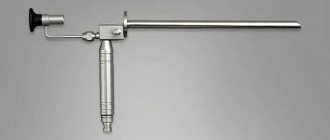
- Contact lithotripsy - an endoscopic device is brought to the stone through the urethra and bladder, the active part of which comes into contact with the stone (which is why the method is called contact). A shock wave is formed at the point of contact.
- Percutaneous lithotripsy - with this technique, a lithotripter is inserted into the kidney through an incision in the lumbar region. Used for crushing giant and coral-shaped stones.
- In cases where the stone cannot be crushed, surgery is performed. Depending on the volume of the operation, the following types of operations for urolithiasis are distinguished:
- Pyelolithotomy - a kidney stone is removed through a small incision in the renal pelvis.
- Nephrolithotomy - an incision is made directly through the kidney. This operation is indicated for stones that cannot be removed by other methods and when lithotripsy is ineffective. It is the most difficult operation for the patient.
- Ureterolithotripsy is an operation to remove a stone from the ureter.
PREVENTION OF URILOSTICAL DISEASE
It is easier to prevent kidney stones than to treat them later. There is a whole range of preventive measures aimed at reducing the rate of stone formation and getting rid of them. It is recommended for all patients who have had at least one attack of renal colic to adhere to simple prevention tips.
Prevention Tips:
- Proper drinking regime. Water consumption per day should be at the level of two liters. In summer, you can increase this volume to three liters. But you should first consult a doctor, since for some heart diseases large amounts of fluid are contraindicated.
- Prevention of dehydration (dehydration). In extreme conditions (in the heat, when playing sports, during illnesses with high fever), you should drink more liquid in small portions of 100–150 grams every half hour to hour.
- Diet for urolithiasis. A balanced diet, in which the ratio of different types of meat, dairy and plant products is selected individually, can reduce the risk of stone formation. Ideally, the diet should be selected by a doctor. Food requires sufficient levels of microelements and vitamins of different groups. If necessary, you can take multivitamin complexes.
Diet option for urolithiasis:
- Limiting salt intake. It is better to under-salt food than to over-salt it. Excess table salt puts a strain on the kidneys, causing urolithiasis.
- Physical activity. The load on the abdominal and back muscles improves renal blood flow, which stimulates metabolic processes in the kidneys and improves their detoxification function.
- Timely treatment of diseases. Pay attention to the gastrointestinal tract and endocrine system - these organ systems should be periodically examined, since disturbances in their functioning are one of the leading factors predisposing to urolithiasis.
- Prevention of diseases of the genitourinary system. Pyelonephritis and urethritis can cause exacerbation of urolithiasis, so it is better not to get sick with them, and if there are symptoms of the disease, start treatment.
- Spa treatment. Patients with kidney stones during remission are recommended to visit resorts where treatment with mineral waters is carried out 1–2 times a year. This is one of the most effective methods of prevention. In Russia, dispensaries specializing in the treatment of this disease are available in Kislovodsk, Pyatigorsk, and Zheleznovodsk. A doctor will help you choose a specific sanatorium, since certain mineral waters are suitable for each type of stone.
COMPLICATIONS OF URILOSTICAL DISEASE
Delayed consultation with a doctor and abuse of traditional methods of treating urolithiasis without the consent of a doctor can lead to serious complications in the urinary system.
Complications often include:
- Infections of the urinary system can not only provoke urolithiasis, they can also be its complication. Often the following diseases occur against the background of the disease: pyelonephritis, cystitis, urethritis.
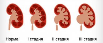
- Kidney block and hydronephrosis - blockage of the ureter by a calculus makes it impossible for urine to flow out. As a result, it accumulates in the kidney, causing it to enlarge. This condition can lead to kidney failure and kidney loss.
- Nephrogenic hypertension. Against the background of renal pathology, an uncontrolled increase in blood pressure is often observed - symptomatic hypertension.
- Nephrosclerosis is the degeneration of kidney tissue due to constant disturbances in urine metabolism. This is the most common cause of chronic renal failure.
- Purulent complications that occur when pathogenic microorganisms enter the kidney (abscess and pyonephrosis). Kidney stones make these diseases worse and can lead to septic shock and death.
The information is provided for informational purposes only. Don't self-medicate. At the first symptoms of the disease, consult a doctor. There are contraindications, a doctor's consultation is required.
Based on materials from the site: www.oceydoc.ru
Folk remedies
At home, in the absence of pain, and also to prevent relapses, you can use traditional methods. For phosphate stones, the effect is observed with regular drinking of rosehip or barberry decoctions.
Combined herbal preparations are also used, consisting of several herbs that have moderate diuretic, antispasmodic and uroseptic effects.
- Combine the ingredients in the indicated quantities: garden parsley herb - 20 g, bearberry leaves, common juniper fruits, field steelhead root, dandelion root - 15 g each; common anise fruits, shepherd's purse herb - 10 g each. Pour 10 g of raw material in an enamel bowl with 1 cup of boiling water, close the lid and heat in a water bath for 30 minutes, leave for 10 minutes, strain, squeeze out the grounds. Bring the volume of the decoction to 200 ml with boiled water. Take 1/2-1/3 cup warm 2-3 times a day.
- Tricolor violet herb - 30 g, horsetail herb - 30 g, St. John's wort herb - 25 g, dandelion herb - 25 g, buckthorn laxative root - 25 g; Brew a tablespoon of the crushed mixture with a glass of boiling water, leave for 30 minutes, strain and take a glass 3 times a day for phosphate and carbonate stones.
- This method of removing stones involves taking two decoctions. The first decoction is prepared from rosehip roots. They need to be crushed using a coffee grinder to end up with 50 g of dry powder. Then add 700 ml of water to the powder and leave to simmer for 15 minutes. After this, prepare an infusion of bearberry. To do this, pour boiling water (300 ml) over dried or fresh herbs (about 30 g), leave for about 2 hours. You need to take the first remedy three times a day after meals, 300 ml. 25 minutes after consuming it, you should take 100 ml of bearberry infusion.
- Mix the ingredients in the indicated proportions: greater celandine herb - 30 g, oregano herb - 20 g, barberry bark - 20 g; Pour a tablespoon of the mixture into a glass of boiling water, leave for 30 minutes and take a glass 3 times a day for uric acid stones.
- Mix the ingredients in the indicated proportions: leaves and roots of stinging nettle - 50 g, licorice root - 30 g; Pour a tablespoon of the mixture into a glass of boiling water, leave until cool, strain and drink in 3 doses during the day for kidney stones with nephritis.
- The dissolution of sand and stones in the urinary organs is facilitated by fresh onions and garlic, strawberries, a decoction of melon seeds in milk, black radish juice with honey or sugar, infusions and decoctions of beans, peas, infusions of shepherd's purse leaves, black currants, fruits (fresh and dry) rose hips, rowan fruits, dandelion roots, calamus rhizomes, corn silks, horsetail grass (contraindicated for nephritis). Recommended are pumpkin, cabbage brine and juice, barberry, strawberries, and rose hips.
The list of traditional medicine recipes is large. It is worth remembering that some herbs have contraindications, so when choosing a method of treatment with folk remedies, consultation with a doctor is required.
Surgical methods
Large urinary stones that cannot be dissolved are broken into small fragments, which either come out on their own or are removed surgically. Stones are destroyed by lithotripsy, affecting them with a shock wave.
There are several types of lithotripsy:
- Contact lithotripsy - an endoscopic device is brought to the stone through the urethra and bladder, the active part of which comes into contact with the stone (which is why the method is called contact). A shock wave is formed at the point of contact.
- Percutaneous lithotripsy - with this technique, a lithotripter is inserted into the kidney through an incision in the lumbar region. Used for crushing giant and coral-shaped stones.
- ESWL - external shock wave lithotripsy - is a non-invasive method in which the impact on kidney stones is carried out without any skin incisions or other invasive techniques.
In cases where the stone cannot be crushed, surgery is performed. Depending on the volume of the operation, the following types of operations for urolithiasis are distinguished:
- Nephrolithotomy - an incision is made directly through the kidney. This operation is indicated for stones that cannot be removed by other methods and when lithotripsy is ineffective. It is the most difficult operation for the patient.
- Pyelolithotomy - a kidney stone is removed through a small incision in the renal pelvis.
- Ureterolithotripsy is an operation to remove stones from the ureter.
Kidney stones (Nephrolithiasis, Kidney stone disease)
Conservative treatment
Treatment of nephrolithiasis can be conservative or surgical and in all cases is aimed at removing kidney stones, eliminating infection and preventing the re-formation of stones.
For small kidney stones (up to 3 mm), which can be passed independently, plenty of water load and a diet excluding meat and offal are prescribed. For urate stones, a dairy-vegetable diet that alkalizes urine and alkaline mineral waters (Borjomi, Essentuki) is recommended; for phosphate stones - take acidic mineral waters (Kislovodsk, Zheleznovodsk, Truskavets), etc. Additionally, under the supervision of a urologist, medications that dissolve kidney stones can be used (for example, citrate therapy for urate stones).
First aid for renal colic
With the development of renal colic, therapeutic measures are aimed at relieving obstruction and pain. For this purpose, injections of platyphylline, metamizole sodium, morphine or combined analgesics in combination with atropine solution are used; A warm sitz bath is performed and a heating pad is applied to the lumbar region. In case of intractable renal colic, novocaine blockade of the spermatic cord (in men) or round ligament of the uterus (in women) and catheterization of the ureter are required.
Surgery
Surgical removal of stones is indicated for frequent renal colic, secondary pyelonephritis, large stones, ureteral strictures, hydronephrosis, kidney blockade, threatening hematuria, stones in a single kidney, coral stones. For nephrolithiasis, remote lithotripsy is used to avoid any intervention in the body and remove stone fragments through the urinary tract. For stones up to 2 cm in diameter, you can use the “flexible retrograde nephrolithotripsy” method, as well as percutaneous nephrolitholapaxy, which allows you to remove the stone through a puncture in the kidney.
Open or laparoscopic interventions to remove stones - pyelolithotomy (dissection of the pelvis) and nephrolithotomy (dissection of the parenchyma) are rarely resorted to, mainly when minimally invasive surgery is ineffective. In case of complicated kidney stone disease and loss of kidney function, nephrectomy is indicated. After removal of stones, patients are recommended spa treatment, lifelong diet, and elimination of associated risk factors.
Nutrition rules
Diet and nutrition for urolithiasis depends on the pH and composition of the stones. Depending on them, doctors have compiled a list of products whose consumption is contraindicated in a particular case.
If the stones are of urate origin, you should not take:
- alcoholic drinks;
- coffee;
- meat broths;
- fried and spicy foods;
- offal;
- chocolate, cocoa;
- protein of animal origin.
If you have phosphate stones, you should not use:
- vegetables with green skin and/or pulp;
- any spices;
- spicy dishes;
- pumpkin, including its seeds;
- legumes;
- potato;
- dairy products.
If you have oxalate stones, you should avoid using:
- dairy products;
- citrus fruits;
- strawberries and wild strawberries;
- lettuce leaves;
- spinach;
- legumes;
- cheeses of any kind;
- nuts;
- sorrel;
- cocoa, coffee and tea.
Compliance with a certain diet is an integral part of the therapeutic program, which allows you to stop further formation of stones in the urinary system, as well as suppress the growth of existing stones.
Nutrition for urolithiasis is based on the following principles:
- Don't overeat. Food that enters the stomach in large volumes will only worsen the situation.
- Systematic consumption of food. Ideally, you should eat around the same time. It is not recommended to skip meals, as this can lead to increased stone formation and poor health.
- Don't eat excessively high-calorie foods. The energy value of products must correspond to the energy costs that occur in reality.
- The diet should be enriched with foods rich in vitamins and amino acids.
- Drink about 2-3 liters of regular still water per day. This will increase the volume of urine excreted.
Prevention
When diagnosed with urolithiasis, prevention should be carried out much earlier than the first signs of the disease appear. People who are at risk or have chronic metabolic diseases should pay special attention to their health.
Prevention of urolithiasis consists of following the following recommendations:
- Consume clean water. In some regions, water contains large amounts of salts, which leads to increased concentrations in the urine and the formation of crystals. It is better to buy bottled water or use highly purified filters.
- Maintain drinking regime. If there are no contraindications, a person should drink about 2 liters of liquid per day. The best option is clean drinking water. It is an ideal solvent and helps dilute the salts, preventing the formation of crystals and the formation of stones from them. People living in hot climates need to increase the volume to 3 liters.
- Eat a balanced diet. Kidney stones are formed both in meat eaters who adhere to a protein diet, and in vegetarians who consume a lot of acidic vegetables and fruits. Therefore, nutrition should be varied and balanced in composition. It is recommended to eat 150-170 g of meat and 50 g of fish per day. It is not necessary to eat them every day, for example, you can fish 2 times a week for 300 g. You also need 300-400 g of vegetables and the same amount of fruits in any form every day. The total amount of cereals and bread should be 300-400 g.
- Stay hydrated. Infectious diseases, burns, hot weather, prolonged physical activity and sports cause significant fluid loss. You must constantly replenish its reserves. To do this, it is advisable to drink frequently (every half hour), or in small portions of 100-150 ml. This will help reduce intoxication, remove harmful substances from the body and protect the kidneys.
- Take vitamins. Deficiency of vitamins, especially E and group B, negatively affects the condition of the mucous membrane of the genitourinary organs and kidney function, and also leads to disruption of metabolic processes. Therefore, it is recommended to take vitamin complexes 2 times a year.
- Don't over-salt your food. For an adult, the daily salt requirement is 5 g or one teaspoon. This amount includes all the salt in the dishes you prepare and in the products (mayonnaise, herring, chips). Excess salt makes it difficult for the kidneys to function.
- Get outdoors. Ultraviolet deficiency has a bad effect on bone health. Minerals are washed out of them, which can take part in stone formation.
- Treat diseases of the urinary system in a timely manner. Any inflammation can provoke the formation of stones and exacerbation of urolithiasis. Therefore, at the first symptoms, seek qualified help and do not self-medicate.
- Lead an active lifestyle. Lack of physical activity contributes to urinary stagnation. And exercises aimed at strengthening the abdominal and lumbar muscles improve kidney function and eliminate congestion. The daily norm should be walking (30-40 minutes) and a set of exercises lasting 15-20 minutes. The best option is to additionally visit the gym or pool 2-3 times a week.
- Take herbal diuretics periodically. Watermelon, pomegranate juice, and a concentrated decoction of dried apricots (100 g per 0.5 liter of water) are suitable. Some medicinal herbs have a diuretic and anti-inflammatory effect: bear's ears, corn silk, horsetail and bearberry. They “wash” the kidneys, prevent salts from precipitating and remove small stones and sand that have already formed.
- Take care of your digestive health. Deficiency of digestive enzymes in gastrointestinal diseases leads to the formation of calcium oxalate stones. Thus, in case of digestive disorders, ascorbic acid turns into oxalate, which is deposited in the kidneys in the form of crystals.
- Avoid hypothermia. Keep your feet and lower back warm. The receptors located in these areas have a reflex connection with the kidneys and bladder. Hypothermia can cause inflammation or spasm of the smooth muscle around the stone.
Particular attention to prevention should be paid to people whose relatives suffer from urolithiasis. Since there is a high probability that the tendency to form stones is inherited.
“NOT the time to collect stones”: what is urolithiasis?
If a stone has been removed from the soul, a person experiences relief. But what if the stones are completely material and are located in the body? The urologist at the Expert Clinic Voronezh, Ellina Nikolaevna Yushina, told us about urolithiasis.
— Ellina Nikolaevna, what is urolithiasis?
— This is a common urological disease in which calculi (stones) form in different parts of the urinary system. Recently, the diagnosis of “urolithiasis” has been made more often. This is probably due not to an increase in incidence, but to improvements in diagnostic methods. Early detection contributes to successful treatment and avoids relapses.
— Are urolithiasis and kidney stones synonymous?
- Not really. The term “renal stone disease” indicates the location of the stone in the kidney. Urolithiasis is a broader concept that can mean the presence of stones in the kidneys, bladder or ureter.
— What are the causes of urolithiasis?
— The mechanism of development of the disease is still not well understood. There are several theories about the origin of this disease, but none of them has become established as the main one. The matrix and physicochemical theories are well known. According to the matrix, the calculus crystallizes around a microscopic protein clot (fibrin, epithelium, leukocytes killed during the inflammatory process). When the underlying substance (matrix) appears, salt crystals are deposited on it, and the calculus grows like a snowball.
The physicochemical theory is as follows: urine contains crystalloids and colloids, the level of which is balanced due to the content of so-called protective colloids. In cases where this balance is disturbed, colloids in a supersaturated urine solution begin to precipitate, followed by their atypical crystallization, which is the beginning of calculus formation.
There are a number of other theories, but as mentioned earlier, it has not yet been possible to develop a single, “leading” theory.
— Who is at risk?
— Urolithiasis can appear at any age. But the peak incidence occurs between 25 and 50 years of age. In children and elderly people, stones more often form in the bladder, in young patients and middle-aged people - in the kidneys.
There are factors that increase the risk of developing stones.
- Heredity. Metabolic features leading to urolithiasis can be genetically determined. Heredity on both the maternal and paternal lines is important. The tendency to form stones is sometimes passed down through generations (from grandparents).
- Certain metabolic diseases (eg, gout, obesity, congenital disorder of cystine metabolism).
- Diseases of the endocrine system. In particular, diabetes mellitus, pathologies of the parathyroid glands that regulate calcium metabolism.
- Chronic, long-term untreated diseases of the gastrointestinal tract (for example, chronic gastritis). This also leads to metabolic disorders.
- Sedentary lifestyle. The consequence is a violation of phosphorus-calcium metabolism. Often, urolithiasis is diagnosed in elderly people and bedridden patients who previously (before the decrease in physical activity) did not have such problems. This is also true for other people who do not have sufficient physical activity.
- A large amount of vitamin C in the diet. Everything is good in moderation. Hypervitaminosis is no less dangerous than hypovitaminosis.
- Unbalanced diet. Oxalates can form due to excess oxalic acid in the diet. The source of excess oxalic acid entering the body is sometimes the consumption of large amounts of spinach, beets, leeks, etc. Another common type of stones is urate stones. The cause of their occurrence may be an excessive intake of purine bases from food, which, in combination with metabolic disorders (such as gout, diabetes mellitus), leads to an increase in the level of uric acid in the blood, which in turn is accompanied by stone formation in the kidneys, bladder, and crystal deposition uric acid on the surface of cartilage tissue in joints. Types of meat such as turkey, goose, rabbit, and so-called “liver” (liver, kidneys, brains, lungs) are rich in purines. Legumes (beans, lentils, peas) are also a rich source of purines. We need purines in moderation for the normal functioning of the body, so these foods can and should be eaten, but should not be abused, especially if you know that you have gout or obesity.
“Long-term overload with protein foods can lead to serious impairment of kidney function. This situation is especially dangerous for people with existing renal pathology (including diabetes mellitus, arterial hypertension, urolithiasis, etc.), and the elderly.” Quote from the material “Rules of healthy eating. What should you eat to stay healthy?
- Geographical factor. There are regions where living in increases the risk of stones in the urinary tract. This is usually associated with the composition and quality of drinking water.
- Long-term use of certain medications, in particular sulfonamides.
- Inflammatory processes in the kidneys.
- Other pathologies of the urinary tract. There is a version that almost all people are prone to the formation of solid sediment in the urine from time to time. But for healthy people this is not dangerous: the sediment leaves on its own and does not linger. The stones do not reach 5 mm. Urolithiasis does not develop. If the outflow of urine is impaired, the sediment cannot leave completely, the crystals increase.
A diameter of 5–6 mm is considered a threshold value. The fact is that crystals smaller than 6 mm come off on their own in 80% of cases. Once the threshold size is exceeded, the chances are sharply reduced to 25 - 30%.
— What are the symptoms of urolithiasis?
— They depend on the location of the stone and the degree of urodynamic disturbance (urine passage). If urodynamics are not impaired, the disease can be asymptomatic for a long time. Characterized by discomfort in the lower back, pain in the urethra, and frequent urination. Since the calculus can injure the mucous membrane, hematuria (blood in the urine) is possible. With complete obstruction (blockage) of the ureter, acute, severe pain in the projection of the kidneys, nausea, and vomiting occur. If an infection occurs, the patient notes an increase in temperature, general weakness, shaking chills, profuse sweating, and pallor. All this points to such a formidable complication of urolithiasis as occlusive pyelonephritis.
You can read more about pyelonephritis in our article
— If urolithiasis is asymptomatic, how can it be detected?
— Timely diagnosis of this disease is a very important problem. After all, stone movement and ureteral obstruction often appear after vibration or changes in atmospheric pressure. An attack takes a person by surprise after a long car ride or flight. This means that with a high probability a person is away from home, in an unfamiliar environment. This situation is especially dangerous when traveling abroad. Not everyone takes out insurance, and not all insurances provide the necessary treatment. And medical care for renal colic is a very expensive service. The worst decision in such a situation is to wait until you get home. This can lead to kidney loss. Therefore, it is important to undergo regular medical examinations and perform ultrasound examinations of the kidneys. If you haven’t done it before, be sure to do an ultrasound of your kidneys before your trip.
— What complications can urolithiasis lead to if left untreated?
— Due to stagnation of urine and injuries to the mucous membrane, inflammatory processes begin. The consequence of which can be pyelonephritis. Obstruction of the ureter threatens hydronephrotic transformation of the kidney, fulminant acute pyelonephritis, the development of pyonephrosis with subsequent necrosis (necrosis) of kidney tissue, cicatricial changes in the parenchyma (secondary wrinkled kidney), which will lead to either immediate loss of the organ (nephrectomy) or severe delayed consequences: chronic renal failure, hypertension of renal origin. Such hypertension is difficult to correct with medication, and blood pressure can reach 200/90 mmHg.
When increased blood pressure is not an independent disease, but a symptom of diseases of other organs, we speak of symptomatic arterial hypertension. You can find out more about it here
— Who should I contact if I need a doctor to treat urolithiasis?
— A urologist is involved in the treatment and prevention of urolithiasis.
— What examinations are prescribed for suspected urolithiasis?
— A urine test with sediment microscopy is required to determine the type of stones, and an ultrasound of the kidneys and bladder is performed. Sometimes CT or MRI of the kidneys and retroperitoneum with intravenous contrast is required for clarification. In some cases, it becomes necessary to perform a CT or MRI of the pelvic organs.
If the examination reveals coral urolithiasis, the function of the parathyroid glands is checked.
Read the material on the topic: “Detecting kidney stones. CT, ultrasound or MRI – what to choose?”
— What types of stones exist?
- Oxalates are the most common - 70 - 80% of cases. 15% – urates. 5 – 10% – phosphates. Struvite and cholesterol stones are also found. Cystine stones are rare - less than 1%: they are typical only for people with hereditary disorders of the metabolism of one of the essential amino acids - cystine.
— Why is it important to know the type of stones?
— Information about the type of stones allows you to choose the right treatment and take measures to prevent relapses, choose a diet that changes the composition and acidity of urine. If surgery is required, you can determine in advance whether the stone can be crushed. This is important for choosing a surgical treatment method
— What methods of treating urolithiasis are common in modern medicine?
— For severe forms of urolithiasis, the method of choice is surgical treatment. In modern urological practice, methods such as DLT (external lithotripsy) and CLT (contact lithotripsy) are most often used: stone fragmentation is performed using ultrasonic waves, laser or pneumatic pulses. Less popular is percutaneous nephrolitholapaxy with the installation of a stent for kidney drainage. Over the past 10 years, methods have become more gentle, endoscopic operations without incisions are possible: access is through the natural openings of the body.
In less severe cases, the metabolism is affected through diet and medications.
An important stage is sanatorium-resort treatment. Treatment at resorts with mineral waters (for example, Truskavets and places with a similar water composition) helps prevent the formation of new stones.
To prevent infectious and inflammatory complications in urolithiasis, antibiotics are used.
How to use antibiotics correctly? Instructions for use
Why don't antibiotics help?
— What should be the diet for urolithiasis?
— The diet depends on the type of stones and the metabolic characteristics of a particular patient. Recommendations are given by a urologist. You can also consult a nutritionist: this makes sense if, in addition to urolithiasis, other conditions need to be taken into account.
For prevention, it is important to drink enough water, maintain physical activity, pay sufficient attention to your health - undergo a medical examination in a timely manner.
Interviewed by Daria Ushkova
You can make an appointment with a urologist here
ATTENTION: the service is not available in all cities
The editors recommend:
Delicate problem: what you need to know about cystitis
What does a urine test according to Nechiporenko show?
Why does my lower back hurt? We are convening a medical consultation
For reference:
Yushina Ellina Nikolaevna
In 2001 she graduated from the Voronezh State Medical Academy named after N.N. Burdenko, where she completed her residency in the specialty “Urology”.
Passes certification every 5 years.
Currently a urologist at the Expert Clinic Voronezh. Receives at the address: Pushkinskaya St., 11.