Urolithiasis disease. We have all heard something about this disease. The phrases “urolithiasis” and “kidney stones” are now widely heard. But how much do we know about the seriousness of urolithiasis and who among us is at risk? In modern urology, this is the most acute, pressing and widespread problem. Nowadays, 25 to 40% of patients who consult a urologist receive a diagnosis of urolithiasis. Every year, doctors observe a steady increase in the incidence, and ignoring the first symptoms threatens serious complications.
Urolithiasis (or urolithiasis) is a chronic pathology caused by metabolic disorders and accompanied by the formation of calculi (stones). The disease develops and disrupts the full outflow of urine, and an inflammatory process begins in the body. The most dangerous thing is that sometimes the disease is completely asymptomatic (in about 13% of cases).
The disease can occur at any age. But more often it is diagnosed in people 30-50 years old. Moreover, men get sick 3 times more often than women.
How do kidney stones develop?
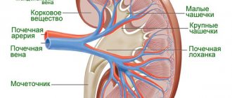
The urinary tract consists of two kidneys, the ureters, the bladder and the urethra. Urine contains substances such as calcium, cysteine, oxalate and uric acid. When the levels of these substances are higher, they crystallize. If these crystals are small, they travel in the urinary tract and exit the body through urine. If this does not happen, then they settle on the kidney and increase in size. As the mass grows, it begins to cause pain and block the flow of urine.
The most important question for us arises - how to treat urolithiasis?
If conservative therapy is ineffective (medicines, physiotherapy, diets), surgical intervention is necessary.
Previously, open surgery was the only option for removing stones from the urinary tract. But these days this method is used extremely rarely, since it is traumatic. The main indications for open surgery are developing renal failure and large stones.
Today, specialists more often resort to minimally invasive and endoscopic operations. They are characterized by low trauma and a short rehabilitation period for the patient. Endoscopic operations are performed through small punctures in the abdominal cavity (laparoscopic operations).
The most advanced and safe method now is minimally invasive procedures that are carried out using electromagnetic waves. Among them there are two types:
- DLT - extracorporeal lithotripsy
- CLT – contact lithotripsy
Lithotripsy is performed using a special device.
External lithotripsy (ESL) is performed without external intervention, and contact lithotripsy (CLT) is performed with minimal invasion through the urethra.
Types of kidney stones
There are four types of kidney stones.
1. Calcium stones
These stones are the most common type among all four. There are two types of calcium stones:
- Calcium oxalate is a combination of calcium and oxalate;
- Calcium phosphate is a combination of calcium and phosphate.
2. Cystine stones
These stones are rare and are caused by genetic disorders. Cystine is a chemical that the body produces naturally. In people with genetic disorders, cystine leaks into the urine through the kidneys.
3. Uric acid stones
If the level of acidity in the urine is high, it can lead to the development of uric acid stones. They can be formed by the acid itself or by its combination with calcium.
4. Struvite stones
These stones develop in people who suffer from recurrent urinary tract infections: bacteria produce ammonia, which in turn can cause stones to form in the urine. They consist of phosphate, ammonium and magnesium.
What causes kidney stones in a child?
Below are some of the causes of kidney stones in children:
- increased mineral content in urine;
- insufficient water intake or dehydration;
- genetic disorders;
- congenital problems with metabolism;
- family history of kidney stones;
- urinary tract defects or infections;
- obesity and decreased physical activity;
- a diet high in sodium, protein, or both.
In some cases, the causes of kidney stones are unknown.
What are the symptoms of urolithiasis?
The first sign is renal colic, that is, acute pain in the lumbar region. The ureter becomes clogged with a calculus, which causes a sharp increase in pressure in the pelvis and stretches it.
Symptoms and their severity depend on where exactly the stone is located in the kidney:
- If the stone is in the collecting apparatus of the kidney, then the patient has aching pain in the lumbar region on the corresponding side. Traces of blood in the urine often appear.
- If the stone is located at different levels of the ureter, then the pain shifts to the groin area. The patient complains of frequent and painful urination.
- When a stone completely blocks the lumen of one of the ureters, the pain syndrome becomes unbearable. Renal colic appears. The pain radiates to the lower abdomen, genitals, perineum or rectum. Frequent and painful urination appears, which may suddenly stop (symptom of “stream interruption”).
How are kidney stones diagnosed in children?
To determine the presence of kidney stones in a child, a comprehensive diagnosis is carried out. It includes:
- studying the child’s medical history;
- physical examination;
- laboratory and imaging tests.
Studying the child's medical history
This helps the doctor understand if someone has a family history of kidney stones or any other genetic disorder that could trigger the condition.
Physical examination
Kidney stones are dealt with by specialists such as a pediatric urologist or nephrologist. During the examination, the doctor will also ask questions regarding the patient’s health, diet, and water consumption.
If the stone is passed in your urine, you can give it to your doctor, who will then send it to the laboratory for further tests.
Laboratory and imaging tests
To accurately diagnose kidney stones, additional laboratory tests may be recommended to the patient. They include:
- Urine and blood analysis
A urine test is important to determine factors (such as high calcium levels) that lead to stone formation.
Blood tests are used to identify other risk factors that may be causing or may affect the baby.
- Genetic tests
Genetic tests help determine the severity of the disease, as well as its further course.
- Scanning tests
Doctors examine the urinary tract for kidney stones using ultrasound scans. If ultrasound results are insufficient, computed tomography (CT) is performed. or x-ray.
- Visualization tests
Imaging tests are used to determine the exact size and location of the stones, which is important for treatment.
Urolithiasis disease
Every year, more than half a million people seek emergency care for kidney stones. According to statistics, every tenth person on Earth has urolithiasis. Conditions such as hypertension, diabetes and obesity can increase the risk of kidney stones.
Do children get kidney stones?
The incidence of urolithiasis in children is growing every year, which is probably due to the two most important reasons: 1) drinking insufficient amounts of fluid (still water), 2) eating foods high in salts (chips, French fries, sausages, sausages) , canned foods, etc.) and carbonated drinks. The presence of urolithiasis in young children is a serious problem, usually indicating the presence of serious metabolic diseases (for example, oxalosis, cystinuria, cystinosis, distal renal tubular acidosis, etc.)
What are kidney stones?
Kidney stones are a hard, crystal-like mass. They consist of salts that are found in normal human urine in small quantities. Kidney stones can vary in composition and size. Depending on the chemical composition, stones can be divided into several types: urate, oxalate, phosphate, carbonate, mixed.
Usually a person does not know about his disease until the stones begin to change their location and move along the urinary tract. Even a small stone leaving the kidney can cause excruciating pain.
A person with a hereditary predisposition is most likely to develop urolithiasis. The causes of salt deposition in the kidneys and the formation of stones can be excess calcium or uric acid in the blood, which are caused by metabolic disorders (for example, hyperparathyroidism, excess intake of vitamin D, gout). An important role is played by insufficient fluid intake into the body or, conversely, large loss of fluid (for example, due to excessive use of diuretics). Both lead to the formation of very concentrated urine, which promotes the formation of stones. With kidney infections, urine stagnation may occur and its acidity may change. High levels of fructose in your diet also increases your risk of developing kidney stones.
What are the main symptoms of having a kidney stone?
In size, stones can be tiny grains that pass unnoticeably into the urine, or they can be large (up to several centimeters) formations of complex shape. Typically, the larger the stone, the more severe the symptoms.
Usually the first symptom of the disease is acute stabbing pain in the back (in the area of the diseased kidney) or in the lower abdomen. The pain may spread to the groin area and may also be accompanied by nausea and vomiting. The pain will continue until the stone is passed in the urine, because the muscular wall of the ureter will try to push it out. Damage to the mucous membrane of the urinary tract may result in the presence of blood in the urine (visible or invisible to the eye). As the stone travels down to the bladder, it will cause a strong urge to urinate and a burning sensation when urinating.
The passage of kidney stones (renal colic) may also be accompanied by fever and chills.
What can be done for renal colic?
To relieve pain and reduce temperature, take antispasmodics, be sure to consult a doctor for examination and treatment.
If the pain continues or you notice blood in your urine, call your doctor.
Surgery is necessary if conservative measures fail, there is severe and prolonged pain, or complications develop
Diagnosis of urolithiasis is based on the following data:
- Typical patient complaints include periodic lower back pain, attacks of renal colic, and urinary disorders.
- Studying family history
- General and biochemical tests of urine and blood.
- Ultrasound of the kidneys and bladder.
- Special blood tests for the level of parathyroid hormone and calcitonin, calcium and phosphorus salts, oxalates, determination of blood pH.
- Survey radiography of the abdominal organs. Computed tomography and other studies as necessary.
Are there any long-term consequences of having a kidney stone?
Kidney stones increase your risk of developing chronic kidney disease; if you have had at least one stone, you are at increased risk of developing another stone in 50% of cases within 5 to 7 years. Urolithiasis, especially in children, is usually accompanied by urinary tract infections, which also damage the kidney. In addition, the reasons that led to the formation of stones could also damage the kidney tissue, and the consequences may appear after some time, therefore, with at least one episode of renal colic, long-term careful monitoring by a nephrologist and urologist is necessary.
What needs to be done to prevent and/or stop the formation of urolithiasis?
- Drink plenty of fluids (6 to 8 glasses of water per day) to help produce enough urine.
- Eat more fruits and vegetables, which make your urine less acidic. Less acidic urine has a lower potential for stone formation.
- You can reduce excess salt in your diet. What foods contain a lot of salt? Everyone will think that salty chips and French fries contain something that is rarely eaten. There are other foods that contain a significant amount of salt: sausages, sausages, cheeses, canned foods, fast food and even carbonated drinks.
- If you are overweight, you need to diet. To do this, you should consult a nutritionist.
Treatment for urolithiasis depends on the type of stone and the severity of symptoms.
Most kidney stones eventually pass through the urinary tract on their own within 48 hours, with sufficient fluid intake. Antispasmodic medications may be prescribed to relieve symptoms. Some patients with severe pain from kidney stones need to stay in the hospital to receive appropriate treatment. Depending on the type of stone, medications may be prescribed to reduce stone formation and help dissolve and remove the material that forms the stone.
Stones up to 4 mm have an 80% chance of passing, while stones measuring 5 mm already have a 20% chance. Stones larger than 9-10 mm rarely go away on their own and usually require treatment.
Surgery for kidney stones is usually required if:
- The stone is too big to come out on its own
- The stone grows
- The stone blocks the flow of urine and causes infection or kidney damage
- The pain is not relieved by painkillers.
Modern medicine offers minimally invasive methods for treating kidney stones, such as:
Extracorporeal shock wave or laser lithotripsy This is used to remove small stones that are located near the kidney or ureter. The method uses ultrasound or laser to break up the stone. After this procedure, the stone, crushed into sand, leaves the body with urine.
Percutaneous nephrolithotomy is a procedure used for large kidney stones, or in cases of kidney abnormalities. The stone is removed using an endoscope, which is inserted into the kidney through a small surgical incision.
Urethroscopy Urethroscopy is performed when there are stones in the lower urinary tract. The urethroscope consists of a hollow metal tube equipped with lighting and optical systems. It is inserted through the urethra and allows you to examine the entire length of the urethra, the bladder, and also perform the necessary surgical and therapeutic manipulations.
Nephrolithotomy Open surgery may be necessary if other methods do not work or are not possible. Nephrolithotomy for the treatment of kidney stones is used today in very rare cases.
Who is at risk?
The following are some of the factors that increase the risk of developing kidney stones in children:
- if the child has had kidney stones in the past;
- insufficient fluid intake;
- low carbohydrate diets (ketogenic diets);
- cystic fibrosis;
- congenital abnormalities in the kidneys, ureters or bladder;
- the use of certain medications: for example, furosemide (Lasix), acetazolamide (Diamox), allopurinol (Aloprim, Zyloprim);
- rare genetic disorders;
- Inflammatory bowel diseases affecting urinary oxalate levels.
Treatment of kidney stones in children
To eliminate kidney stones in children, one of the following procedures is usually prescribed.
Extracorporeal shock wave lithotripsy is one of the common treatment methods used in children. A lithotripter is used to transmit acoustic shock waves through the skin, breaking up the stones and then leaving the body. This procedure is performed without incisions, but under anesthesia.
A ureteral stent is needed if there is a blockage or narrowing in the ureter. The ureter is the tube that carries urine from the kidney to the bladder. A stent is a soft tube that is placed in the ureter to help the flow of urine. A child with a stent can move around, but should avoid strenuous physical activity.
Percutaneous nephrolithotripsy is chosen for the treatment of large kidney stones. During this procedure, a tube is inserted into the kidney through an incision in the back, and the doctor uses a nephroscope to find the stones and remove them.
During the nephrostomy procedure, a small tube is used to drain urine. If a kidney stone is blocking urine drainage, this tube is used so that kidney damage and infection can be cleared. This procedure uses an external bag to drain urine.
With ureteroscopy, the doctor first finds stones using a microscopic camera that moves through the urethra into the bladder, then into the ureters and kidneys, and then removes them.
What are the benefits of this treatment?
- low-traumatic (surgery without incisions)
- Absolutely painless treatment - without anesthesia (quick rehabilitation of the patient, the procedure is carried out without hospitalization).
Today, DLT and CLT are a reliable and safe method of curing urolithiasis.
At the SOVA clinic, the DLT procedure is carried out using innovative equipment - the Compact Sigma device from Dornier MedTech , manufactured in Germany.
The company is a recognized world leader in the market of urological equipment and surgical lasers.
Dornier Compact Sigma is a high-quality device that is characterized by high efficiency, a wide range of applications and safety for the patient. When performing lithotripsy, the harmful effects of the shock wave on the human body are practically eliminated due to precise focusing on the stone. The procedure is performed with minimal pain and does not require anesthesia.
Home prevention of kidney stones
Below are some home remedies:
- More fluid intake helps increase urine production and reduce the mineral components that form stones.
- Basil is known to help stabilize uric acid levels, which can prevent the formation of kidney stones. It also contains acetic acid, which helps dissolve some stones.
- The antioxidant properties of pomegranate reduce the likelihood of kidney stones.
How to prevent your child from developing kidney stones?
Kidney stones can occur several times. However, to minimize the chances of recurrence of stones, you should take certain precautions, namely:
- increase drinking water consumption;
- Carry out constant monitoring to prevent the appearance of new stones.
On the Vikids platform you can:
Read similar articles
on the forum
Find a doctor