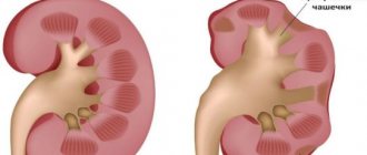
Kidney agenesis in a child is a disorder of kidney formation during fetal development. Renal agenesis can be unilateral, with one kidney, or bilateral. The two types of renal agenesis have very different clinical courses, with unilateral agenesis being more favorable.
The kidneys perform many important functions in the body. Having at least one kidney is necessary for life. The kidneys filter waste and excess fluid from the blood, maintain healthy levels of electrolytes and minerals in the blood such as sodium, phosphorus, calcium and potassium, help maintain normal blood pressure, and secrete hormones that are important for body function. There are usually two fist-sized kidneys present, one on each side of the spinal column at the back just below the rib cage.
Unilateral renal agenesis in a child
Unilateral renal agenesis in a child is the result of fetal developmental defects that cause the formation of only one kidney. Having a single kidney instead of two is not necessarily life-threatening. A single kidney usually expands and is capable of performing most body functions to a degree sufficient for life. When a solitary kidney negatively affects health, it occurs through very subtle and gradual changes that may not be noticed initially. Having kidney agenesis in a child also means that if injury or disease leads to kidney failure, there will be no spare kidney. In this case, the consequences of disease of a single kidney can be life-threatening.
What is urolithiasis
Urolithiasis is a common disease characterized by the formation of stones in the organs of the urinary system (kidneys or bladder).
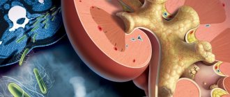
The largest number of hospitalizations of patients with urological diseases is due to urolithiasis. The formation of stones occurs in the kidneys, much less often in the bladder. Subsequently, stones can migrate and be localized at any level of the urinary system: ureter, bladder, urethra. In addition to renal colic, the most serious complication is the development of obstructive pyelonephritis (acute inflammation in the kidney due to blockage of the ureter with a stone).
Bilateral renal agenesis in a child
Bilateral renal agenesis is a genetic disorder involving the failure of both kidneys to form during fetal development. Fetal kidney development begins between five and seven weeks of pregnancy. Fetal urine production begins early in pregnancy and is responsible for the formation of most of the amniotic fluid present in the second and third trimesters of pregnancy. The fetus continually ingests amniotic fluid, which is reabsorbed in the fetal gastrointestinal tract and excreted back into the amniotic cavity by the kidneys. Because amniotic fluid is maintained by fetal urine, a fetus without kidneys has little amniotic fluid to surround and protect it in the intrauterine environment. This condition is known as oligohydramnios and results in physical compression of the fetus in the womb. Bilateral renal agenesis causes a number of physical complications known as Potter's syndrome. The amniotic membrane adheres to the fetus and the compression causes further physical malformations, including constricted facial features.
Compression and lack of amniotic fluid also lead to serious defects in lung development. Fetal urine is critical for proper lung development. Fetal urine helps dilate the airways and supplies the amino acid proline, a critical amino acid for lung development. After leaving the womb, the baby must rely on the lungs and breathe air to obtain oxygen to live. Alveoli are many small sacs in the lungs that are responsible for exchanging oxygen with the blood. If the alveoli are underdeveloped at birth (pulmonary hypoplasia), the baby will not be able to breathe normally and will go into respiratory failure. In most cases, the condition is fatal within the first few months of postnatal life. If the lungs of a child with bilateral renal agenesis are sufficiently developed, the baby can be saved with dialysis and possible kidney transplantation.
Signs and symptoms of renal agenesis
- Unilateral renal agenesis
Most people born with a solitary kidney do not experience any noticeable signs or symptoms that would indicate unilateral renal agenesis. A solitary kidney is usually discovered when a patient undergoes an ultrasound or surgery for some other problem. However, unilateral renal agenesis in a child can cause gradual changes in body functions that lead to unhealthy clinical conditions. A potential complication of a solitary kidney is high blood pressure.
The kidneys typically help maintain healthy blood pressure in two ways. The kidneys regulate how much fluid remains in the bloodstream and how much fluid is eliminated from the body. The more fluid that flows through the bloodstream, the more your blood pressure rises. The kidneys also secrete the hormone renin, which works as part of a group of hormones to dilate or contract blood vessels. The greater the contraction of blood vessels, the higher the blood pressure.
In addition, one kidney doing the work of two kidneys may be subject to more wear and tear than normal. The single kidney may end up being slightly damaged, causing the body to lose excess protein through urine. This condition is known as proteinuria and is a sign of kidney damage. Another potential complication of having a solitary kidney is the decreased efficiency of removing waste from the bloodstream. The reduced ability to filter blood is known as reduced glomerular filtration rate (GFR), a potential complication of unilateral renal agenesis in a child. As long as these complications are managed, people with a solitary kidney often experience no symptoms.
- Bilateral renal agenesis
There are many signs of bilateral renal agenesis in newborns (first six weeks of life). Potter facies are facial features associated with Potter syndrome in bilateral renal agenesis. Facies include a compressed facial appearance, a flattened nose, a recessed chin, prominent epicanthal folds (a fold of skin from the root of the nose to the eyebrow), and low-set abnormal ears. Poor or absent urine output during the first 48 hours of life, respiratory failure, and poorly developed lungs are telltale signs.
Diagnosis of renal agenesis in a child
- Unilateral renal agenesis
The diagnosis of unilateral renal agenesis in a child can be made using various imaging techniques, such as ultrasound, or directly through exploratory surgery. The diagnosis is usually made in adults without symptoms while they are being diagnosed with some other health problem. Complications of unilateral renal agenesis may include proteinuria and decreased glomerular filtration rate. Diagnosis of proteinuria is made using a urine test. High levels of protein in the urine indicate kidney damage.
- Bilateral renal agenesis
Urinalysis, if urine is present, is used to detect proteinuria. Ultrasound imaging can detect renal agenesis both before and after birth, but the prenatal condition is not as easily visualized. A chest x-ray, although not recommended for an infant, may be used to assess lung development. In newborns who die from this condition, an autopsy is recommended to confirm the diagnosis.
Treatment of urolithiasis
When renal colic develops, it is necessary to relieve pain. For this purpose, the doctor prescribes non-steroidal anti-inflammatory drugs, less often narcotic analgesics.
Surgical treatment is performed in the case of a large stone, the inability to relieve pain with medications, recurrent attacks of renal colic, inflammatory processes in the organs of the urinary system, or impaired urine outflow.
- Draining the kidney using a special internal catheter-stent or performing percutaneous puncture nephrostomy (creating an artificial path for urine outflow) often precedes other surgical interventions.
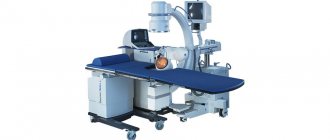
- External shock wave lithotripsy is a non-invasive treatment method aimed at fragmenting stones under X-ray or ultrasound guidance with high-intensity pulses focused on the stone.
- Ureteroscopy is a minimally invasive endoscopic method for removing stones from the ureters. The destruction of the stone occurs using laser, pneumatic or ultrasonic energy.
- Percutaneous nephrolithotripsy is an invasive endoscopic removal of kidney stones. This treatment method is used for stones larger than 1.5 cm. Laser, ultrasonic or pneumatic energy is used to destroy the stone. Fragments of destroyed stone are evacuated with tongs.
Treatment of renal agenesis in a child
Unilateral renal agenesis does not require any treatment other than to prevent complications if they occur. Kidney function is monitored by regular physical examinations that check blood pressure and blood and urine tests. These tests are usually performed once a year and are designed to evaluate whether the solitary kidney is functioning properly. Regular monitoring is critical because if a single kidney becomes diseased or damaged, there is no backup kidney available to support life.
People with unilateral renal agenesis are advised to keep their blood pressure levels below 130/80 to prevent kidney damage. If medications are needed to maintain normal blood pressure levels, blood pressure medications that also protect kidney function may be needed. Angiotensin-converting enzyme (ACE) inhibitors and angiotensin receptor blockers are two classes of blood pressure medications that may also protect kidney function and reduce protein loss. Diuretics can also be used to help lower blood pressure by removing excess fluid from the body.
People with unilateral renal agenesis are often advised to avoid contact sports unless protective equipment is worn. Damage to a single functional kidney can quickly become life-threatening.
Diagnosis of urolithiasis
Diagnosis of urolithiasis begins with a general medical examination, collecting an anamnesis about the patient’s health condition and complaints, laboratory and instrumental diagnostics are performed.
- Clinical and biochemical blood test. Shifts in these tests make it possible to identify the inflammatory process (increased blood leukocytes), the phenomenon of renal failure (increased creatinine and urea);
- A general urine test in the presence of an infectious agent in the urinary system reflects inflammatory changes (presence of leukocytes, nitrite-forming bacteria);
- A survey X-ray of the abdominal organs allows you to visualize stones in the kidneys, ureters and bladder;
- Excretory urography. It is performed on the same principle as a survey photograph, only with the use of an X-ray contrast agent;
- Ultrasound of the kidneys and bladder with the orifices of the ureters;
- Computed tomography of the retroperitoneal organs with contrast enhancement. It is the most effective diagnostic method, allowing a detailed assessment of the location, number of stones and their density, as well as obtaining additional information about the anatomical and functional state of the organs of the urinary system. The data obtained helps in choosing the optimal method of stone removal.
Forecasts
In the absence of trauma or kidney disease, the prognosis for unilateral renal agenesis is good. People usually lead normal, healthy lives. Avoiding injury and basic preventive health care are important to maintain quality of life.
Bilateral renal agenesis has a very poor prognosis. This usually leads to death in the first days of life. The usual cause of death is respiratory failure and acute renal failure in the neonatal period. If survival progresses into early childhood, patients may have chronic pulmonary disease or chronic renal failure. If the lungs have sufficient development, a kidney transplant is necessary for survival. After a kidney transplant, the prognosis improves.
Prevention of urolithiasis
Important in the treatment of urolithiasis and the prevention of repeated attacks of renal colic is the elimination of risk factors, changes in lifestyle and eating habits. After stone removal, patients are advised to undergo regular follow-up with a urologist. Correction of metabolic disorders should be carried out with the participation of endocrinologists, gastroenterologists, and nutritionists.
You can prevent the development of urolithiasis by following a number of recommendations:
- follow a diet: minimize the consumption of spicy, salty and sour foods, control the amount of protein foods and vitamins;
- give up bad habits: smoking, drinking alcohol;
- drink enough water (2-3 liters of fluid per day);
- avoid stress and nervous tension;
- regularly undergo a medical examination, including a general urine test and ultrasound of the kidneys and bladder, and also promptly treat diseases of the urinary system;
- get rid of excess weight;
- move more;
- don't get too cold.