Features of the development of hypoplasia
The content of the article
Uterine hypoplasia can develop at any age. The main reason leading to the progression of uterine hypoplasia is a deficiency of hormones responsible for the development of the organs of the reproductive system.
Secondary causes of progression of uterine hypoplasia:
- malfunction of the thyroid gland;
- developmental delay in the prenatal period;
- chronic intoxication of the body;
- past infectious diseases, as well as their complications;
- surgical interventions on the organs of the reproductive system.
Additional reasons indicated by scientists are nicotine or drug addiction, mental disorders of various types, anorexia, hypovitaminosis and others.
Hereditary and external unfavorable factors
Hereditary enamel hypoplasia. Its cause is an inherited defect of a gene (or genes) and this hereditary pathology is often combined with general dysplasia of dental tissues. It usually appears on baby teeth. There are hereditary enamel hypoplasia associated with a violation of its protein matrix, caused by impaired maturation of enamel tissue and associated with hypocalcification.
Hypoplasia of enamel of primary teeth. Unfavorable external factors, if there are no hereditary ones, before birth and immediately after birth include: toxicosis during pregnancy, some infectious diseases (such as rubella, influenza, ARVI) and diseases of the gastrointestinal tract (for example, gastritis) in a pregnant woman, prematurity of the child, birth injuries in a child, congenital allergies in a child, the use of artificial feeding, congenital pathologies of the cardiovascular system.
Enamel hypoplasia of permanent teeth. External unfavorable factors that can provoke this pathology and affect during the first years of life include: artificial feeding, severe forms of infectious diseases, pathological disorders of metabolic processes (for example, rickets and similar pathologies), pneumonia, diseases of the thyroid gland and kidneys, anemia (iron deficiency), allergic reactions in a complicated form. Moreover, it is believed that the negative impact of external factors on the development of the rudiments of permanent teeth can occur even in the first months of a child’s life, when the body’s protective capabilities are still limited and any diseases can lead to metabolic disorders.
Symptoms
The first sign indicating pathology is a delay in sexual development. The first menstruation occurs only at the age of 15 years. It should be noted that in women with this diagnosis, not only the uterus, but also the vagina, oviducts and ovaries may be underdeveloped. There are also external signs of uterine hypoplasia - women with this pathology have short stature, a narrow chest, underdeveloped pelvis and breasts. Upon reaching puberty, patients may experience decreased libido and anorgasmia (lack of orgasm).
ENAMEL HYPOPLASIA
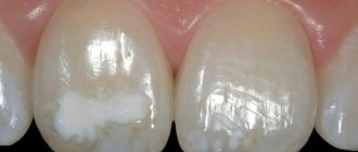
Hypoplasia is the insufficient development or disruption of the structure of the enamel of primary and permanent teeth. This pathology is often referred to as speckled teeth, but it is an independent disease. It appears in the form of pronounced grooves, pits or point defects on the surface of the teeth (mainly on symmetrical ones), as well as areas of reduced enamel thickness. Most cases affect the lower incisors and canines, and the depth of damage and coverage may vary. Hypoplasia is diagnosed in almost 40% of children of preschool and school age. Most often, the disease affects permanent teeth, which can lead to serious complications. The most dangerous consequence is aplasia - enamel atrophy and its complete absence.
For information about prices and treatment times, call:
+7
or fill out the feedback form:
Main reasons
Factors causing hypoplasia are divided into hereditary and acquired. In the first case, the main provoking mechanisms are “triggered” during the prenatal period. These may be developmental pathologies or adverse effects on the fetus, as well as birth injuries. Studies have shown that the risk of developing hypoplasia increases many times in children whose mothers suffered from severe toxicosis during pregnancy or suffered from infectious and inflammatory diseases. An unbalanced diet and bad habits of a pregnant woman have a negative impact on the formation of a child’s dental tissue. If the cause of hypoplasia is metabolic disorders of the fetus, then damage to the enamel is noticeable along the entire length of the crown.
This pathology can develop not only during intrauterine development, but also after illnesses suffered in infancy (in particular, rickets). The location and coverage of the teeth depend on the age at which the child had the disease. If these are the first months of life, then the enamel of the central incisors and cusps of the sixth teeth, which are formed before six months, is destroyed. At 8-9 months, hypoplasia may affect the lateral incisors and canines. Subsequently, when the teeth have already formed, the pathology covers other areas without any clear system. The causes of hypoplasia in older age can be chronic diseases and unbalanced nutrition, iron deficiency in the body and excess fluoride, impaired phosphorus metabolism and injuries to the buds of the teeth.
Classification of hypoplasia
Depending on the degree of severity, two forms are distinguished - systemic and local.
- Systemic (SGE) is caused by disturbances during intrauterine development and is manifested by the appearance of chalky spots on the vestibular surface of symmetrical teeth. The enamel, even on the affected area, retains its smoothness and shine, and the stain itself also does not change shape or color. Pathology can affect several teeth, and in advanced cases, the entire dentition.
Depending on the nature of the lesion, systemic hypoplasia is classified into spotted, pitted, grooved and cup-shaped. In the first form, the enamel changes color, in the second, horizontal pits appear, in the third, grooves with one or two walls are formed, and in the fourth, cup-shaped depressions are formed. There are also such varieties as Pfluger’s cone-shaped teeth with undeveloped cusps, Hutchinson’s barrel-shaped teeth - front incisors with a crescent-shaped recess at the cutting edge, and Fournier’s teeth, similar to the second form, but without a crescent-shaped notch.
- Local hypoplasia (LHE) is an acquired pathology and develops on permanent teeth due to mechanical trauma to their buds or infection. Typically, one or two teeth are affected. Characteristic clinical manifestations are multiple pinpoint depressions and whitish or yellow-brown spots - mainly on the premolars.
Treatment of hypoplasia
In the presence of shallow lesions and single spots, treatment is usually not carried out, as this can further damage the enamel. In this case, you need to take care of caries prevention and follow the rules of oral hygiene. Treatment methods include resurfacing of the damaged area followed by remineralization. If spots or dots are located on a visible surface, then this cosmetic defect can be eliminated by filling. In advanced stages, only restoration with veneers or prosthetics with metal-ceramic crowns is possible.
Depending on the extent of staining and enamel damage, treatment may include professional teeth cleaning, enamel remineralization, and the use of fluoride varnish. To eliminate aesthetic defects at later stages, whitening and restoration procedures (veneers, crowns) are performed. To treat mottling, a method such as grinding off the pigmented layers of enamel and restoring it through filling can also be used. Prevention of speckled teeth of endemic or non-endemic form involves monitoring the fluoride content in consumed water and normalizing nutrition.
Diagnostics
Primary diagnosis is based on identification and assessment of external signs:
- small labia;
- lack of hair in the genital area;
- short vagina;
- the cervix is elongated...
The main way to determine the infantile uterus is an ultrasound of the uterus. This study helps to establish the degree of development of the pathology. Additionally, the gynecologist prescribes blood tests for hormones (estradiol, testosterone, LH, prolactin, FSH, progesterone, T3, T4, TSH).
Additional techniques are hysterosalpingoscopy, showing the degree of patency of the fallopian tubes, MRI.
Causes
The main problem at the origins of enamel hypoplasia is serious disturbances in the metabolism of the fetus. Usually this can be associated with pathologies of germ cells or with certain factors that have a detrimental effect on the intrauterine development of the child.
However, not only disruptions in mineral metabolism and demineralization of tooth germs play a role.
The version that infectious processes and toxicosis also cause developmental abnormalities is being confirmed.
Toxoplasmosis, rubella or ARVI suffered by a pregnant woman can affect anomalies associated with teeth in general and enamel in particular.
In addition, the causes of enamel hypoplasia can be:
- birth injury by a premature baby;
- calcium metabolism disorder;
- atopic dermatitis;
- encephalopathy.
Enamel hypoplasia is systemic, which is expressed in the damage to several dental units at once.
Every second clinically healthy child is diagnosed with this disease. It can affect permanent and baby teeth, with the former being more susceptible to pathology.
Degrees of hypoplasia
Uterine hypoplasia has three degrees of development:
1st degree.
The size of the uterus corresponds to adult parameters, and the ratio of the cervix to the body of the uterus is 1:3. The fallopian tubes have individual elements of tortuosity, which can provoke an ectopic pregnancy. In most cases, pregnancy occurs naturally. The ovaries are not always developed, so women experience irregular menstruation.
2nd degree.
It is characterized not only by the organ’s too small size (2 times smaller than required), but also by a disproportion between the body of the uterus and the cervix. It normally makes up 1/3 of the length of the uterus, but with grade 1 hypoplasia, the cervix makes up 2/3 of the size of the organ.
In addition to abnormal parameters, the physiological characteristics of the uterus are also changed. Due to the small size of the organ, the endometrium does not reach the thickness required for embryo implantation. It is very thin, so girls' periods are scanty and short-lived. Often there is no menstruation at all. Hypoplasia of the 1st degree is complemented by abnormal development of the appendages. They have an abnormally large length and insufficient functionality of the ciliated epithelium. It is possible to get pregnant with stage 1 pathology, but IVF remains the only method.
3rd degree
. The length of the uterus is 3.5-5 cm, which is typical for childhood. Menstruation occurs later, at 15-16 years old. It is painful due to the narrowness of the cervical canal and increased intrauterine pressure during menstruation. The rush of blood causes a painful enlargement of the organ. The appendages are very tortuous and narrow.
Clinical manifestations of enamel hypoplasia
Structural disorders can occur in one group of teeth formed in the same period (systemic hypoplasia), in several adjacent ones of the same or different periods of development (focal) or on a single tooth (local).
Systemic hypoplasia is characterized by discoloration, underdevelopment or complete absence (aplasia) of enamel.
Focal hypoplasia is a fairly rare form of the disease that affects both primary and permanent teeth. Fangs, incisors and molars affected by the pathological process acquire a yellow color and a rough surface, have enlarged canals and shortened roots.
Local hypoplasia is not a congenital anomaly. It is a consequence of damage to the enamel or infection entering the tooth germ. This form of pathology affects exclusively permanent teeth.
How is uterine hypoplasia treated?
Pregnancy with uterine hypoplasia is possible only after hormonal treatment. The main task is for the uterus to achieve normal size and functionality of the endometrium and fallopian tubes. Hormones are selected based on test results.
A woman who has been diagnosed with hypoplasia should be very careful. In case of pregnancy with such a pathology, abortion is extremely undesirable.
If you find an error, please select a piece of text and press Ctrl+Enter
Causes of hypoplasia
Many factors can contribute to improper enamel development. Pathology is a manifestation of improper mineral metabolism. The rudiments of teeth are formed during intrauterine development, so the impact of negative factors on the body of a pregnant woman can cause disease.
Reasons that can lead to enamel hypoplasia:
- rubella, toxoplasmosis, acute viral infections during pregnancy, especially in the early stages;
- birth injury;
- prematurity;
- pregnancy with severe toxicosis4
- rickets;
- diseases of the central nervous system in the first year of a baby’s life;
- deficiency of vitamins and microelements;
- severe somatic diseases.
The pathology is polyetiological, so in many cases it is difficult to reliably determine the cause of the lesion. The depth and localization of enamel defects suggest the period during which the unfavorable factor was affected. This is due to the fact that the rudiments of teeth are formed gradually.
According to ICD-10, neonatal, prenatal and postnatal enamel hypoplasia are distinguished depending on the period in which the factor that caused the disturbance of mineral metabolism was active.
There is a theory according to which hypoplasia of the enamel of permanent teeth is associated with inflammatory processes in milk teeth. The English doctor Turner observed such a picture in his patients. The inflammatory process that progresses in baby teeth affects the rudiments of permanent teeth and causes disruptions in mineral metabolism. As a result, teeth immediately after eruption have an abnormal enamel structure.