Classification of pituitary adenoma
In neurology, an extensive classification of pituitary tumors has been developed. Depending on the size of the tumor, the following types are distinguished:
- microadenoma (no more than 10 mm);
- macroadenoma (10-30 mm);
- mesoadenoma (10-20 mm);
- giant adenoma (from 30 mm).
According to the nature of staining, adenoma can be chromophobic, basophilic and eosinophilic. According to the criterion of hormonal activity, active and inactive tumors are distinguished.
According to the nature of tumor spread in neurology, the following types are distinguished:
- endosellar;
- with suprasellar growth;
- with infrasellar growth;
- with parasellar distribution;
- with retrosellar distribution.
Depending on the etiology, the neoplasm can be primary pituitary, secondary, or caused by hypothalamic disorders. The classification of cerebral pituitary adenoma also provides for the identification of several of its varieties, depending on the hormones produced in vivo and in vitvo: GH-secreting (somatotropinoma), PRL-secreting (prolactinoma), ACTH-secreting (corticotropinoma), LH and FSH-secreting (gonadotropinoma) , TSH-secreting (thyrotropinoma), alpha-subunit secreting, mixed.
Principles of disease treatment
Let’s make a reservation right away: with this diagnosis, the patient needs highly qualified medical care and constant monitoring. Therefore, there is no need to rely on chance, believing that the tumor will resolve and everything will pass. The outbreak cannot go away on its own! In the absence of adequate therapy, the danger of becoming disabled with irreversible functional impairment is too great, and deaths from the consequences also occur.
Depending on the severity of the clinical picture, patients are recommended to solve the problem surgically and/or conservative methods. Basic therapy procedures include:
- neurosurgery
- removal of adenoma by transnasal access (through the nose) under endoscopic control or transcranial method (standard craniotomy is performed in the frontal part) under the control of a fluoroscope and microscope;
90% of patients are operated on transnasally, 10% require transcranial ectomy. The latter tactic is used for massive tumors (more than 3 cm), asymmetric growth of newly formed tissue, extension of the lesion beyond the sella, and tumors with secondary nodes.
- treatment with medications
- the use of drugs from a number of dopamine receptor agonists, peptide-containing agents, targeted drugs for hormone correction; - radiotherapy (radiation treatment)
– proton therapy, remote gamma therapy using the Gamma Knife system; - Combination treatment
– the program course combines several of these therapeutic tactics.
The doctor may not use surgery, but recommend observation of a person diagnosed with pituitary adenoma in the absence of focal neurological and ophthalmological disorders and the tumor is hormonally inactive. Such a patient is managed by a neurosurgeon in close collaboration with an endocrinologist and an ophthalmologist. The patient is systematically examined (1-2 times a year), sent for MRI/CT, eye and neurological examinations, and measurement of hormones in the blood. In parallel with this, the person undergoes courses of targeted maintenance therapy.
Since surgery is the leading method of treating pituitary adenoma, we will briefly highlight the course of the surgical process of endoscopic surgery.
Causes of pituitary adenoma formation
The exact reasons for the formation of pituitary adenoma have not yet been established in neurology. However, there are hypotheses that prove the appearance of a tumor due to infectious phenomena in the nervous system, traumatic brain injuries and the negative impact of various factors on the fetus. The most dangerous neuroinfections that can lead to tumor formation include neurosyphilis, tuberculosis, brucellosis, encephalitis, polio, brain abscess, meningitis, and cerebral malaria.
Currently, neuroscience research is being conducted to establish a connection between the formation of pituitary adenoma and women’s use of oral contraceptives. Scientists are also exploring a hypothesis that proves that a tumor may appear due to increased hypothalamic stimulation of the pituitary gland. This mechanism of tumor occurrence is often observed in patients with primary hypogonadism or hypothyroidism.
Ophthalmo-neurological syndrome
The manifestations of this syndrome depend on the direction and extent of the adenoma. The most characteristic symptoms of ophthalmo-neurological syndrome are changes in the visual field, headache and oculomotor disturbances. Headache occurs due to pressure of the adenoma on the sella turcica. Typically, headaches in patients do not depend on their body position, are not accompanied by nausea or vomiting, and are dull in nature.
The pain syndrome is localized in the temporal regions and behind the orbit. If the growth of the tumor accelerates, a sudden increase in pain may occur, which cannot be eliminated even with the help of strong analgesics. If the adenoma begins to actively grow, it will sooner or later lead to compression of the optic nerves. As a result, the patient will not be able to avoid the visual field limitation. If the adenoma develops over a long period of time, the patient may even experience atrophy of the optic nerves.
No less serious complications of the disease are also considered to be decreased visual acuity and impaired oculomotor function. If a pituitary adenoma grows through the floor of the sella turcica and begins to spread into the sphenoid or ethmoid sinuses, the patient may experience nasal congestion, the symptoms of which will be similar to those of a nasal tumor or sinusitis. Active growth of a pituitary adenoma upward can cause damage to the hypothalamus, which will lead to disorders of consciousness.
Hormonally inactive pituitary adenomas are pituitary adenomas that occur without clinical manifestations of hypersecretion of pituitary hormones. The combination of all these processes differing in etiology is dictated by the fact that they cause similar clinical manifestations and syndromes.Causes
Currently, the development of both hormonally inactive pituitary adenomas and other pituitary adenomas is associated with monoclonal somatic mutations. The influence of hypothalamic hormones and neurotransmitters is assumed to be the factors initiating cellular transformation. Many hormonally inactive pituitary adenomas, which do not clinically manifest themselves as overproduction of any hormone, can actually produce glycoprotein hormones (gonadotropins, the α-subunit of glycoprotein hormones), which are detected by immunohistochemical examination of a removed tumor. The growth pattern of hormonally inactive pituitary adenomas varies from very slow, frozen at the microadenoma stage, to rapid, with rapid progression of pituitary insufficiency and neurological symptoms.
Craniopharygioma is a hypothalamic tumor originating from the remnants of Rathke's pouch (an epithelial protrusion of the posterior wall of the embryo's pharynx, which is the rudiment of the adenohypophysis). Tumor development is associated with impaired embryonic differentiation of Rathke's pouch cells. The tumor can be localized in the hypothalamus, third ventricle, sella turcica and more often has a cystic structure. Craniopharyngiomas are hormonally inactive; the clinical manifestations of the tumor are based on mechanical compression of the surrounding brain structures.
Among the tumors of the hypothalamic region, in addition to craniopharyngioma, there are gliomas, hemangiomas, dysgerminomas, hamartomas, ganglioneurinomas, ependymomas, medulloblastomas, lipomas, neuroblastomas, lymphomas, plasmacytomas, colloid idermoid cysts, sarcomas. Involvement of the hypothalamus in the pathological process is possible with a disseminated specific or nonspecific infectious process, as well as with dissemination of systemic diseases.
Pathogenesis
The pathogenesis of hormonally inactive pituitary adenomas and infiltrative pathology of the hypothalamic-pituitary region is determined by the speed and prevalence of the destructive process, as well as the age at which a particular disease develops. At the same time, as a result of tumor growth and destruction of the hypothalamic-pituitary structures, several typical syndromes can develop, the severity and set of which varies significantly in individual patients.
- Adenopituitary insufficiency, the severity of which can vary, ranging from loss of one function (deficiency of growth hormone or gonadotropins) up to panhypopituitarism.
- Diabetes insipidus. With low damage to the pituitary stalk, the secretion of vasopressin by axons of the median eminence is preserved, and diabetes insipidus does not develop. With a destructive process in the hypothalamus or high damage to the pituitary stalk, vasopressin production decreases.
- Hyperprolactinemia. The most common hormonal phenomenon in hormonally inactive pituitary adenomas is hyperprolactinemia of varying severity. The cause of hyperprolactinemia when the pituitary stalk is compressed by a tumor or during an infiltrative process is the cessation of the supply of dopamine, which suppresses the production of prolactin. Along with hyperprolactinemia, complete compression or destruction of the pituitary stalk is accompanied by a deficiency of all other hormones of the adenohypophysis (luteinizing hormone (LH), follicle-stimulating hormone (FSH), growth hormone, thyroid-stimulating hormone (TSH), ACTH). This phenomenon is known as isolated pituitary gland syndrome.
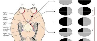
- Chiasmal syndrome (bitemporal hemianopsia), caused by compression of the optic chiasm by a large pituitary adenoma.
Epidemiology
Hormonally inactive pituitary adenomas account for 25% of all pituitary adenomas (the most common pituitary adenoma); among tumors with suprasellar localization, 70% of all adenomas are hormonally inactive pituitary adenomas. According to autopsy data, the prevalence of pituitary microincidentalomas reaches 10-25%. The frequency of new cases of clinically significant hormonally inactive pituitary adenomas is approximately 6 cases per 1 million population per year.
Craniopharygioma is a rare disease, but the most common suprasellar tumor in children (5-10% of pediatric brain tumors).
Hemangiomas with damage to the hypothalamus are detected during the perinatal period and up to 2 years.
Dysgerminoma and hamartroma are usually diagnosed between the ages of 2 and 25 years. At the age of 10 to 25 years, dermoid tumors, lipomas, and neuroblastomas may appear.
The time of manifestation of glioma is quite extensive - from the neonatal period to 50 years.
Symptoms
Clinical symptoms are determined by the set and severity of the syndromes mentioned above.
- Adenohypophyseal insufficiency. The most common and early symptom in women is menstrual irregularities, including amenorrhea. This forces women to consult a gynecologist, which allows diagnosis to be made at relatively earlier stages of the tumor process than in men, who are not inclined to seek medical help when erectile dysfunction occurs. Less often, other manifestations of pituitary insufficiency come to the fore: progressive general weakness and hypotension as manifestations of secondary hypocortisolism and hypothyroidism. In some cases, a detailed clinical picture of panhypopituitarism develops. With the development of hormonally inactive pituitary adenomas, craniopharyngioma or other destructive process of the hypothalamic-pituitary region in childhood, as a rule, there is a delay in sexual and physical development (stunted growth, absence of puberty).
- Diabetes insipidus. Clinically manifested by severe polyuria (without glucosuria) and polydipsia. Disorders of water metabolism can have a three-phase character: first, polyuria develops acutely, then at about 7-10 days in young patients there is a phase of normal water metabolism and, finally, persistent development of diabetes insipidus. This triad is explained by the fact that at first the supply of ADH to the posterior lobe of the pituitary gland is acutely lost, then its autolysis occurs with the release of hormones into the bloodstream and, finally, a complete cessation of the supply of ADH into the blood occurs. The lower the cortisol level, the lower the degree of polyuria. As ACTH production decreases (as part of progressive hypopituitarism), the severity of polyuria (Hanna syndrome) also decreases. This is due to the antagonistic interaction between cortisol and ADH.
- Hyperprolactinemia. As a rule, it is asymptomatic, but a number of manifestations (amenorrhea) can be caused by both increased prolactin production and hypogonadotropic hypogonadism.
- Neurological symptoms: chiasmal syndrome (with hormonally inactive pituitary adenomas in 75% of cases - bitemporal hemianopsia, in 15% of cases - quadryanopsy), cranial nerve palsy, headache, nausea, vomiting.
Diagnostics
In patients with appropriate symptoms, an MRI of the pituitary gland is performed. Usually, by the time the diagnosis is made, hormonally inactive pituitary adenomas are of significant size and have pronounced neuro-ophthalmological manifestations. With cranopharyngioma, in 80% of cases, radiography reveals calcifications in the tumor. Hormonal studies reveal varying degrees of deficiency of pituitary tropic hormones and hyperprolactinemia (usually mild or moderate).
Differential diagnosis
In case of hyperprolactinemia, differential diagnosis of hormonally inactive pituitary adenomas with prolactinoma is necessary, which is of fundamental clinical importance, since in the latter case the patient is only indicated for conservative treatment with dopaminomimetics. Rapid tumor growth and a slight (up to 200 μg/l) increase in prolactin levels are more typical for hormonally inactive adenoma. As a marker of hormonally inactive pituitary adenomas, it is proposed to study the levels of chromogranins and secretogranin, as well as beta-hCG.
Craniopharyngioma must be differentiated from other diseases that occur with delayed sexual and physical development and hypopituitarism, as well as from other tumors of the pituitary gland and brain. Tumor processes in the hypothalamic-pituitary region often have to be differentiated from systemic and genetic lesions.
Treatment
Surgical treatment (tumor removal) for hormonally inactive pituitary adenomas is indicated for macroadenomas of suprasellar localization, as well as in the presence of neurological symptoms (chiasmal syndrome) and pituitary insufficiency. In addition, surgical treatment is indicated in most cases of craniopharyngiomas. Most hormonally inactive pituitary adenomas and craniopharyngiomas are radioresistant. For partial and total hypopituitarism, hormone replacement therapy is indicated. In case of accidentally detected hormonally inactive pituitary adenomas (incidentalomas) and the absence of data on hormonal activity, dynamic observation is indicated: for formations less than 1 cm, control MRI is carried out after 1.2 and 5 years, for formations more than 1 cm - after 6 months, and then after 1, 2 and 5 years.
Forecast
After operations for large hormonally inactive pituitary adenomas and craniopharyngiomas, visual function is restored in most cases. Complete restoration of adenohypophysis function after surgery for hormonally inactive macroadenomas and large craniopharyngiomas occurs only in 10% of cases, and therefore most patients continue to receive replacement therapy to some extent after surgery. Craniopharyngioma is characterized by recurrences after removal of the primary tumor. Most accidentally detected hormonally inactive microadenomas (incidental) are characterized by constant size or very slow growth and very rarely acquire clinical significance with the development of pituitary insufficiency, hyperprolactinemia or neurological symptoms.
www.eurolab.uaIllustrations from the site: © 2011 Thinkstock
Endocrine metabolic syndrome
- Somatotropinoma - this type of pituitary adenoma in children is accompanied by signs of gigantism (height above two meters, imbalance in body proportions, elongation of limbs with a relatively small head). In adult patients, the disease can manifest itself as acromegaly - enlargement of certain parts of the body. Signs of this tumor also include obesity, enlarged thyroid gland, diabetes mellitus, hirsutism, and the formation of warts on the skin.
- Prolactinoma - this type of pituitary adenoma activates the secretion of prolactin. In women, the disease manifests itself with symptoms such as amenorrhea, problems with the menstrual cycle, galactorrhea and even infertility. In rare cases, women with prolactinoma complain of acne, seborrhea, and hypertrichosis. In men, the most characteristic manifestations of the tumor are ophthalmic and neurological symptoms, as well as impotence, gynecomastia and decreased libido.
- Corticotropinoma - this tumor always accompanies Itsenko-Cushing's disease. It is characterized by signs of hyperkerticism and pronounced skin pigmentation. Some patients also experience mental disorders. This tumor is considered the most dangerous because it often transforms into malignant. However, in comparison with other types of pituitary tumors, it is much easier to identify, since its signs appear already in the early stages.
- Thyrotropinoma is the rarest type of pituitary tumor, as it occurs in only 1-2.8% of all cases of pituitary adenoma. In most patients, tumor formation is accompanied by a malfunction of the thyroid gland. Due to the active secretion of thyroid-stimulating hormone, patients experience secondary hyperthyroidism.
- Gonadotropinoma is a benign tumor, accompanied by increased secretion of FSH and LH hormones. In rare cases, the pathology occurs without symptoms. The most characteristic signs of the tumor include ophthalmic-neurological disorders, hypogonadism and galactorrhea.
Treatment of pituitary adenoma
Treatment options may include medications, radiation and neurosurgical methods. The choice of the most optimal methods for treating a tumor depends on several factors: the patient’s vision state, the nature of the growth and size of the tumor, the patient’s age, and the presence of other serious diseases.
The type of tumor is also important. For example, radiation therapy to eliminate prolactinoma is ineffective. It is more suitable for the treatment of corticotropinoma smaller than 15 mm. Surgical techniques are used to remove corticotropin and somatotropin.
- Drug treatment. Prescribing medications to a patient with a pituitary tumor is indicated to eliminate the symptoms of the disease and improve health. For this purpose, the doctor prescribes general health-improving drugs and vitamin complexes. Conservative treatment is indicated for small tumors. The selection of medications also depends on the type of tumor. For somatotropinomas, somatostatin agonists (somatulin and sandostatin) are prescribed, for prolactinomas - dopamine agonists and ergoline drugs, for corticotropinoma - steroidogenesis blockers (nizoral, mammomit, orimethene).
- Radiation therapy. This technique is especially effective for microadenomas. Proton therapy, gamma therapy, and teletherapy are often used to treat tumors. Using this treatment method, it is possible to stop the growth of the tumor, and sometimes even achieve its regression.
- Stereotactic radiosurgery. This is an innovative method of treating pituitary tumors, which involves the introduction of a radioactive substance into the pathological process. Compared to radiation therapy, this technique is more effective, as it avoids irradiation of the entire body.
- Surgery. Surgery for pituitary adenoma is prescribed by a neurosurgeon after a comprehensive diagnosis. Indications for surgery may include large tumor sizes or complications such as cyst formation, hemorrhage, and visual impairment. Microadenomas are usually removed using the transcranial method, which involves craniotomy. The operation can also be performed transnasally using endoscopic techniques.
Therapeutic measures
For any brain tumor, including pituitary adenoma, when prescribing a treatment plan, the possibility of surgery is first considered. If for some reason surgical removal of a pituitary tumor is not possible, drug therapy is often prescribed with drugs designed to reduce the effect of excess hormones on the body, and radiotherapy can also be used.
The development of modern technologies makes it possible to actively and effectively use stereotactic radiation therapy, in particular, with the use of the CyberKnife apparatus, for pituitary adenoma. It has the highest precision and allows you to remove the tumor using a non-surgical method. The procedure is practically painless and does not require hospitalization, and its cost is more affordable than similar procedures in clinics in Europe and Israel. You can make an appointment for a consultation in Moscow by phone. +7 (495) 085-79-02 or 8 (800) 5-000-983.
Prognosis of pituitary adenoma
Pituitary adenoma is a benign neoplasm. However, in the case of active growth, the tumor can transform into malignant. The size of the tumor affects the possibility of complete removal of the pituitary adenoma. It has been proven that after removal of a tumor measuring two centimeters or more, there is a high risk of its recurrence over the next five years.
The prognosis of pituitary adenoma will also depend on the type of tumor. For example, in 85% of patients with microcorticotropinomas, complete restoration of endocrine function is observed after surgery. In patients with prolactinoma and somatotropinoma, these figures are lower - about 20%. In rare cases, patients with prolactinoma experience self-healing.
Treatment prognosis
Pituitary adenomas are benign formations, but with active growth they can cause many problems and even degenerate into a malignant process.
If the tumor is large (more than 2 cm), then there is a high risk of its recurrence in the next 5 years after surgical removal.
The nature of the adenoma is also of no small importance in predicting such formations. For example, with prolactinomas or somatotropinomas, a quarter of patients experience complete restoration of endocrine activity; with microcorticotropinomas, 85% of patients recover completely.
Average relapse rates are approximately 12%, and recovery ends in 65-67% of cases. But such forecasts are justified only if you turn to highly specialized specialists in a timely manner.