Due to the choroid of the organs of vision, accommodation, adaptation and nutrition of the retina are carried out. We are talking about a very important structural component of the eye. It includes several elements, one of the main ones is the ciliary body, consisting of muscles and blood vessels.
In newborn children, this structural element of the eye is not sufficiently developed: it is very subtle. When a child reaches the beginning of the second year of life, the muscle acquires the skill of accommodation. The structural element of a child's eye will be able to perform all its functions only when the child is 7-10 years old.
Structure
Functions
Symptoms
Treatment
Structure
The structural element consists of a couple of parts:
- A flat zone located up to the dentate line of the eye (size – 4 mm).
- Ciliary zone (hence the additional name of the element – ciliary body). The width of the layer is 2 mm, a special feature is the presence of ciliary processes.
At the cellular level, the element includes the mesoderm layer (connective and muscle tissue), as well as the neuroectodermal layer (retina and non-functional epithelium).
In cross-section, the structural element in question is a triangle, the angle of which penetrates into the organ of vision.
If we consider the element, starting from its most distant internal part, then it is immediately worth noting the muscle tissue, followed by a layer of blood vessels and basal plates. The structural layers end with non-functional epithelium and membrane membrane.
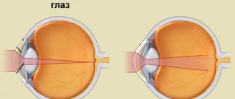
The layer of muscle tissue is connected to the lens of the eye through the ciliary girdle; due to this special formation, the function of accommodation is ensured.
The vascular layer of the body is a collection of mostly venous vessels that are located in the muscular layer of the local area of the organ.
In addition to the basic structural elements, the anatomical part of the eye in question includes nerve endings. It is due to them that a person feels pain during developing pathological processes.
CILAR BODY
CILAR BODY
[
corpus ciliare
(PNA, JNA, BNA);
syn. ciliary body
] - part of the choroid of the eyeball, passing anteriorly into the iris, posteriorly into the choroid itself. In the posterior two-thirds of the eye, the eye is flat and has a smooth surface facing the inside of the eye. In the anterior third of the R., the t. is thickened and on its inner surface there are from 70 to 80 ciliary processes (processus ciliares) with a height of approx. 1 mm and up to 2 mm long, forming the so-called. ciliary crown (corona ciliaris); the spaces between the processes are filled with numerous ciliary ridges (ciliary folds, T.).
In R. t. there are two layers: the outer muscular layer, represented by the ciliary, or ciliary, muscle (m. ciliaris), and the inner, vascular layer, or ciliary circle (orbiculus ciliaris), which is a continuation of the choroid itself.
Schematic representation of the structure of the ciliary body (in section): 1 - cornea; 2 - iris; 3 - ciliary crown; 4 - vascular layer of the ciliary body; 5 - sclera; 6 - meridional fibers - Brücke muscle; 7 - radial fibers - Ivanov muscle; 8 - circular fibers - Müller's muscle; 9 - large arterial circle of the iris; 10 - scleral spur; 11 - Schlemm's canal; 12 - trabecular apparatus.
The ciliary muscle is a group of smooth muscle fibers running in different directions, which begin from the inner surface of the sclera near the limbus from the so-called. corneal-scleral part or scleral spur and are lost in the choroid. In that part of the muscle, the edge lies closer to the sclera (see), the fibers run meridionally (meridional fibers, fibrae meridionales) - Brücke's muscle; deeper located muscle fibers go partly in the radial direction (radial fibers, fibrae radiales) - Ivanov's muscle, and partly in the circular direction (circular fibers, fibrae circulares) - Müller's muscle (Fig.).
A feature of the ciliary muscle is the abundance of elastic fibers. Physiol. its role is to ensure accommodation of the eye (see). The muscle is innervated by parasympathetic fibers of the oculomotor nerve, and sympathetic fibers from the cervical nodes of the sympathetic trunk.
The inner layer of the R. t. (ciliary circle) consists of many vessels (mainly small veins and wide capillaries), which are enclosed in loose tissue containing many pigment cells - chromatophores (see Pigment cells), and go inside ciliary processes. The epithelium covering the inner surface of the vascular layer of R. t. is represented by two rows of cells: the outer, richly pigmented, and the inner, devoid of pigment; genetically, the inner layer represents a continuation of the reduced retina into the R. region. The fibers of the ligament of cinnamon (ciliary girdle, zonula cillaris), which suspends the lens, are attached to the processes of the R. t. and its flat part (see). Sensitive nerves of R. t. originate from the trigeminal nerve. The processes of R. t. produce intraocular fluid.
In the clinic, inflammation of R. t. is often observed - cyclitis and inflammation of R. t. and the iris at the same time - iridocyclitis (see), the cause of which is most often infections or trauma. Among benign tumors of R. t., rare epithelial cysts can be noted, and among malignant ones, melanoblastoma (see Melanoma).
Bibliography:
Krasnov M. JI. Elements of anatomy in the clinical practice of an ophthalmologist, p. 59, M., 1952; Multi-volume guide to eye diseases, ed. V. N. Arkhangelsky, vol. 1, book. 1, p. 152, M., 1962; Hogan M. J., Alvarado J. A. a. Weddell J. E. Histology of the human eye, Philadelphia, 1971; W o 1 1 ff E. The anatomy of the eye and -orbit, p. 57, L., 1948.
M. L. Krasnov.
Functions
The ciliary body is called upon to perform a number of the following functions:
- involvement in the process of accommodation due to the variability of the shape of the lens capsule through the muscle tissue of the element in question;
- providing the organ with the proper amount of intraocular fluid (due to the established blood supply process in the body);
- regulation of intraocular pressure, which ensures clarity and clarity of vision;
- nutrition of the retina through the local vascular system;
- acting as a support for the iris of the organ of vision.
Ciliary body - what is it?
The ciliary
or
ciliary body
is a part of the choroid of the eye, which consists of blood vessels and muscles. Tension or relaxation of these muscles changes the shape of the lens, thanks to which we see well both far and near (accommodate). Blood vessels form special dense plexuses and nourish the iris, as well as the ciliary body. The capillaries of the ciliary processes produce intraocular fluid, which in turn regulates intraocular pressure. The body is attached to the protrusion of the sclera and serves as a support for the iris of the eye, another component of the choroid.
Symptoms
The development of pathological processes affecting the anatomical structure in question is accompanied by the following symptoms:
- fluctuations in intraocular pressure;
- blurred vision;
- failure in the production of intraocular fluid;
- redness of the eyeball;
- deterioration of near vision;
- soreness in the eye:
- cloudiness of the moisture in the anterior chamber.
If initial alarming symptoms appear, the patient should immediately contact an ophthalmologist to determine the cause of the phenomena and plan treatment.
Definition of ciliary tenderness
The ciliary (ciliary) body is a part of the choroid (vascular tract) of the eye. It is a ring 6 – 7 mm wide. The ciliary body is not accessible for inspection, because an opaque sclera covers it on the outside. The projection of the ciliary body onto the sclera is represented by a zone around the limbus 6–7 mm wide. Innervation of the iris and ciliary body is provided by short ciliary nerves, which include sensory fibers from the nasociliary nerve (branch of the ophthalmic nerve - 1 branch of the trigeminal nerve), autonomic parasympathetic fibers from the oculomotor nerve (postganglionic fibers after switching in the ciliary node) and autonomic sympathetic fibers from the plexus of the carotid artery. Long ciliary nerves also take part in the sensory innervation of the anterior part of the choroid.
Pain is one of the main symptoms of acute iridocyclitis (anterior uveitis. As a result of irritation of the ciliary nerves, sharp pain occurs in the eyeball and the corresponding half of the head. Increased pain at night can be explained by the predominance of the tone of the parasympathetic nervous system, increased passive hyperemia of the ciliary body. Increased pain intensity occurs upon palpation of the eye through the eyelids in the area of the projection of the ciliary body (ciliary soreness) ... A painful reaction is also characteristic of accommodation. Ciliary soreness, among other signs, is important when carrying out differential diagnosis with other diseases manifested by redness of the eye.
Clinical significance.
The test allows you to determine one of the clinical signs of iridocyclitis.
Research algorithm.
1.Ask the patient to look up or down.
2. Using two index fingers, alternately, lightly press through the eyelids on the eyeball in the projection area of the ciliary body (approximately 6-7 mm from the limbus).
Criteria for evaluation:
If pain appears or intensifies during the test, the symptom of ciliary pain is considered positive.
In the absence of this symptom, the sample is considered negative.
Section 2. MANIPULATIONS FOR DEVELOPMENT.
Instillation of eye drops into the conjunctival sac
Clinical significance.
Instillation (instillation) of drops is one of the main methods of administering drugs for the local treatment of most diseases of the organ of vision, as well as for a number of diagnostic studies. To instill eye drops, use a dropper bottle or a traditional pipette.
Manipulation algorithm.
1. Position the patient facing a window or next to a source of artificial light.
2. Pull back the lower eyelid using a sterile cotton ball with the left hand and ask the patient to look up.
3. Place a dropper or pipette in front of the eyeball in an inclined position at a distance of 3-5 mm from the conjunctiva, without touching the eyelashes. For convenience, you can fix the palm with the pipette on the patient’s face using your little finger .
4. Place 2-3 drops of the drug into the area of the lower fornix of the conjunctiva.
5. Remove excess drops with a sterile cotton ball from the lower eyelid.
Criteria for evaluation.
Visual control of the “hit” of the drug in the conjunctival sac.
Diagnostics
At the initial stage of disease diagnosis, an analysis of the manifestations of pathology is performed. In order to make an accurate diagnosis, they resort to special examination methods:
- Palpation. The specialist presses the apple of the eye with a finger and detects pain.
- Examination of the anatomical structure using a microscope (using a special lens).
- Transillumination. The procedure is performed to identify a tumor or foreign body in the eye. Due to the rays of the diaphanoscope, a uniform red glow of the pupil is created. In the presence of pathologies, it is absent or reduced.
- Ultrasonography.
- Biomicroscopy. A non-contact examination of the organ is performed using an ophthalmic slit lamp.
- Tonometry, tonography. The techniques allow you to find out the pressure inside the eye, identify disruptions in the process of secretion and movement of intraocular fluid.
Functions of the ciliary body
Its second medical name is ciliary body. It contains multiple branches of vessels and cells, having a similar structure to smooth muscle tissue. The entire checkered structure is arranged in layers, so that each layer has its own direction. Thanks to this anatomy, the ciliary body performs its functions. The main tasks of the ciliary body are the following functions:
- Provides constant access for the supply of nutrients to the organ of vision, in particular to muscle structures;
- Allows the eyes to focus at different distances (accommodation);
- Stabilizes and maintains the necessary pressure inside the eyeball.
Thanks to the above functions, the functionality of the visual organ is ensured. In order to understand the full functioning of the human eyes, it is necessary to examine their structure in detail.