Accessory apparatus of the eye, its protective parts
The auxiliary apparatus of the eye is of great importance.
The human eye is a thin, complex and fragile organism that needs protective devices. In order to efficiently perform its functions of ensuring a clear perception of the surrounding world in colors, an auxiliary device is needed.
Protective parts, conjunctiva
The protective mechanisms of the eye consist of the eyelids, eyebrows, eyelashes, and conjunctiva. Eyebrows prevent salty sweat from getting into your eyes. Eyelashes are located along the edge of the eyelids; they protect the eyelids from dust and weather conditions. The eyelids consist of lamellar connective tissue, the structure resembles cartilage, they are capable of performing the following functions:
- protect from external damage;
- promote washing the eyes with tear fluid;
- when blinking, the cornea and sclera are cleared of foreign particles;
- help to focus vision;
- help regulate pressure inside the eye;
- reduce the intensity of the light flux.
On the outside there is a thin covering of skin that gathers into folds, under which the eyelid muscle is located. Inside it is covered with a thin tissue structure - the conjunctiva.
The conjunctiva moves from the eyelid to the eye organ, bypassing the cornea. Among the conjunctiva of the eyelid and eye there is a conjunctival sac, where foreign bodies mainly settle. The upper eyelid differs from the lower eyelid by the presence of several folds:
- supra-sulcata;
- sulcinate;
- tarsal.
Structure of the eye
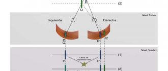
The human visual apparatus has a stereoscopic structure. This means that it contains two eyes, capable of transmitting a more three-dimensional picture of the environment. This vision is also called binocular.
Thanks to the system of transparent lenses that make up the eyeball, a person is able to catch rays of light reflected from objects. It can create an impression of the location of objects in the surrounding space. The eye consists of the following structures:
- Cornea. This is a strong connective tissue plate that covers the eyeball and transmits light well.
- Pupil. Contrary to the common misconception that it is something material, this component of the visual organ is a hole in the iris through which light enters the retina.
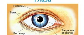
- Iris. This is a cluster of cells around the pupil, which is saturated with multi-colored pigments. It is the iris that gives the eyes blue, grey, green, brown or black.
- Lens. It is a double-convex lens that transmits and refracts light rays.
- Ciliary body. This structure is the small muscle that guides the lens. It is thanks to her that the latter changes its curvature and allows you to see distant or close objects.
- Sclera. They are called proteins. The outside of the sclera is covered with the conjunctival membrane.
- Vascular network. Small capillaries perform a trophic function, supplying nerve cells with nutrients and removing metabolic products.
- Retina. This is a collection of rods and cones that perceive light and color.
- Optic nerve. It is he who conducts electrical impulses to the brain, where in the optical centers they are converted into visual signals.
Accessory apparatus of the eye, its components
Protective is the main function of the eyelashes.
The auxiliary devices of the human eye are the lacrimal apparatus and the motor muscles of the eyeball. They are created by nature to control the eye organ and help to functionally carry out its work.
This also includes the fascia of the orbit and the fatty body. The eyeball is placed in the orbit, the bottom of which is covered with a membrane - the vagina - and surrounds the eye. Fascia includes the vagina, tendons, ligaments, and blood vessels.
Around the muscles, the optic nerve, between the periosteum and the vagina, there are fatty partitions, and on the posterior plane of the eye organ there is the substance of the orbit - the fatty body, it is elastic, creates a place for the free position of the visual apparatus.
Muscles of the eyeball
Six are attached to the eyeball
striated muscles:
four rectus
- superior, inferior, lateral and medial, and
two obliques
- superior and inferior.
Start
all muscles of the eyeball, except for the inferior oblique - the common tendon ring, annulus tendineus communis, fixed around the circumference of the optic canal of the sphenoid bone.
From the common tendon ring begins the muscle that lifts the upper eyelid, m. levator palpebrae superioris. It is located in the orbit above the superior rectus muscle of the eyeball, and ends in the thickness of the upper eyelid.
Attachment:
sclera in front of the equator.
1.
Lateral rectus muscle, m. rectus lateralis, turns the eyeball outward, towards the temple.
2.
Medial rectus muscle. m. rectus medialis, rotates the eyeball inward, towards the nose.
3.
Superior rectus muscle, mm. rectus superior, turns the eyeball upward, towards the brow ridge.
4.
Inferior rectus muscle, mm. rectus inferior turns the eyeball downwards towards the cheek.
5.
Superior oblique mouse, m. obliquus superior.
Start:
the common tendon ring, its thin round tendon passes through the trochlea, trochlea, which looks like a ring, turns outward, lies on the floor with the superior rectus muscle and attaches to the eyeball in its superolateral part, behind the equator.
Functions:
rotates the eyeball
down
and
out.
6.
Inferior oblique muscle, m. obliquus inferior.
Start:
the lower wall of the orbit
near the opening of the nasolacrimal duct.
It is directed backward and outward, passing between the inferior wall of the orbit and the inferior rectus muscle, and is attached to the lateral surface of the eyeball, behind the equator.
Function:
rotates the eyeball
upward
and
outward.
Visual apparatus
At the inner corner of both eyelids there are points called lacrimal, from which the lacrimal canaliculi originate. The gland that produces tear fluid is located under the upper eyelid in the fossa of the orbit. About 15 ducts emerge from it, which drain fluid from the gland into the lacrimal sac at the border of the lower eyelid and eye.
The composition of the lacrimal substance includes the substance lysozyme, which has antibacterial properties. Near the inner corner of the eye, in a small depression called the lacrimal lake, this liquid substance is concentrated.
It has already washed the surface of the organ and moves through the excretory canals into the duct connecting to the nasal sinuses. There it dries. Functions of tear fluid:
- nutrition and hydration of the cornea;
- prevents drying of the conjunctiva and cornea;
- promotes cleansing of foreign bodies;
- plays the role of a lubricating fluid when flashing;
- releases emotions in the form of crying.
Development of the visual system with age
In the embryo, the visual organs are formed as outgrowths of the diencephalon. Information from the embryo's eyes is processed in the midbrain region. At birth, the child's vision is blurred and unable to distinguish colors. This ability is formed during the first months of life. Vision becomes clearer as the eyeball matures, and the baby is already able to distinguish between images and form a complete picture from them. Visual acuity peaks during adolescence and then begins to decline slowly. This is due to the gradual degradation of visual structures. The ciliary muscle wears out, becomes weak and unable to sufficiently change the curvature of the lens. The latter, while remaining flatter, focuses a clear image only from closely located objects. This is how myopia develops.
Motor muscles
The auxiliary apparatus of the eye is a complex “mechanism.”
The apparatus of eye movement consists of four rectus muscles, originating from the fibrous ring:
- The external muscle is connected to the lateral wall of the ocular organ and turns the eye outward.
- The intrinsic muscle is attached to the medial wall of the eye and rotates inward.
- The lower muscle connects to the lower wall of the eye organ, lowers and slightly retracts inward.
- The superior muscle is attached to the upper wall of the eyeball, lifts it upward and slightly directs it inward.
Of the two oblique muscles:
- The lower muscle originates from the plane of the jaw above, is connected to the wall at the bottom of the eye organ, moves upward, and slightly moves outward.
- The superior muscle starts from the surface of the frontal bone, lowers, and slightly abducts outward.
The movements of the muscles of the left and right eyes are not chaotic, but strictly simultaneous, aimed at ensuring that the paired organs look at one point.
Functions of structures
The structure of the eye and the entire visual system perform important tasks to protect the organs. The lacrimal glands have a cleansing value. Thanks to them, the blinking process takes place and dirt is removed from the visual apparatus. Thus, the body resists infections. Medial and lateral muscle tissues rotate the eye in different directions. The superior straight line is responsible for the upward and outward movement of the organ of vision, and the inferior straight line is responsible for the upward and downward movement of the organ of vision. The protective functions of the auxiliary apparatus of the eye are provided by the conjunctiva: it is sensitive to all irritants. Therefore, with any unwanted touch, the eyelids quickly close. In addition, it blocks the path of allergens, chemicals and infections to the visual apparatus.
The structure of the main functional apparatus of the eyeball, their age-related changes
The eye is an organ of vision, which is a peripheral part of the visual analyzer
, in which the receptor function is performed by the neurons of the retina.
Includes:
eyeball, optic nerve, extraocular muscles, eyelids, lacrimal apparatus. 4 parts by function:
1) Light refractive apparatus of the eye (dioptric) apparatus of the eye (includes the cornea, lens, vitreous body, fluids of the anterior and posterior chambers of the eye)
2) Accommodative apparatus of the eye (iris, ciliary body with ciliary belt)
3) Receptive apparatus of the eye (retina)
4) Accessory apparatus of the eye (ocular muscles (ocular muscles), eyelids, lacrimal glands)
There are 3 membranes in the wall of the eye.
1. The outer shell is fibrous. In the posterior part it is represented by the sclera (tunica albuginea),
in the anterior part -
the cornea
.
2. The middle layer is vascular. In the anterior part, its derivatives are the ciliary body (ciliary) and the iris.
.
3. The inner shell is the retina. The visual retina is located in the posterior wall, and the anterior
- the mixed part that covers the inside of the ciliary body and the iris.
Scheme of the meridional section of the eye A - Sclera, B - Choroid,
B - Retina, D - Optic nerve,
D - Vitreous body, E - Lens,
F - Cornea, Z - Iris, I - Ciliary (ciliary) body, K - Radial ligament, L - Aqueous humor of the anterior and posterior chambers of the eye, M - Serrated edge, N - central fossa, O - Blind spot
There is a lens and vitreous body
, which occupies the main cavity of the eye. The anterior chamber of the eye and the posterior chamber are distinguished - between the iris and the lens; the cavity is filled with aqueous humor.
The retina , the inner sensitive layer of the eyeball, consists of: an outer pigment layer
internal photosensitive nerve.
Functionally there are:
1) posterior (larger) visual part
retina (in contact with the vitreous body, photoreceptor cells). In the posterior pole of the eye:
blind spot - the exit point of the optic nerve,
The macula lutea is the place of best vision with a small depression - the central fovea; there are only photoreceptor cells, mainly cones, and the other layers seem to be moved apart.
2) ciliary
covering the ciliary body
3) iris
covering the posterior surface of the iris.
SCLERA
It is formed by lamellar connective tissue, in which collagen fibers form strong connective tissue plates, between which fibroblasts are located. The sclera performs a protective, formative function and light flows do not pass through it; it is not transparent. The sclera contains blood vessels. In front, the sclera merges into the cornea. In this place there is a part of the sclera in the form of a ring - the limbus.
The sclera contains venous sinuses - Schlemm's canal, through which intraocular fluid passes. It flows from the anterior chamber of the eye. Damage to Schlemm's canals leads to glaucoma (clouding of the cornea).
Dioptric apparatus of the eye: structure, functions.
The light refractive apparatus of the eye (diopter) of the eye includes: the cornea, lens, vitreous body, fluids of the anterior and posterior chambers of the eye.
CORNEA
Includes five components:
1 — Stratified squamous non-keratinizing epithelium
. The first layer is basal, then spinous
layer and layer of flat cells. Regenerates well and is constantly moisturized with liquid.
2 — Anterior limiting membrane
– wide, contains a large number of collagen fibers.
3 — Proprietary substance of the cornea
is a continuation of the sclera. Consists of connective tissue plates and fibroblasts. There are no blood vessels, so nutrition is diffuse due to the vessels of the limbus and intraocular fluid.
4 — Posterior border plate
has the same structure as the anterior one.
5 — Single layer squamous epithelium
. It limits the cornea from the anterior chamber of the eye.
6 — Choroid
– the choroid itself and its derivative parts: the ciliary body with processes and the iris.
The choroid itself contains a supravascular lamina, a vascular lamina, a hemocapillary lamina, and a basal lamina.
· The supravascular plate is represented by loose, unformed connective tissue with fibroblasts and a large number of pigmentocytes.
• The vascular plate - loose connective tissue - contains fairly large vessels running parallel to the surface.
• Hemocapillary plate - micro circular bed - carries out tissue trophism.
• The basal lamina contains collagen fibers. Adjacent to it is the retinal pigment epithelium.
