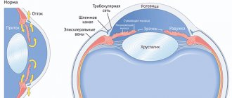
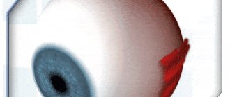
The structure of chamber formations
The anterior chamber of the eye is located directly behind the cornea. The peculiarity of the “element” is that it has different depths along its entire length. The greatest depth is in the area of the pupil, the indicator reaches three and a half millimeters, it decreases towards the periphery. Aberration in camera parameters can become a symptom of an eye disease. For example, the depth increases after removal of the lens or decreases when the mucous membrane is detached.
The posterior chamber of the eye is located directly behind the anterior chamber and contains many tiny zonules of Zinn, which act as a “connecting” element between the lens and the ciliary body. They are also responsible for the contraction of the ciliary muscles, whose task is to transform the shape of the lens. Thanks to this, a person sees well at any distance.
Both chambers are filled with a liquid identical in composition to blood plasma. Moisture contains a large amount of useful substances that are transferred to the organ of vision and ensure its smooth operation. The liquid also accepts metabolic products from the eye, after which it “redirects” them to the circulatory system. Moisture is produced through the ciliary processes of the ciliary body, and outflow occurs through the drainage system.
| The volume of the posterior and anterior chambers holds from 1.23 to 1.32 cubic centimeters of intraocular fluid. It is important to maintain a balance between the decrease and increase of moisture inside the eye. When the indicator shifts, the smooth operation of the visual apparatus is disrupted. |
If more fluid is produced than comes out, glaucoma develops. In the opposite situation, there is a high risk of eye subatrophy. Any slight imbalance has a negative impact on the functioning of the eyes and can even lead to blindness.
The volume of chamber formations in the visual apparatus must be stable; this is the only way to ensure uninterrupted production of intraocular moisture and its outflow.
The structure of the eye chambers and their functions
The anterior chamber of the eye is located just behind the cornea. Therefore, on the outside it is limited by the endothelium of the cornea, consisting of a single layer of flat cells.
On the lateral sides, the angle of the anterior chamber of the eye is limited. And the reverse surface of the cavity represents the anterior surface of the iris and the body of the lens.
The depth of the anterior chamber is variable. It has its maximum value near the pupil and is 3.5 mm. With distance from the center of the pupil to the periphery (lateral surface) of the cavity, the depth uniformly decreases. But when the crystal capsule is removed or the retina is detached, the depth can change significantly: in the first case it will increase, in the second it will decrease.
Below the anterior chamber is the posterior chamber of the eye. It is shaped like a ring, since the central part of the cavity is occupied by the lens. Therefore, on the inside of the ring, the chamber cavity is limited by its equator. The outer part borders on the inner surface of the ciliary body. In front is the posterior layer of the iris, and behind the chamber cavity is the outer part of the vitreous body - a gel-like liquid whose optical properties resemble glass.
Inside the posterior chamber of the eye there are many very thin threads called zonules of cinnamon. They are necessary to control the lens capsule and ciliary body. It is thanks to them that contraction of the ciliary muscle is possible, as well as ligaments, with the help of which the shape of the lens changes. This structural feature of the visual organ gives a person the opportunity to see equally well both at short and long distances.
Both chambers of the eye are filled with intraocular fluid. Its composition resembles blood plasma. The liquid contains nutrients and transfers them to the eye tissues from the inside, ensuring the functioning of the visual organ. Additionally, it receives metabolic products from them, which are subsequently redirected into the general bloodstream. The volume of the chamber cavities of the eye is in the range of 1.23-1.32 ml. And it is all filled with this liquid.
It is important that a strict balance is maintained between the production (formation) of new and the outflow of spent intraocular moisture. If it shifts to one side or another, visual functions are disrupted. If the volume of fluid produced exceeds the volume of moisture that has left the cavity, then intraocular pressure develops, which leads to the development of glaucoma. If more fluid goes into the outflow than it is produced, the pressure inside the chamber cavities drops, which threatens subatrophy of the visual organ. Any imbalance is dangerous for vision and leads, if not to loss of the visual organ and blindness, then at least to deterioration of vision.
The production of fluid to fill the eye chambers is carried out in the ciliary processes by filtering the blood flow from the capillaries - the smallest vessels. It is secreted in the posterior chamber space, then enters the anterior chamber. Subsequently flows through the surface of the anterior chamber angle. This is facilitated by the pressure difference in the veins, which seem to absorb waste fluid.
Physiological role of eye cameras
Their main purpose is to “monitor” the circulation of intraocular fluid. The production of moisture occurs in the ciliary processes by filtering the capillary blood flow. First it appears in the posterior chamber (secreting), then moves to the anterior one. Afterwards, due to low blood pressure, moisture leaves through the UPC, which is responsible for the outflow of fluid.
Also, chamber formations have a number of additional functions:
- Responsible for the conductivity of light rays;
- Form “good relationships” between all structures of the eye and intraocular tissues;
- On “their shoulders” lies light refraction. Thanks to this, the rays are focused on the retina, i.e. cameras act as conductors of sorts.
Organ functions
The main task of the cameras is to regulate the relationships between the tissues of the eyeball. Thanks to them, light rays enter the retina of the eye. Together with the cornea, the anterior and posterior chambers of the eye provide refraction of rays: the optical properties of the cornea and intraocular fluid allow the visual apparatus to capture and form images. In addition, in the second part, with the help of ciliary processes on the celiac body, intraocular aqueous fluid is produced. It then travels through the drainage systems to other parts of the eyeball. The front part is responsible for the outflow of moisture from the organ.
Angle of the anterior chamber - general structure
The UPC is a peripheral plane where the cornea smoothly flows into the sclera, and the iris into the ciliary body. The main value of the anterior chamber angle is the drainage system, which is responsible for the outflow of intraocular moisture into the circulatory structure.
It includes:
- Venous sinus, located in the white of the eye;
- Trabecular diaphragm, which is a network with a porous-layered structure. It decreases in size closer to the outside, this has a positive effect on the outflow of intraocular fluid;
- Collecting tubules.
First, the moisture released in the eyes enters the trabecular diaphragm, then it is “directed” into the lumen of Schlemm’s canal (located near the limbus in the sclera of the eyeball).
In some cases, the outflow of intraocular fluid occurs in a different way, through the uveoscleral pathway. Thus, approximately fifteen percent of the total volume of moisture penetrates into the bloodstream. In this case, it first enters the ciliary body, then moves in the direction of the muscle fibers and penetrates the suprachoroidal space. From here, the fluid passes through the veins into Schlemm's canal or the white membrane of the eye.
Collecting tubules in the sclera remove moisture in three directions:
- In the episcleral veins;
- In the vessels of the ciliary body;
- In the venous plexus located on the surface of the white membrane of the eyes.
You will learn more about the structure of the anterior chamber and its functions from the video
Return to contents
Angle-closure glaucoma: symptoms, diagnosis and treatment. Clinics. Consultation with an ophthalmologist
The development of a pathology such as primary angle-closure glaucoma accounts for up to 40% of all cases of glaucoma. Most often, this type of glaucoma affects people of the Eastern race, mainly women.
The causes and nature of the course of angle-closure glaucoma are determined by the special structure of the anterior segment of the eyeball in this disease. Due to the large size of the lens, the iris moves forward towards the cornea, which determines the structure of the angle.
A closed, narrow or wedge-shaped angle of the anterior chamber prevents the flow of intraocular fluid, which provokes an increase in intraocular pressure.
The initial stages of the disease occur without symptoms. The primary manifestation is an acute attack of glaucoma, which may be preceded by rainbow circles when looking at bright light, short-term pain in the area of the affected eye, as well as in the temples and eyebrows (subacute attack of glaucoma).
Each subsequent subacute attack leads to a deterioration of the optic nerve, and the peripheral field of vision gradually narrows.
During the period between attacks, normal intraocular pressure levels are observed. An acute or subacute attack causes adhesions to appear in the angle of the anterior chamber, which deprives the drainage network of access to moisture and leads to a sharp increase in intraocular pressure.
Risk factors
The main factors that increase the likelihood of angle-closure glaucoma are the following conditions:
- the patient’s age is more than 40 years (along with degenerative processes, age-related changes also affect the development of thickening of the lens, as a result of which the root of the iris is pressed even more against the drainage network, the narrow angle of the anterior chamber turns into a closed one);
- being female (the development of this disease occurs more often in women);
- race (angle-closure glaucoma develops more often in people of Eastern nationality);
- the presence of farsightedness (if the patient has been wearing reading glasses and distance glasses since youth);
- presence of a shallow anterior chamber;
- presence of peripheral and central circulatory disorders;
- irregularity of preventive examinations by an ophthalmologist.
Anterior chamber closure mechanism
The development of angle-closure glaucoma is due to the special structure of the eye. The narrowness of the space between the anterior surface of the lens and the posterior surface of the iris, caused by the presence of a closed angle and a large lens, prevents the normal outflow of aqueous humor through it into the anterior chamber.
The accumulation of fluid in the posterior chamber contributes to the bulging of the iris forward - pupillary block, which means a violation of the circulation of aqueous humor between the anterior and posterior chambers through the pupil.
Progression of the pupillary block leads to contact of the iris root with the cornea and, as a consequence, blockade of the anterior chamber angle, a sharp decrease in fluid outflow and the development of an acute attack of glaucoma.
The normal process of dilation of the pupil sometimes leads to obstruction of the outflow tract by the fold of the iris root. This is facilitated by flat iris syndrome, which is characterized by a special attachment of the root of the iris to the ciliary body near the drainage network.
With this syndrome, dilation of the pupil and the occurrence of a subacute or acute attack of glaucoma can be triggered by prolonged exposure to poor lighting, instillation of eye drops that dilate the pupil, and taking certain medications (for example, psychotropic drugs).
In extremely rare cases, closure of the anterior chamber angle occurs due to the displacement of the vitreous body simultaneously with the lens forward, which is caused by the accumulation of fluid in the posterior part of the eye.
There are also a number of other reasons that lead to the closure of the anterior chamber angle: tumors inside the eyes, inflammatory diseases, etc. However, these pathologies can cause the development of secondary glaucoma, treatment of which is aimed at removing the immediate causes of glaucoma and reducing intraocular pressure in several ways.
Acute attack of glaucoma
The onset of an acute attack usually occurs suddenly. The reasons for this phenomenon remain not fully understood. There are a number of factors that can trigger an attack:
- emotional stress;
- stressful situations;
- prolonged stay in a room with insufficient lighting;
- instillation of eye drops, as a result of which the pupil dilates.
Symptoms of an acute attack of glaucoma
An acute attack of glaucoma is characterized by the following manifestations:
- the occurrence of sharp pain in the eye;
- redness of the eye;
- a sharp decrease or blurred vision;
- pupil dilation;
- the appearance of headache with accompanying nausea or vomiting.
Due to the fact that the bulging root of the iris blocks the outflow pathways, as a result of which the rate of outflow of intraocular fluid decreases, intraocular pressure increases sharply.
This process causes irreversible damage to the optic nerve fibers.
In this situation, it is necessary to immediately seek medical help followed by hospitalization in a hospital - these measures will help save vision.
Treatment is carried out using conservative therapy: eye drops, tablets, injections that reduce intraocular pressure. Additionally, hirudotherapy and laser iridectomy are prescribed. If conservative therapy is ineffective, surgical intervention is chosen as a treatment method.
Angle-closure glaucoma: diagnosis and treatment
An ophthalmologist can assume the presence of angle-closure glaucoma after conducting a routine examination with gonioscopy. Using optical coherence tomography of the anterior segment and ultrasound biomicroscopy, the structures of the anterior chamber angle, the size of the lens and other equally important details are assessed.
Patients with a closed anterior chamber angle are most predisposed to an acute attack of glaucoma.
Due to the fact that there are no exact signs that can be used to predict the risk of developing an acute attack, great importance is given to prevention to prevent irreversible blindness and subsequent disability.
Persons over 40 years of age need to undergo regular preventive examinations by an ophthalmologist to identify a closed anterior chamber angle.
Drug therapy for angle-closure glaucoma begins with the use of drugs whose action is aimed at narrowing the pupil: pilocarpine, Fotil.
These drugs help control intraocular pressure and are a prophylactic agent that prevents the occurrence of an acute attack of glaucoma.
However, with prolonged use of pilocarpine, a sluggish inflammatory process in the ciliary body may develop, as well as the progression of cataracts, the formation of an adhesive process between the iris and the lens, which subsequently complicates surgical interventions aimed at removing cataracts.
To treat angle-closure glaucoma, which proceeds according to the principle of pupillary block, laser iridectomy is used, which ensures unhindered access of intraocular fluid to the outflow tract, equalization of pressure between chambers, and reduction of intraocular pressure. In addition, iridectomy is used as a prophylactic measure. If there is no effect from this ophthalmological operation, glaucoma drops are prescribed: beta blockers, Azopt or prostaglandin preparations.
The results of the studies suggest that it is inappropriate to use medications that constrict the pupil after laser iridectomy.
In some cases, this laser intervention is not effective in eliminating blockade of the anterior chamber angle, which can occur with large thickness of the lens, flat iris syndrome and other pathological conditions.
In case of inadequate reduction of intraocular pressure, the use of conservative or laser therapy, surgical treatment of angle-closure glaucoma is recommended.
Since the development of this pathology is associated, first of all, with the massive size of the lens, which leads to a blockage of the outflow tract, if even the slightest opacities are detected in it, it is recommended to perform an operation such as phacoemulsification of cataract and implantation of an intraocular lens.
With the help of this microsurgical surgical intervention, the angle of the anterior chamber is opened, and, as a result, the outflow of intraocular fluid is facilitated.
The likelihood of an acute attack of glaucoma after the lens has been removed is significantly reduced, and the need for instilling drops that narrow the pupil disappears.
Moscow clinics
Below are the TOP 3 ophthalmological clinics in Moscow, where you can undergo diagnosis and treatment of angle-closure glaucoma.
Source: https://mosglaz.ru/blog/item/1176-zakrytougolnaya-glaukoma.html
Posterior chamber of the eye
It is protected in front by the iris and on the back by the vitreous body. On the outside it has a boundary, the role of which is played by the ciliary body; on the inside, a part of the lens acts as a limiter. The entire space of the chamber is filled with connecting threads, which are responsible for the weakening and tension of the ciliary muscles.
Thanks to this function, a person can equally well distinguish between objects located at close and far distances.
You will learn a lot of interesting things about the structure of the rear camera and its functions by watching the video
Diseases affecting chamber formations
Ailments that affect the back or front of the eye are classified into congenital and acquired. The first category includes:
- Lack of angle in the “facade” chamber;
- Abnormal attachment to the iris from the anterior edge;
- Blocking of the UPC by rudiments of embryonic matter.
The group of acquired pathologies includes:
- Failure in the outflow of intraocular fluid due to blocking of the anterior angle by a pigment spot or enlarged iris;
- Accumulation of purulent discharge;
- Uneven change in the depth of the “facade” chamber. The cause of the deviation may be previous damage to the visual apparatus, displacement of the lens or asthenopia of the zonules;
- Blood condensation (hyphema);
- Formation of connecting cords;
- CCP cracking;
- A hard deposit forms on the endothelial layer of the cornea;
- Glaucoma occurs due to a disruption in the circulation of moisture produced by the eyes;
- Rupture of the anterior part of the ciliary body;
- Injury to the lens or damping of the ligaments responsible for its support ultimately changes the depth of the anterior chamber;
- Reduction in the size of the anterior chamber, the cause of the pathology most often becomes a closed pupil.
To maintain visual acuity, do not ignore visits to the ophthalmologist. Only a professional doctor, with the help of special tests and examinations, will be able to identify the pathology and select the optimal therapy to prevent its progression. As a preventative measure to prevent eye diseases, visit an ophthalmologist once every twelve months.
| If eye problems begin suddenly, pain appears, or you notice the appearance of blood clots on the whites, visit the doctor unscheduled. |
Glaucoma: causes, symptoms, diagnosis and treatment. Surgery for glaucoma
Glaucoma is an insidious disease of the visual organs, which is one of the main causes of blindness in the world.
Translated from Greek, “glaucoma” means “color of the sea” or “azure”. The disease received this name because during an acute attack of glaucoma, the pupil becomes motionless and acquires a greenish color.
Glaucoma is an eye disease characterized by increased intraocular pressure, damage to the retina and optic nerve, loss of visual fields, which leads to complete blindness.
Glaucoma includes almost 60 eye diseases that develop according to the same pattern:
- impaired circulation of aqueous fluid in the eye;
- an increase in intraocular pressure above the individual permissible level;
- deterioration of blood supply to the tissues of the eye, the appearance of tissue hypoxia at the exit site of the optic nerve;
- compression of nerve fibers;
- dystrophy of optic fibers, decay of retinal cells;
- death (atrophy) of the optic nerve.
According to the World Health Organization, every minute 1 person on the planet goes blind from glaucoma. Currently in Russia there are about 1 million people suffering from this disease.
Today there are no methods that completely and permanently eliminate glaucoma, but early detection and timely treatment can stop the progression of the disease.
Glaucoma occurs in men and women of any age, starting from birth:
- congenital glaucoma is detected in 1 baby per 10-20 thousand newborns;
- glaucoma at the age of 40-45 years is diagnosed in 0.1% of the population;
- at the age of 50-60 years – in 1.5% of people;
- Among 75-year-olds, more than 3% suffer from glaucoma.
Symptoms of glaucoma
It is difficult for a person with glaucoma to recognize the disease themselves, since there may not be any special signs. At the onset of the disease, slight discomfort occurs in the affected eye, blurred vision, and rainbow circles when looking at a light source.
Then, even with good visual acuity, a narrowing of the fields of peripheral vision begins to appear. In some cases, loss of vision for a short period.
In an acute attack of glaucoma, pain begins in the eye and temples, nausea, blurred vision, and the eye becomes red with a bluish tint, the pupil becomes wide and does not respond to light.
Types of glaucoma
The following types of glaucoma are distinguished:
- congenital glaucoma;
- young age glaucoma;
- primary adult glaucoma;
- secondary glaucoma.
Congenital glaucoma is a fairly rare disease that is caused by genetic causes or trauma to the fetus during embryonic development. It may appear in the first days, months or first year after birth.
Young age glaucoma (or juvenile glaucoma) occurs between the ages of 3 and 35 years.
Primary glaucoma in adults
Primary glaucoma most often affects people over 40 years of age.
The following clinical forms of primary glaucoma are distinguished:
- open angle glaucoma;
- angle-closure glaucoma;
- glaucoma with low intraocular pressure.
Open angle glaucoma
The appearance of open-angle glaucoma may be preceded by diabetes mellitus, myopia, and atherosclerosis, since all these diseases lead to a decrease in the blood supply to the vessels of the brain and eye and a deterioration in the hydrodynamics of the eye.
With this type of disease, both eyes are affected, but asymmetrically. One of the main symptoms of open-angle glaucoma is a gradual increase in intraocular pressure, which imperceptibly narrows the boundaries of peripheral vision, up to tunnel vision or blindness.
Angle-closure glaucoma
Angle-closure glaucoma accounts for every fourth case of primary glaucoma. This type is more common in women than in men.
Primary narrow-angle glaucoma can appear when the following factors coincide:
- anatomical feature of the eye (small eye size, small anterior chamber with a narrow angle, large lens, farsightedness);
- closure of the anterior chamber angle (impaired outflow of intraocular fluid, increased intraocular pressure, impaired blood supply, optic nerve atrophy);
- age-related changes in the eye.
When the angle of the anterior chamber is completely closed, an acute attack of angle-closure glaucoma occurs, which can occur due to nervous tension, overwork, prolonged exposure to darkness, prolonged tilting of the head, or drug-induced dilation of the pupil.
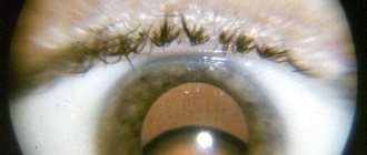
Patients report headache and eye pain, blurred vision and the appearance of rainbow circles, in some cases nausea and vomiting.
Upon visual examination, it is noticeable that the eye has become red, with a cloudy cornea and a dilated pupil that does not respond to light. Intraocular pressure rises to 60-80 mm Hg. pillar Visual acuity decreases. If an acute attack of glaucoma is not treated, a person may face permanent loss of vision.
In some cases, after surgery on the eyeball, shallow anterior chamber syndrome , which is characterized by a decrease in the distance between the anterior surface of the iris and the posterior surface of the cornea. If the complication is not corrected, it will also lead to glaucoma.
Glaucoma with low intraocular pressure
Glaucoma with low intraocular pressure characterized by all the typical signs of primary glaucoma: changes in the visual field and partial atrophy of the optic nerve. However, the level of intraocular pressure remains normal. This type of disease is combined with vegetative-vascular dystonia, which occurs according to the hypotonic type.
Secondary glaucoma
Secondary glaucoma occurs due to impaired outflow of intraocular fluid.
Here we can highlight such causes of glaucoma as eye trauma, iridocyclitis, uveitis (and other inflammatory eye diseases), as well as intraocular tumor and changes in the position of the lens.
Despite the apparent similarity of primary and secondary glaucoma, they have differences.
Features of secondary glaucoma:
- only 1 eye is affected;
- vision decreases rapidly;
- intraocular pressure increases in the evening;
- up to a certain stage, pathological changes are reversible.
When treating secondary glaucoma, it is necessary to remove the provoking factor and compensate for the condition of the eye.
Doctors distinguish several forms of secondary glaucoma: traumatic, postoperative, inflammatory, vascular, degenerative, neoplastic (appears as a result of eye growth), etc.
One of the most dangerous forms of the disease is neovascular glaucoma , in which blood vessels begin to grow in the iris of the eye. At a certain period, this tumor begins to bleed, which leads to an increase in intraocular pressure.
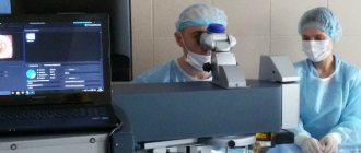
Diagnosis of glaucoma at MedicCity
Diagnosis of glaucoma must be carried out at the initial stage of the disease. The treatment tactics for glaucoma and the preservation of vision depend on this.
Diagnosis of glaucoma is carried out using the following methods using high-precision ophthalmological equipment:
Glaucoma treatment
Depending on the type, stage and severity of the disease, your doctor may suggest eye drops, laser treatment or surgery.
Treatment for glaucoma usually begins with eye drops that reduce eye pressure. They must be used as prescribed by a doctor and strictly according to the hour.
In more advanced stages of the disease, laser pressure correction is used using iridectomy and laser trabeculoplasty.
Laser iridectomy is used to treat angle-closure glaucoma and to prevent recurrent attacks of the acute form of the disease.
Laser trabeculoplasty is effective in the early stages of treating secondary glaucoma. With its help, the circulation of intraocular fluid improves and eye pressure decreases.
If treatment of glaucoma by these methods is ineffective, surgical intervention is used. Glaucoma surgery is performed using penetrating surgery, deep non-penetrating sclerectomy , or the use of drainage devices.
But remember: any operation is a big risk and the need for subsequent rehabilitation of the body! Therefore, pay special attention to the prevention of glaucoma!
To identify the disease at the very beginning, try to undergo a full examination by an ophthalmologist at least once a year. Take care of your eyesight and come to our clinic for the prevention of glaucoma and other ophthalmological diseases!
Source: https://www.mediccity.ru/directions/631
Symptoms of damage to the chambers of the eye
If there are deviations in the functioning of the “elements” of the organ of vision, the following signs are observed:
- Cloudiness of the cornea;
- Painful sensations;
- The appearance of new growths or spots on the eyes;
- Fear of bright light;
- The resulting image is unclear and has blurry contours;
- Problems with visual acuity;
- With hemorrhage in the anterior chamber, a change in the color of the iris is observed.
If dangerous symptoms appear, consult a doctor immediately to identify the disease at an early stage. Return to contents
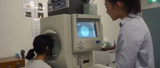
Diagnosis of eye chamber pathologies
If the development of a particular disease is suspected, the ophthalmologist sends the patient for several examinations:
- Biomicroscopy. It is carried out using a slit lamp;
- Microscopy of the anterior chamber. Helps detect glaucoma;
- Analysis of intraocular moisture, study of its circulation process;
- Coherence optical tomography;
- Ultrasonography;
- Pachymetry. Used to measure the depth of the anterior chamber;
- Automated tonometry. It is used to determine the level of pressure exerted by intraocular moisture.
| Modern equipment and innovative technologies will help detect any abnormality at an early stage, and the doctor will be able to select treatment to block its progression. |