One of the rare, but still occurring pathologies of the reproductive and reproductive systems is the third testicle in a man. This anomaly is called polyorchidism. An additional testicle can be localized either next to two full ones, or remain in the abdominal cavity without descending into the scrotum. In most cases, the accessory element has an underdeveloped structure, shape, size, and also the vas deferens. Otherwise, if a man has one or two fully functioning testicles, then the sexual and reproductive functions of the body do not decrease against the background of the third. What then threatens the presence of a 3rd egg in a man and how the anomaly manifests itself, we will discuss below.
Testicular polyorchidism: what is it, causes of pathology, clinical picture, treatment
Men are sometimes diagnosed with a rare abnormality of the reproductive system, in which one of the testicles appears double in the scrotum. This pathology is called polyorchidism. Even less common is duplication of both testicles. Experts note that it is superfluous, and in most cases underdeveloped.
Etiology of pathology
Polyorchidism is the appearance of a third testicle in the scrotum or inguinal canal. There are almost isolated cases when it is located in the abdominal cavity. In this case, it is extremely difficult to detect, because the presence of two normal testicles in the scrotum does not in any way indicate the presence of a third, less developed one in the cavity.
The accessory testicle is less developed, and its vas deferens is just as imperfect. The system performs its sexual function as usual with the normal development of the scrotal organs. The pathology is detected completely by accident during routine examinations or is palpated by the patient himself. An additional formation is found most often on the left side.
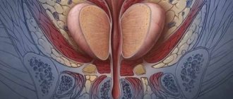
The photo shows the third testicle with polyorchidism
Reasons for the formation of an accessory testicle
The main cause of the pathology is considered to be the division of the genital fold even during the formation of the fetus in the womb by the abdominal ligaments, called mesenteric ligaments. This happens at 4-6 weeks of pregnancy. Duplication affects one or two testicles at once. Similarly, the epididymis or vas deferens can double.
Factors provoking pathology are:
- Genetic predisposition;
- Stressful state of a woman during pregnancy;
- Previous viral diseases of a pregnant woman in the early stages.
Much less often, an anomaly in the intrauterine formation of the reproductive and reproductive systems of the fetus can be established without the influence of negative external factors.
It is impossible to determine the presence of three testicles in a fetus or even an infant. Their descent into the scrotum occurs some time after birth, and the size of the testicles is so small that the doctor simply cannot determine the pathology upon palpation.
The additional testicle does not cause any discomfort or inconvenience. A man can live his whole life without realizing the presence of this pathology. There is no special effort to detect it, since cases of its formation are extremely rare. An extra testicle can degenerate into a tumor, and when identified, the cause of the tumor is determined. Then this pathology can be identified.
Prerequisites for degeneration into a tumor can be:
- Regular hypothermia of the body;
- Viral or bacterial infections of the genitourinary system;
- Exposure to radiation;
- Failure to maintain intimate hygiene;
- Genetic failure against the backdrop of the negative influence of the environmental situation.
Polyorchidism develops asymptomatically. An additional examination, which may reveal pathology, is carried out if a hernia is suspected or if there is a defect in testicular descent. Degeneration into a tumor is accompanied by an increase in body temperature and pain in the area where the third testicle is located.
Diagnostic measures
Testicular polyorchidism is most often detected in patients during adolescence with the onset of routine medical examinations. The main type of diagnosis is palpation of the scrotum. If the additional testicle is located in the abdominal cavity, then such an examination will not reveal it. If pathology is detected, a biopsy is performed.
No matter how harmless the presence of a third testicle may seem, its presence still poses some threat to the patient. There remains a risk of its transformation into a tumor.
If polyorchidism is suspected, several instrumental diagnostic methods are used:
- Ultrasound examination of the scrotum and abdominal cavity.
- Computer or magnetic resonance imaging.
- Laparoscopy.
During diagnosis, it is important to exclude the presence of other pathologies (varicocele, spermatocele, tumor, cyst, scrotal disease, testicular inflammation). When the accessory testicle is located in the scrotum, its size may be visually larger, which will also attract medical attention to the organ.
When palpating the scrotum by the patient himself or the attending physician, three testicles are felt, one of which is smaller in size than the other two. A noticeable difference in size and underdevelopment of the extra testicle will also be visible on ultrasound.
Using this type of study, it is difficult to identify pathology localized in the abdominal cavity. Blood or urine tests, as well as spermogram results, do not show polyorchidism.
Palpation of a bifurcated testicle or its display on the monitor are the most accurate when making a final diagnosis.
Treatment
When pathology of the development of the third testicle in men is detected, the only treatment method is surgical intervention. The operation can be performed immediately after diagnosing the deviation. The sooner this is done, the lower the risk of the excess testicle degenerating into a malignant neoplasm.
There are several methods of surgical intervention for polyorchidism:
- Standard (using a scalpel).
- Bloodless.
In the second case, the intervention is performed using modern equipment, and the patient is sent home the next day or every other day. During the operation, not only the third testicle, but its appendages and seminal ducts are removed.
The intervention is performed under local anesthesia and takes up to two hours. With standard surgery, the patient is discharged after 4 days. Medical monitoring of the patient lasts about another month.
At this time, men should avoid excessive physical activity, eat nutritiously and spend more time outdoors. If necessary, the time of physical rest from exercise can be increased.
The bloodless operation takes no more than an hour. A catheter is inserted into a small incision in the scrotum, through which a mini-video camera and the necessary instruments are inserted. The operation is performed under local anesthesia. The patient experiences pain after the intervention for 2 days.
Forecast
After treatment of polyorchidism, men fully retain their sexual and reproductive functions. Surgical intervention performed using any of the methods has a favorable prognosis for the health of patients. After the operation, a month of sexual abstinence is recommended. Adolescence is considered the most favorable age for detection and treatment.
Source: https://gidmed.com/urologiya/zabolevaniya-urolog/moshonki/poliorhizm-yaichka.html
Preventive measures
There are no methods of prevention for an already formed extra testicle. Precautionary measures for the development of the disease include the lifestyle of a pregnant woman:
- pregnancy planning (it is worth treating or preventing exacerbations of chronic diseases)
- do not come into contact with harmful substances of chemical origin
- correct work and rest schedule (light work or early maternity leave in hazardous work)
- exclude uncontrolled medication use
- give up bad habits (smoking, alcohol) when planning pregnancy
- do not engage in excessive physical activity
- avoid stressful situations
- follow all individual recommendations of the doctor leading the pregnancy
Testicular hypotrophy: how the disease manifests itself, what a man should do
The importance of the testicles in a man’s body is difficult to overestimate; thanks to these gonads, boys first grow up and then become fathers. Testicles are the central organ of the entire male reproductive system.
Therefore, the health of the testicles is considered a matter of paramount importance, because any pathological situation in this part of the male body affects reproductive function and can lead to infertility. It happens that the stronger sex does not even suspect the existence of a problem until the question of childbearing arises.
For example, testicular hypotrophy is often first discovered during a doctor’s examination and examination for infertility.
Information from anatomy
The testicles are paired organs with a rather complex structure. They are located in a special container - the scrotum and are separated from each other by a partition.
On the outside, each testicle is covered with a fibrous membrane, under which there is parenchyma, divided by connective tissue fibers into many lobules, each of which contains seminiferous tubules with maturing sperm. Hormones called androgens are produced in the surrounding tissues.
At the upper pole of each testicle there passes the spermatic cord with vessels and the vas deferens, and at the bottom the appendages located on the lateral surfaces of the testicles flow into it.
The activity of the testicles is carried out under the influence of the central nervous system.
They simultaneously perform the functions of an endocrine gland (produce the hormone testosterone) and an exocrine gland (produce sperm).
The testicles initially form in the abdominal cavity of the fetus; a few weeks before birth, they descend into the scrotum through the inguinal canal. Sometimes this process in boys is delayed or does not occur at all. This situation is called cryptorchidism and requires surgery so that the man does not have problems with his health and conceiving a child in the future.
The testicles can be felt with your hand; they are normally oval-shaped, painless, softly elastic, one is usually located slightly higher than the other. Dimensions vary from 5-6 cm in length to 3-3.5 cm in width.
The difference in size between the two testicles is allowed, but it should not exceed more than 0.6-0.7 cm, otherwise you need to contact a urologist to find out the causes of hypotrophy (decreased volume) of one or both gonads.
Reasons for the development of pathology
Experts distinguish two conditions in which the testicles are reduced in size:
- hypoplasia (ICD-10 code Q55.1) - congenital underdevelopment of an organ on one or two sides (from slight reduction to complete absence), detected in children at an early age - usually after 2-3 years;
- hypotrophy (ICD code -10 N0) is a secondary decrease in the size and volume of the testicle that occurs during life due to insufficient nutrition or the formation of a large number of scar adhesions that replace functionally active tissue and tighten the organ.
Source: https://oprostatite.info/urologiya/poroki-razvitiya-muzhskix-polovyx-organov/gipotrofiya-yaichka
Gradual development of varicocele
Damage to the veins occurs gradually, the clinical picture is constantly changing.
- Initially, there are no external signs of varicocele in men; it can be determined at the first stage only through palpation when the patient’s body is placed vertically.
- At the second stage, the size and consistency of the testicles remain the same, but the enlarged veins are clearly visible.
- At the third stage, the cluster-shaped dilatation of the veins is pronounced, the testicles become denser to the touch and decrease in size.
Testicular disease in men: types, signs and treatment features
Diseases of the reproductive organs are a common cause of infertility. Infectious and oncological diseases, characterized by severe consequences, are especially dangerous.
Testicular disease in men can be asymptomatic and not bother the patient in any way. During routine diagnostics, dystrophic changes and disturbances of venous outflow in the scrotum area are often detected.
General information
Testicles produce male sex hormones and sperm
Testicles are the internal reproductive organs of men located in the scrotum. These paired organs have two very important functions: the release of sex hormones and the formation of sperm necessary for procreation. Testicular diseases are a common cause of male infertility.
The scrotum is a kind of bag that protects the testicles. The main function of the scrotum is to maintain a certain temperature necessary for the formation of sperm.
The fact is that the temperature in the abdominal cavity is too high for the reproductive organs, so in the process of evolution, the testicles in men began to descend into the scrotum. Each testicle consists of small tubes and cells that synthesize hormones.
The seminiferous tubules contain germ cells (gametes) with 23 chromosomes. Spermatogenesis occurs constantly and depends on many internal and external factors.
On average, the testicles secrete 200 thousand sperm per minute.
The release of male sex hormones called androgens by the testicles is necessary for the formation of sexual characteristics.
Testosterone and its derivatives begin to be actively synthesized during puberty and affect the development of internal organs, muscles and other body systems.
A lack of sex hormones not only causes impaired sexual development, but also often causes mental disorders. If the testicles are unable to produce the required amount of testosterone on their own, hormone replacement therapy may be required.
To perform their functions, the testicles must receive sufficient blood supply. Timely outflow of venous blood is also important. Impaired blood supply and blood flow often lead to infertility in men.
Possible testicular diseases
Varicocele – varicose veins of the spermatic cord and testicle
Diseases of the reproductive organs can be congenital or acquired. During intrauterine development, two testicles form in the abdominal cavity and gradually descend into the scrotum. This occurs during the seventh month of pregnancy.
Any negative influences at this stage of development can cause improper formation of the testicles. Sometimes the testicles do not descend or cannot perform their functions.
Acquired testicular diseases are more common. Pathologies can be associated with injuries, impaired blood supply, excessive exposure to heat and other factors. The presence of a large number of nerve endings in the scrotum causes pronounced symptoms of many ailments, especially injuries.
Main diseases:
- Varicocele is an enlargement of the veins of the scrotum, leading to impaired blood flow from the testicles. This is a type of varicose veins. Varicocele is a common cause of insufficient sperm production and decreased sperm quality in general. A long-term consequence of the disease is male infertility. Long-term progression of the disease (5-10 years) and frequent asymptomatic progression complicate diagnosis.
- Incomplete testicular descent is a pathology characterized by abnormal position of the testicle. Often the testicles descend after birth, but sometimes the process remains incomplete and the organs do not enter the scrotum. Increased temperature in the abdominal cavity can cause testicular dystrophy. It is also a common cause of infertility.
- Testicular torsion is a pathology in which the testicles move relative to the spermatic cord and compress surrounding structures. This can lead to disruption of the blood supply and innervation of the organ. Patients typically experience severe pain. If the natural position of the testicle is not restored in time, significant damage to the organ is possible.
- Testicular cancer is an oncological disease characterized by the growth of a malignant tumor in the testicular area. This is a fairly rare form of cancer, most often found in men between 15 and 35 years old. Timely treatment prevents further spread of the tumor.
Oncology is considered the most dangerous disease of the male reproductive organs, but this pathology can also be successfully cured. Testicular torsion and varicocele are more common and less dangerous ailments.
Causes
Testicular diseases can be caused by various factors and causes.
The formation of testicular diseases can be influenced by a variety of internal and external factors.
The most common causes of pathologies of the male genital organs include:
- Mechanical injury. External impact on the testicle can not only damage the tissue, but also provoke torsion of the spermatic cord. Such injuries cause impaired blood supply and loss of innervation.
- Vascular pathology. Deterioration of blood supply may be due to compression of blood vessels, altered anatomy of the arteries of the spermatic cord, trauma and other factors. Impaired blood flow with varicocele usually occurs due to dysfunction of the internal valves of the scrotal veins.
- Defects in the development of reproductive organs. Incomplete prolapse and incorrect temperature conditions can provoke oncology and testicular dystrophy.
- Consequences of surgery. Quite often, the blood supply to the testicles is disrupted after surgery for inguinal-scrotal hernias.
- Inflammatory and infectious diseases. Such pathologies are risk factors for testicular cancer.
Most diseases of the male genital organs have different mechanisms of occurrence. Risk factors for diseases include a sedentary lifestyle, a family history of pathologies of the reproductive organs, disorders of intrauterine development and trauma.
Signs and symptoms
Lumps, enlargement and pain in the testicular area are a reason to consult a urologist!
Testicular disease in men is often asymptomatic. For example, with the development of varicose veins of the scrotum, the first signs of the disease may appear in the later stages.
The same applies to such a dangerous pathology as testicular cancer. Torsion and incomplete subsidence, in turn, almost always have severe symptoms.
Possible symptoms and signs of disease:
- Pain in the lower abdomen and scrotum.
- Discomfort, tension in the abdominal muscles and pulling sensations.
- Ejaculation disorders.
- Deterioration in sperm quality.
- Scrotal swelling.
- Nausea and vomiting.
- Increased body temperature.
- Painful urination.
- Feeling of heaviness in the scrotum.
- Back pain.
Most of the symptoms listed are not specific, so an examination is necessary for an accurate diagnosis. Pain in the abdomen and back may not be associated with testicular pathology, but these are also possible signs of an oncological process.
Diagnostics
An accurate diagnosis can be established only after examination, examinations and tests.
A man can independently suspect that he has a disease. Swelling, pain, swelling of the scrotum and other signs clearly indicate one or another ailment. In this case, it is necessary to contact a urologist or andrologist for consultation as soon as possible.
The doctor will interview the patient, review the patient's history for risk factors, and perform a physical examination.
To clarify the diagnosis, the following laboratory and instrumental studies may be required:
- Ultrasound is a visualization method that allows you to observe organs on a monitor and assess their condition. Changes in the size and structure of the testicle are easily detected during ultrasound. This method also allows you to assess the state of blood flow, which may be necessary when diagnosing varicocele.
- Blood analysis. First of all, it is necessary to determine the concentration of hormones to assess testicular function. Also, during the oncological process, organ cells release tumor markers. Tumor markers are the most reliable way to pre-diagnose cancer.
- Urinalysis to detect infection.
- Biopsy – sampling of testicular tissue followed by histological examination. This is the optimal method for diagnosing cancer.
Diagnosis can also be carried out after surgery for a tumor disease. Tumor examination allows us to clarify the type of oncology and stage of the disease.
Features of treatment and prognosis
Treatment depends on the type and severity of the disease
Treatment depends on the underlying cause of the disease. Sometimes you can get by with medication, but with oncology and varicocele, surgery is most often required.
Treatment of testicular diseases:
- To treat varicose veins, an operation is performed to remove the affected vein and redistribute blood flow in the scrotum. This operation helps prevent progressive testicular dystrophy and infertility. The doctor may choose open surgery, laparoscopy, or percutaneous embolization. Less invasive interventions are preferable.
- Incomplete testicular descent is treated surgically. An operation called orchiopexy aims to free the organ from surrounding tissue and move it into the scrotum. Hormone replacement therapy may be required as additional treatment.
- Testicular torsion does not always require surgical intervention. Sometimes the doctor can “set” the testicle himself. If there is significant twisting of the spermatic cord, impaired blood flow and testicular dystrophy, intervention may be required.
- In oncology, treatment should be aimed at removing the malignant tumor, preventing further growth of abnormal cells and, if possible, restoring the functions of the organ. The sooner the operation is performed, the lower the risk of the disease spreading to other organs. During the intervention, the doctor usually removes the affected testicle completely. Chemotherapy and radiation therapy help prevent further tumor growth and recurrence.
- Antibiotics for the infectious nature of the disease.
In severe illnesses, an important task is to preserve the functions of the second testicle, since this is the only way to prevent infertility. Patients often require lifelong hormone replacement therapy.
More information about testicular inflammation can be found in the video:
Source: https://organserdce.com/vessels/varikoz/varicocele/bolezn-yaichek-u-muzhchin.html
There are three testicles in the scrotum - is it dangerous?
During self-examination of the scrotum, which men are recommended to do regularly, since it is thus possible to independently identify an additional formation in the scrotum at an early stage, an additional testicle may also be detected.
Most often, it does not manifest itself in any way and is discovered by the patient by chance or during a medical examination.
An abnormal development of the genital organs, as a result of which three testicles appear in the scrotum, is called polyorchidism. Cases have been described in medicine where patients even had 4 testicles.
Diagnosis and treatment of polyorchidism
As a diagnosis, if a doctor or patient suspects that there is an additional formation in the scrotum, palpation, ultrasound of the scrotum, abdominal organs, MRI, and CT are used. In some cases, laparoscopy is even prescribed.
Differential diagnosis is also necessary, which is carried out to exclude the possibility of developing an ovarian cyst (spermatocele) or tumor tumors in the scrotum.
| For the most part, the patient does not even feel that he has three testicles in his scrotum. It does not manifest itself in any way and does not affect sexual function. |
You can live your whole life and not suspect the presence of such an anomaly.
There are no special symptoms for this pathology. Usually the accessory testicle is smaller.
In rare cases, when the size of all three testicles is the same, an enlarged scrotum may cause suspicion to a specialist.
Despite its apparent harmlessness, the detected third testicle in a man indicates the need for a more attentive attitude to one’s health.
This is because:
- the presence of “extra” organic formation in the body increases the risk of a malignant tumor;
- penetration of bacteria into the third testicle, hypothermia, cause difficult to diagnose diseases.
The standard procedure offered by medicine when this pathology is detected is surgical removal of the accessory testicle.
Possible additional formations in the scrotum
During self-examination of the scrotum, the patient may identify other additional space-occupying formations. After examination by a doctor, it turns out that it is an epididymal cyst (spermatocele). This is a round, voluminous, fluid-filled formation, manifesting itself only as rare pain sensations, in the form of a nagging pain in the scrotum. And then, this manifests itself only with large cysts.
An extremely rare occurrence in adult men is a cyst of the spermatic cord, which is also accompanied by the appearance of additional space-occupying formations.
Inflammation of the epididymis (epididymitis) also manifests itself in the appearance of additional space-occupying formations in the scrotum. In the case of acute epididymitis, the increase in the size of the scrotum occurs quickly, is accompanied by sharp pain, temperature, and the skin of the scrotum loses folds, stretches, and becomes red.
| In all cases of discovering additional formations in the scrotum, the patient should consult a doctor as soon as possible to make an accurate diagnosis and begin treatment. |
Source: https://urolog.pw/st/v-moshonke-tri-yaichka