Glaucomocyclic crises - clinical picture (symptoms)
The disease is characterized by frequent attacks accompanied by blurred vision, the appearance of rainbow circles when looking at a light source, a feeling of heaviness in the eye and sometimes slight pain in the eyeball. Objectively, there is a small injection of the eyeball, swelling of the corneal epithelium, corneal precipitates. The anterior chamber angle is open. Iphthalmotonus increases significantly - up to 40–50 mm Hg. Art. At the height of the attack, there is a sharp increase in the resistance to the outflow of aqueous humor with a decrease in the coefficient of ease of outflow to 0.01–0.02 with a normal or reduced secretion rate (0.24–2.4). Subsequently, outside the attack, there is also a persistent decrease in the coefficient of ease of outflow in both eyes, which indicates uveopathy. In patients with glaucomocyclic crises, rheoophthalmography can reveal a decrease in blood supply to the vessels of the ciliary body.
Visual function does not decrease and glaucomatous atrophy of the optic disc usually does not develop. During the interictal period, there are no subjective or objective symptoms of the disease. The duration of glaucomocyclitic crises ranges from several hours to 5–7 days. Crises recur with varying frequencies - from 1–2 months to 5 years.
Acute attack of glaucoma
An acute attack of glaucoma is a condition of a sharp and significant increase in intraocular pressure above 50 mmHg.
The reasons for the development of the attack are still not well understood. It is known that an acute attack of glaucoma often develops following unpleasant worries. The news of the death of a loved one, a family quarrel, material damage and a number of other troubles can serve as an impetus for the development of an acute attack of glaucoma. An acute attack of glaucoma can also be caused by the action of atropine or drugs containing it.
That is why atropine can be administered to older people only after preliminary measurement of intraocular pressure.
An acute attack of glaucoma in a healthy eye often occurs without any reason. An acute attack of glaucoma begins suddenly, most often at night or in the morning
Glaucoma develops in older people. Rarely does it begin before the age of 50. Most often, an acute attack of glaucoma occurs in people 60-70 years of age.
Glaucoma can occur at any age, but most often the disease develops in old age.
| Age groups | Disease frequency |
| Newborns | 1 case of glaucoma in approximately 10,000 newborns |
| 40–50 years | Experts diagnose primary glaucoma in approximately 0.1% of the population |
| 60–75 years | In this age group, glaucoma occurs in approximately 1.5–2% of cases. |
According to the World Health Organization, glaucoma is a major disease that, if not treated promptly, irreversibly causes blindness. More than 5 million people have lost their sight due to glaucoma, which is 13.5% of all blind people in the world.
Causes of glaucoma development
In a healthy eye, a certain pressure is constantly maintained (18–22 mm Hg) due to the balance of fluid inflow and outflow. With glaucoma, fluid circulation is impaired. Fluid accumulates and intraocular pressure begins to rise.
The optic nerve and other structures of the eye experience increased stress, and the blood supply to the eye is disrupted. As a result, the optic nerve atrophies and visual signals stop reaching the brain.
A person begins to see worse, peripheral vision is impaired, as a result of which the visibility area is limited; and eventually blindness may occur.
Precursors of an acute attack of glaucoma
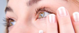
There are warning signs of an acute attack of glaucoma. These include blurred vision and seeing rainbow-colored circles around lights. These phenomena are caused by a sudden and rapid increase in intraocular pressure, which disrupts metabolic processes in the cornea.
As a result, the cornea becomes cloudy, which results in blurred vision. This also explains the vision of rainbow circles. When looking at a light source, the patient sees around it the same rainbow circle that a healthy person sees around a light source on the street in foggy weather.
Patients who have never had an acute attack may not know that these are warning signs of an acute attack of glaucoma. Sometimes, over the course of many years, glaucoma manifests itself only as warning signs.
However, patients who have visited the doctor know that after the warning signs an acute attack may appear, so they seek medical help as soon as the first symptoms of glaucoma appear.
However, an acute attack of glaucoma is not always preceded by its precursors. Often it develops completely unexpectedly both for the patient himself and for those around him.
Symptoms of an acute attack of glaucoma
In typical cases, pain in the eye and photophobia first appear. The pain quickly intensifies, covers the entire half of the head, and sometimes spreads to the entire head, to the upper or even lower jaws.
Headaches usually reach such intensity that they are accompanied by nausea or even vomiting. Chills, increased body temperature, slow pulse and other symptoms of dysfunction of the cardiovascular system appear. In some cases, headaches are unbearable.
They literally make patients lose their minds.
And the presence of vomiting, signs of disorders of “cardiovascular activity” gives grounds for an incorrect diagnosis, and such patients are sent to the therapeutic or neurological, and sometimes even to the infectious diseases department of the hospital with diagnoses of “poisoning”, “cerebral hemorrhage”, “infectious hepatitis” and others.
Meanwhile, even a superficial examination of the eyes shows that one of them (rarely both) is significantly changed.
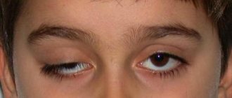
The signs of an acute attack of glaucoma are so obvious that even the average medical professional can detect them. This is redness of the eye, dilation of the pupil and lack of reaction to light, as well as a change in the color of the pupil. Instead of black, during an acute attack of glaucoma, the pupil appears greenish.
But the most important sign of an acute attack of glaucoma is a sharp hardening of the eye. The eye, when felt with the index fingers of both hands, seems hard as a stone.
To be sure, you can compare the hardness of the sick and healthy eyes, and in case of a bilateral attack, the hardness of the eyes of the sick and healthy person.
Types of glaucoma
Open-angle glaucoma accounts for more than 90% of cases of this disease. In open-angle glaucoma, access to the natural drainage system is open, but its functions are impaired. The result is a gradual increase in intraocular pressure.
As a rule, open-angle glaucoma is characterized by an asymptomatic, almost imperceptible course of the disease. Since the field of vision narrows gradually (the process can continue for several years), a person sometimes accidentally discovers that he can see in only one eye.
In some cases, one can identify complaints about the periodic appearance of rainbow circles when looking at a light source, “fogging,” asthenopic complaints associated with weakened accommodation.
In angle-closure glaucoma, intraocular fluid accumulates due to the fact that there is no access to the natural drainage system of the eye - the iris blocks the angle of the anterior chamber. As a result, the pressure increases, and this can lead to an acute attack of glaucoma, which is accompanied by:
- Sharp pain in the eye and in the corresponding half of the head;
- Obvious visual disturbances (blurred vision or a sharp decrease in vision up to complete blindness);
- Redness of the eye (dilation of the vessels of the anterior segment of the eyeball), corneal edema, decreased depth of the anterior chamber, dilation of the pupil and lack of its reaction to light;
- The appearance of halos around light sources.
Emergency care for acute glaucoma
Emergency care for an acute attack of glaucoma should be carried out by an ophthalmologist or other medical personnel with sufficient qualifications.
If this diagnosis is suspected, the patient should be immediately sent to the eye department of the hospital or a specialized ophthalmology clinic, because
Surgery may be necessary if drug treatment is ineffective.
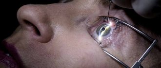
If possible, intraocular pressure should be measured with a tonometer. The pressure measurements in one and the other eye should be indicated in the certificate with which the patient must be urgently sent to the eye department of the hospital.
Before sending the patient, it is necessary to take all possible measures to reduce intraocular pressure and eliminate pain: give the patient 0.5 g of diacarb (or any other diuretic) orally.
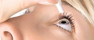
Drop a 1% solution of pilocarpine or any other pupil-constricting drops with a hypotensive effect into the eye several times.
Treatment of an attack of acute glaucoma
1. Pilocarpine 1-2% instilled into the eye, 1 drop every fifteen minutes, for one hour. Then 1 drop every half hour (4 times), then every hour (3 times), then 6 times a day. 2.Timolol (0.5% solution), instillation 1 drop twice a day.3.
Diacarb tablets 0.25-0.5 g twice a day. 4. Mannitol 15% solution intravenously drip (30 minutes). 10ml per 10kg. body weight. 5. Furosemide intramuscularly (IM) or intravenously (IV) 20 mg/day.
If there is no improvement in vision after the above treatment within 4 hours, a lytic mixture is administered (single dose): chlorpromazine 2.5% -1ml IM, diphenhydramine 2% -1ml IM, trimeperidine 2% -1ml IM. After this, lie in bed for 3 hours.
Then a laser iridectomy is performed (removal of part of the iris). If the attack does not stop within 24 hours, surgical treatment is necessary.
Save on social networks:
Related publications:
Source: //krasgmu.net/publ/ostryj_pristup_glaukomy_prichiny_i_lechenie/7-1-0-817
Glaucomocyclic crises - prevention
Prevention. Identifying the cause of the attack and eliminating it.
Glaucomocyclic crises - treatment
During a crisis - mydriatic drugs (1% solution of atropine sulfate, 0.25% solution of scopolamine hydrobromide, 0.1% solution of adrenaline hydrochloride, etc.), corticosteroids in the form of instillations (2.5% hydrocortisone emulsion, 0.1% solution dexamethasone, etc.), diacarb orally 0.25 g 2-3 times a day, hyperosmotic drugs - 50% glycerin solution or glycerin solution with ascorbic acid and fruit syrup (see Glaucoma, treatment). At the same time, butadione (0.15 g each) or reopirin, as well as calcium chloride (10% solution, 1 tablespoon) or diphenhydramine (0.03 g each) are prescribed orally. Usually the attack goes away under the influence of drug therapy. Surgical treatment is usually not performed.
Posner-schlossmann syndrome and Fuchs syndrome
Posner-Schlossmann syndrome is a glaucomocyclic crisis (A. Posner, modern American ophthalmologist; A. Schlossmann, 1867-1932, German pediatrician and biochemist). Synonyms: paroxysmal benign ocular hypertension, cyclic glaucoma, glaucomocyclic crisis syndrome). The syndrome was first described in 1929, but it is named after Posner and Schlossman, who described this syndrome in 1948.
This type of uveopathy occurs in middle age (30-50 years), mainly in men. Typically, only one eye is affected. The disease manifests itself in the form of a peculiar, sudden attack and is expressed in blurred vision, seeing rainbow circles around a light source, and an increase in intraocular pressure to 40-50 mm Hg. A characteristic difference from the inflammatory process (acute iridocyclitis) and an acute attack of primary glaucoma is the absence of severe pain and the presence of an injection of the eyeball of a mixed or stagnant nature.
An obligatory symptom of a crisis is precipitation on the posterior surface of the edematous cornea, which indicates the participation of ciliary body uveopathy in the pathogenesis. Precipitates in the amount of 2-5 elements of white color, with clear boundaries, are usually quite stable, remaining for some time after the crisis has stopped, which lasts up to 2 weeks. Precipitation can be observed for up to a month from the time of its appearance. The syndrome is characterized by moderate mydriasis, in which pupillary reactions are preserved. Other anatomical structures of the eyeball and optical environments are usually not changed. The angle of the anterior chamber of the eye is usually open.
According to Ts.M. Ioffe and A.V. Suprun (1968), against the background of a crisis, the resistance to the outflow of intraocular fluid sharply increases, with a decrease in the coefficient of ease of outflow to 0.01-0.02. The authors associate this fact with allergic swelling of the tissues of the anterior tract of intraocular fluid outflow.
The possible allergic nature of the crisis, which is believed to be angioneurosis of the ciliary body, is indicated by N.S. Gontoire and E.A. Lebedinskaya (1967). Despite the retention nature of the resulting hypertension, the authors do not exclude allergic edema of the ciliary body and an element of hypersecretion in its pathogenesis.
As the crisis resolves, all the above symptoms disappear, and the patient feels practically healthy until the next crisis occurs. The intervals between crises are calculated in weeks, months and years, lengthening noticeably with the age of the patient. Over time, apparently as a consequence of repeated attacks of the disease, degeneration of the iris occurs, expressed by its discoloration.
The relevance of monitoring this pathology lies in the insufficient knowledge of etiopathogenesis and diagnosis, on the one hand, and different views on the treatment of glaucomocyclic crisis, on the other, which causes difficulties in the management of patients with this pathology.
Target -
description of the clinical course and management of Posner-Schlossman syndrome.
Material and methods.
Patient V., born in 1964, was observed. (50 years old), complaining of blurred vision on the left, discomfort in the left eye, by the time of examination the complaints had been bothering him for 1 day. The disease was not associated with anything, it began in the morning after waking up. He has never had any such complaints before and wears +3.00D reading glasses.
Ophthalmological status: visual acuity of the right eye is 0.3, with correction +1.75D=0.8, of the left eye – 0.4, with correction +1.25D=1.0. Intraocular pressure (non-contact pneumotonometer): OD – 15 mm Hg, OS – 65 mm Hg. Art. Gonioscopy of both eyes: the anterior chamber angle is open. The visual fields of both eyes are within normal limits. Biomicroscopy: OD – intact. OS – painless on palpation, moderate corneal injection, the cornea is transparent, spherical, sensitivity is preserved. The anterior chamber is of medium depth, the moisture is transparent. The iris drawing is saved. The pupil is round, dilated, diameter 5 mm. The photoreaction is preserved. The lens is in the posterior chamber, initial opacities. Destruction of the vitreous body. Fundus of the eye: the optic disc is pale pink, the boundaries are clear, the contours are smooth. The arteries are narrowed in caliber, the veins are full-blooded and tortuous. The macular area is without features.
Done in a clinic: epibulbar - 1 drop of timmal, diacarb 250 mg per os. IOP of the left eye after drops after 20 minutes was 31 mm Hg. Recommended: Azarga 1 drop 2 times a day in the left eye for 3 days. After 3 days, the patient’s complaints persisted. Biomicroscopy: OD – intact. OS – painless on palpation, moderate corneal injection. Cornea: precipitates on the endothelium are clearly contoured, spherical, sensitivity is preserved. The anterior chamber is of medium depth, the moisture is transparent. The iris pattern is saved. The pupil is round, diameter 3 mm. The photoreaction is preserved. The lens is in the posterior chamber, initial opacities are destroyed. Vitreous body. Fundus of the eye: the optic disc is pale pink, the boundaries are clear, the contours are smooth. The arteries are narrowed in caliber, the veins are full-blooded and tortuous. The macular area is without features. IOP on a pneumotonometer: OS – 21 mm Hg, OD – 16 mm Hg. The patient was diagnosed with glaucomocyclic crisis, Posner-Schlossman syndrome. The patient was prescribed treatment: local instillation of dexamethasone solution 0.1% 3 times a day for 7 days; per os indomethacin 25 mg, 1 tablet 3 times a day for 7 days.