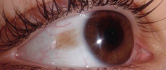
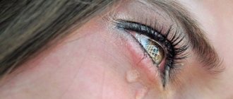
Pterygium (pterygium) is a pathological growth of the conjunctiva on the cornea. Pterygium gradually progresses and reaches the cornea to the central zone, closing the pupil and, as a result, impairs vision.
The part of the pterygium that is fused to the cornea is called the head. The other part, coming from the conjunctiva and penetrated by vessels, is called the body of the pterygium.
Most often, pterygium grows on the nasal side, but it happens that the pterygoid hymen grows on the cornea on both sides simultaneously.
What is pterygium of the eye?
This pathology is not the most common. It usually affects one eye. It has the shape of a triangle, but with a wing-shaped form it can have the same appearance. Pterygium usually develops in adulthood. The risk group includes residents of the southern regions. This leads to the main factors influencing the development of pathology:
- The abundance of ultraviolet rays and their effect on the organ of vision;
- Strong wind and dust;
- Hereditary predisposition;
- Chronic conjunctivitis;
- Chronic infectious diseases of the eyes and ENT region;
- Hormonal disorders;
- Working at a computer;
- Eye injuries.
But in general, the exact causes of development could not be determined, since pterygium can develop in completely different conditions, albeit much less frequently. The disease progresses quite quickly if irritating factors are not eliminated.
We advise you to read: Features of scleritis and its dangerous consequences.
With pathology, the tear film gradually ceases to form, which causes chronic irritation. It then gradually turns into inflammation, in which there is a risk of getting an infectious disease. Patients usually consult a doctor already at the stage when the pterygium becomes visually noticeable. If you seek help at the first sign, you can prevent the spread of the disease, as well as eye infection.
Reasons for development
The exact cause of the disease is not known. However, it has been established that most often pterygium forms in people whose activities involve prolonged exposure to strong UV or infrared radiation. Also, pathology is often diagnosed in people who are constantly outdoors for a long time (agricultural workers, fishermen, hunters). These factors lead to drying of the mucous membranes and the development of constant irritation.
In addition, high levels of dust can contribute to the development of pterygium. Thus, carpenters, painters, and plasterers often suffer from the disease, since constant contact with dust leads to chronic inflammation of the conjunctiva.
Thus, the main reasons for the development of the disease are:
- Chronic conjunctivitis.
- Dry eyes.
Over the past years, ophthalmologists have recorded cases of pterygium in people who spend a lot of time at the computer. In this case, the pathology develops due to the development of chronic dry eye syndrome.
The inflammatory process in the conjunctiva leads to a decrease in its natural protection and disruption of microcirculation, as a result of which new vessels begin to form.
Other reasons for the development of pathology include:
- Genetic predisposition.
- Infectious eye lesions.
- Injury to eye tissue.
- Frequent allergic conjunctivitis.
Stages of pterygium
In total, there are three stages in the pterygium:
- At the initial stage there are no manifestations. There may only be a slight lump on the white of the eye.
- The second stage is already manifested by dryness, redness and discomfort in the affected area, as well as other manifestations. At this stage, the pterygium is already more expanded.
- The third stage is usually characterized by the manifestation of complications of pterygium, including thickening of the cornea with the subsequent development of astigmatism, deterioration of vision with blocking of the pupil, inflammation of the conjunctiva, the appearance of scars in the affected area, as well as tissue degeneration into a malignant neoplasm.
The third stage is considered the most dangerous, which is visually easily determined by the presence of a number of complications. In this case, there is a risk of getting cancer or losing vision in one eye.
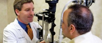
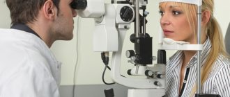
Diagnostics
The diagnosis is made based on an external examination of the eye. To assess the degree of growth, the ophthalmologist performs biomicroscopy. Visometry, refractometry, and ophthalmoscopy will help determine the stage of pterygium.
High-precision studies used as additional diagnostic methods:
- Keratotopography (to determine the extent of the disease).
- Tear fluid examination (to assess the likelihood of progression).
- Morphological study (allows you to evaluate vascular proliferation).
- Fluorescein angiography (vascular assessment).
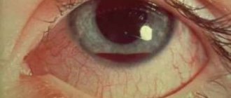
Symptoms
The eye disease pterygium does not appear immediately. At the first stage, there are no visual signs, and therefore at this stage patients do not go to the hospital for help. As the defect grows and spreads, the patient exhibits the following symptoms:
- Dry eyes;
- Increased lacrimation;
- Discomfort and feeling of the presence of a foreign body in the eye;
- Chronic irritation;
- Decreased vision;
- Itching;
- Astigmatism.
These are the main manifestations. They may be similar to the symptoms of other eye diseases, which is why patients often begin to self-medicate. As a result, the condition worsens and the disease progresses faster. Gradually, pterygium passes into the stage of an inflammatory process, in which swelling of the tissues and redness of the whites of the eyes appear.
Causes of the disease
At the moment there are no exact reasons. Despite this, factors have been found that can increase the risk of the disease:
- excessive exposure to ultraviolet rays;
- dust getting into your eyes;
- irritation caused by wind;
- the disease can be inherited;
- excessive computer use;
- predisposition to frequent inflammation of the conjunctiva;
- signs of pterygoid hymen.
Most often, the disorder can be detected based on symptoms such as:
- In the first stages, slight clouding appears on the periphery of the cornea.
- A noticeable manifestation of conjunctival clouding that grows from the side of the nose (developing stage).
- Feeling of a foreign object in the eye. A person begins to experience a similar feeling due to the fact that the pterygium begins to touch the nerve endings. Irritation appears. Feeling dry.
- Severe irritation, xerosis.
- Decreased vision clarity. This happens if the disease is already advanced. The pterygium penetrates the center of the cornea, obscuring vision.
- If the disorder becomes inflamed, constant itching, swelling appears, and the eyes begin to water.
- Due to these symptoms, a change occurs in the outer shell of the eye and blood vessels appear. Due to this, a growth grows on the cornea - pterygium.
After some time, the pterygium may increase in size or remain the same size. The disease is very noticeable and usually develops from the nose. Two eyes can be affected at once.
Treatment
Any therapy begins with diagnosis. Since pterygium manifests itself visually in the form of an increase in the conjunctiva, making a diagnosis even for an inexperienced specialist does not cause difficulties. To confirm the diagnosis, an examination is performed using a slit lamp.
Surgery
Drug treatment for this type of ophthalmological disease is not provided due to its ineffectiveness. If the pathology does not progress and affects only the edge of the cornea, then the doctor may decide not to perform any intervention in this area. But with the development of the disease, it is assumed that the pterygium is removed by excision of its body.
If the pathology progresses, then it is better to perform surgery in the early stages, when visual manifestations have just begun to make themselves felt. Excision of the pterygium is performed with wrapping of the body. When a defect is removed, areas of cloudiness often appear on the cornea. Therefore, the pathology is disposed of before it reaches the center of the optical zone.
Important! After surgery, relapses of the pathology often develop, and therefore it is necessary to consult a doctor on how to prevent new growth.
The pterygium is removed on an outpatient basis using local anesthesia. To do this, the appropriate drug is dripped into the conjunctival sac. Next, the growth is excised using one of three methods:
- By connecting the healthy edges of the conjunctiva;
- By replacing the pterygium with an amniotic membrane. An autograft can be used, which is a section of the membrane taken from under the upper eyelid.
- By suturing the conjunctiva in the form of folds to the sclera, retreating approximately a few millimeters from the cornea.
The last two techniques are considered more effective than conventional excision. With such methods, relapses rarely develop, and the risk of developing complications in the form of visual impairment is reduced. This factor is relevant when the pterygium grows strongly.
To hold the tissue together, a special suture material is used, which dissolves on its own in about 7-10 days. Sometimes biological glue may be used. Upon completion of the manipulations, the eye is covered with an aseptic bandage. All manipulations take about half an hour, and after a couple of hours the patient is sent home.
Laser excision
Removal of pterygium with a laser requires the availability of equipment in the clinic. This procedure is less traumatic for the organ of vision. It is carried out in the same way as during surgical intervention, but the use of a modern method eliminates the influence of the human factor on the area of influence. This eliminates the possibility of infection and the development of complications after surgery.
The cost of such a procedure averages about 15 thousand rubles. Clinics can also offer autoplasty, in which a piece of cornea is sutured onto the remaining area of the pterygium. This procedure is also called excimer corneal resurfacing. This procedure allows the eye to return to its previous appearance. Accordingly, the price of this procedure is added to the cost of laser excision, which amounts to a total of about 21 thousand rubles for all manipulations performed with the pterygium.
Drug treatment
This type of treatment without excision does not produce positive results. In general, medications are used only after surgery. Often, after surgery, complications and even relapses of the pathology develop. Therefore, appropriate treatment with drug prophylaxis is used.
To suppress the growth of the conjunctiva, the site of surgical intervention is treated with the pterygium of the eye after surgery with either liquid nitrogen or Miramistin. After removal, an appointment is scheduled:
- Antibiotics that will prevent infection of the operating area with various pathogens. Drops like Tobrex or Albucid are mainly used.
- Anti-inflammatory drugs of the drop type or in the form of an ointment, for example, Dexamethasone, Indocollir.
- Be sure to use moisturizing drops such as Clean Tear or Visine.
It is necessary to understand that drug therapy is preventive measures against re-growth, and not a complete treatment of an eye defect. They will not replace surgery to remove the body of the pterygium.
Folk remedies
Folk remedies are also used as a prophylactic against pterygium. Any mention that the pathology went away on its own and the defect decreased under the influence of any prescription is a lie. Folk remedies are not an alternative to surgical treatment. But to strengthen the cornea, prevent conjunctival growth and improve the functioning of the organ of vision, the following techniques can be used:
- Eating berries and blueberry juice. It has been proven quite a long time ago that this product is a panacea for tired and sore eyes. With regular use of berries, visual acuity is not only preserved, but also improved.
- Carrots and juice from them. This root vegetable also enjoys constant success among traditional healers. Carotene is very important for visual acuity. If you mix blueberry and carrot juice, you can get a cocktail that is not only tasty, but also very healthy for your eyesight.
- Make chamomile decoction according to the classical scheme - 2 tbsp. add the herbs to a glass of water, then boil in a water bath for half an hour. Cool until warm, then strain and bring the liquid to its original volume. Please note that you need to strain carefully, since you need to wash your eyes with the decoction three times a day. It is a natural anti-inflammatory agent.
- Black tea is also used as an eye wash. Brew one bag of black tea in a cup by pouring boiling water over it. Let it infuse and cool. Rinse your eyes with the solution. This recipe, if used twice a day, can relieve fatigue and redness of the eyes. At the same time, the look is significantly refreshed.
- If you feel dry eyes, fatigue and irritation, you can brew two tea bags, then cool them and apply them to your eyelids as compresses until they cool completely. The product also helps when work requires you to read a lot and work with your eyes and not get enough sleep.
- Juice from raw beets. Daily consumption of just 100g of the juice of this root vegetable helps prevent the development of neoplasms in the body, not only pterygium, but also oncological pathologies. Take it daily on an empty stomach.
Adviсe
These are the main folk remedies that are used for pterygium. It is better, of course, to use those that are drunk or eaten, that is, juices, berries, root vegetables. These are the safest recipes for vision, which, in addition to their therapeutic effect, also enrich the body with microelements. Other methods of exposure must be previously discussed with the attending physician.
After surgery, eye rinsing with decoctions or other liquids other than medications is not performed. Such manipulations are considered an additional risk, leading to the introduction of a secondary infection or foreign body into the area of manipulation. In addition, no one has canceled allergic reactions. Since after surgery the immune system may react inappropriately to any substance, it is better to refrain from topical use of such recipes.
Classification
The choice of treatment regimen usually depends on the type and stage of the disease. To determine drug therapy for pterygium, the ophthalmologist must take into account the types and forms of development of this pathology, since in the initial stages and mild forms conservative methods are still acceptable, and in advanced cases only surgical intervention will be effective. In medical practice, it is customary to divide pterygium into two types:
- progressive (spreading over the surface of the eyeball over time);
- stationary (stopped in its growth).
Depending on the degree of development of the disease, there are five stages of pterygium, namely:
- Stage I is considered initial, is localized at the edge of the eyeball and does not cause any inconvenience to the person.
- Stage II occurs when the pterygium has reached the middle of the distance between the edge of the orbit and the pupil with a small percentage of vision loss.
- Stage III is diagnosed when the pterygium reaches the pupil, and visual acuity may deteriorate to 0.5.
- Stage IV is noted when the hymen of the pterygium grows to the center of the pupil with a sharp decrease in vision to 0.2 - 0.3.5.
- Stage V is considered to be the maximum in terms of the area of growth of the pterygium and its penetration into the tissue of the eyeball. This stage threatens the patient with almost complete loss of vision, and the operation is fraught with great difficulties.
Based on the condition of the pterygium episclera, this pathology can be conditionally ranked according to the following degrees:
- 1st degree of pterygium development is characterized by a transparent thin hymen in which the vessels are clearly visible; this degree, as a rule, is not progressive;
- at grade 2, the growth becomes thicker and rises above the eyeball, its structure is translucent;
- Grade 3 is characterized by an opaque structure of the pterygium; the vessels are not visible at all.
Rehabilitation after surgery
To ensure that the rehabilitation process goes without complications and risks, it is recommended to follow the following rules:
- Wearing an aseptic bandage on the operated eye for 24 hours. Changes as it gets dirty
- One day after surgery, the use of medications begins. The average duration of therapy is a month, unless the doctor decides otherwise.
- For severe pain, analgesics are used in tablet form.
- For the first two weeks, you need to wear glasses that have ultraviolet filters.
- You can’t get your eyes wet for a week after surgery, which means swimming pools, sea swimming, and other factors that carry risks are excluded.
- Two weeks after the operation should pass without physical activity.
The first time after removal of the pterygium, vision may be impaired. If the operation was successful, visual acuity returns. This may happen in 5 days, or maybe in a month. Any eye irritation can trigger new growth of pterygium, and at a faster rate. The sooner you go to the clinic, the smaller the area of intervention will be, and, therefore, the risk of complications.
Sources used:
- Emergency ophthalmology / Edited by E.A. Egorova. - M.: GEOTAR-Media, 2005.
- Eye diseases / N.A. Pletneva. — M.: State Publishing House of Medical Literature
- Trachoma / V.V. Chirkovsky. — M.: State Publishing House of Medical Literature
- Volgograd State Medical University
Consequences
If treatment for pterygium is not carried out, the development of this pathology can lead to severe and sometimes irreversible consequences, including the following:
- irritation of the mucous membrane of the eye, constant sensations of sharpness and pain;
- decrease in visual acuity, and over time, as the tissue grows, its loss;
- poor circulation in the eyeball;
- in rare cases, this neoplasm can develop into a malignant form.
During appropriate therapy, certain complications may occur, which are usually associated with the stage of the disease at which treatment is started. In advanced cases, corneal tissue completely covers the pupil, and the person loses objective vision. In this condition, it is important to understand that vision will not be fully restored, since during surgical intervention the hymen, which is fused with the cornea, is removed and the transparency of the latter is somewhat lost. In addition, the pterygium is saturated with blood vessels; when it is removed, they are naturally damaged, therefore, after surgery, hemorrhages are observed in the eye, which resolve within a couple of weeks.
Prevention
Each pet needs to be provided with proper care: a balanced diet, regular hygiene procedures. Timely vaccination is important to keep your pet healthy.
It is not recommended to let your domestic cat outside; contact with stray animals is undesirable.
Timely deworming is the key to good health for your pet. The procedure is performed on kittens and adult animals.