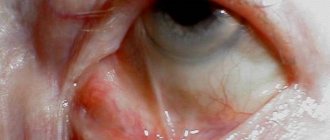
Hyphema in ophthalmology is a pathological condition of the eyes that occurs due to hemorrhage in the anterior chamber of the eyes. Often the iris that bleeds is the one whose tissue is characterized by a large number of blood vessels. It is impossible to say about the exact volume of blood.
Sometimes a thin strip appears, which can only be seen under a microscope, and in some cases a spot forms in the area of the eyeball.
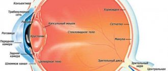
This area is limited on the outside by the cornea, and on the back by the membrane and lens. The blood is always in the lower part, since it is heavier than the eye fluid.
People over 40 years of age, as well as those who have injured their visual organs, are more susceptible to the development of hyphema (that is, the disorder can be diagnosed in any person, even a child). The peculiarity of the disease is its severe course and the high probability of relapse. If proper treatment is not started on time, the person will become completely blind. Reference! The code for hyphema in the international classification ICD-10 is H21.0.
Symptoms
There are 4 degrees of severity of hyphema:
- 1st degree – the anterior chamber is blocked by blood by 1/3:
- 2nd degree – the volume is filled with blood from 1/3 to half;
- 3 degree – more than 1/2;
- Grade 4 – the anterior chamber is completely filled with blood.
Depending on the severity, symptoms will vary. As a rule, patients present the following complaints:
- general eye soreness;
- sensitivity to bright light;
- blurred vision, “veil” before the eyes;
- neurological symptoms: dizziness, headaches.
If these signs appear, you should contact an ophthalmologist.
Main stages and photos
Signs of the disease depend on how much blood is visible in the eye. Basically, the disease is characterized by a thin short strip with clear edges. Depending on the size of the spot, the course of hyphema is divided into three stages, each of which is characterized by certain symptoms.
- Stage 1 – the degree of filling of the anterior eye chamber with blood is no more than one third. The stage is characterized by the appearance of fog in the eyes. Pain occurs when hyphema develops after injury.
- Stage 2 – the chamber is approximately 50% filled with blood. The clarity of vision deteriorates, the same veil remains, and sometimes the effect of “goosebumps” appears before the eyes. Dizziness and headaches begin.
- Stage 3 - in this case, the eye is filled with blood by more than half. The symptoms persist, only their manifestation becomes brighter.
- Stage 4 – This is sometimes called “black eye” or total hyphema. Blood completely fills the chamber of the eye. Most often, this stage of the disease develops due to penetrating injuries to the eyeball. In this case, a person loses the ability to see; he can only see light and darkness.
To preserve your vision, you should immediately consult a doctor when the first symptoms appear.
Degrees and types
In accordance with the amount of collected blood, doctors distinguish 3 degrees of hyphema of the visual organ:
- The blood level does not exaggerate 2 mm. There are blood smears on the iris.
- The degree of blood ranges from 2–5 mm.
- Total hyphema.
If there are no noticeable changes, ophthalmologists talk about the presence of microhyphema, which can only be seen under a microscope.
Blood accumulations can form in the anterior chamber, retina, and vitreous body. When hemorrhage occurred in the first variant, a uniform formation of a scarlet hue appears. In a lying position, the liquid spreads over the surface. When standing, blood settles at the bottom of the eyeball.
Bleeding that occurs in the retina of the eye is almost invisible. And this does not depend on the degree of damage. Patients are tormented by a disorder of clear vision, the formation of floaters over the visual organ. Excessive bleeding can lead to blindness.
Hemorrhages in the orbit occur due to injury, blood pathologies, and vasculitis. Patients experience bulging of the eyes, decreased visibility of the conjunctiva, difficulty in motor activity, and blood draining under the skin of the eyelids.
The formation of blood (hemophthalmos) in the area of the vitreous body implies a brown accumulation behind the lens. Expressed in the form of flashes of light, darkened spots. Total impairment causes complete blindness.
Classification
According to the nature of the prevalence of the pathological process, its degrees are distinguished:
- microhyphema - the presence of blood can only be diagnosed using an ophthalmological microscope;
- limited - the blood level is no more than 2 mm, there are blood smears on the iris;
- average hyphema of the eye - blood level from 2 to 5 mm;
- total hyphema - more than 5 mm of blood in the anterior chamber of the eye.
The severity of the pathological process is determined by the doctor by carrying out the necessary diagnostic measures.
Causes
The most common cause of hyphema formation is mechanical trauma to the eyeball, especially penetrating wounds. Hemorrhage occurs due to damage to the integrity of the blood vessels. If the injury was non-penetrating, it could provoke a sharp rise in intraocular pressure, which also damages the blood vessels. Also reasons are:
- inflammatory ophthalmological diseases: inflammation of the choroid, iris, ciliary body;
- retinal detachment.
Patients with diabetes are at risk. Provoking factors are autoimmune diseases, problems with blood clotting, and cancer.
Reasons for development
Experts talk about four main factors leading to the occurrence of a violation:
- Traumatic hyphema - the most popular cause of development is mechanical in nature. A through puncture, in which all eye membranes are injured, leads to rupture of blood vessels, from which blood spills into the anterior wall. But with blunt trauma, hemorrhage occurs due to a sharp drop in eye pressure, which provokes rupture of blood vessels in the membrane of the visual organ. This most often occurs with concussions and wounds.
- Hyphema of the eye after surgery , for example, cataract removal - when performing laser or strip depilation, the plexus of blood vessels (ciliary body or iris) is sometimes damaged. If hemorrhages occur during the rehabilitation period, this is due to a blood clot and unstable geodynamics.
- Diseases of the organs of vision . Some pathological processes in the eyes can lead to the fact that new vessels grow greatly and at the same time have thin walls. Because of this, the walls break even with slight vibrations.
- General systemic diseases of the body . Diseases that are accompanied by blood clotting disorders also lead to hemorrhage in the organs of vision. In addition, it is worth noting that frequent consumption of alcoholic beverages and drugs provokes disruptions in the process of blood clotting and fragility of blood vessels. All this contributes to the development of hyphema.
Hemorrhage into the sclera under the conjunctiva (hyposphagma)
Hypophagma, or scleral hemorrhage, or subconjunctival hemorrhage, is a condition where blood accumulates between the thinnest outer layer of the eye (conjunctiva) and the tunica albuginea. People also often say “the vessel has burst” and this is true: the root cause is damage to the smallest vessels of the conjunctiva, from which blood flows. But the reasons that caused this condition are extremely diverse:
- Direct traumatic effects on the eyeball: impact, friction, sudden change in barometric pressure, foreign body, chemical effects;
- Increased arterial and venous pressure: hypertensive crisis, sneezing, coughing, physical overload, bending over, choking, labor during childbirth, tension during constipation, vomiting and even intense crying in the child;
- Reduced blood clotting: congenital and acquired hemophilia, use of anticoagulant and antiplatelet medications (aspirin, heparin, ticlid, dipyridamole, Plavix and others);
- Diseases caused by infection (hemorrhagic conjunctivitis, leptospirosis);
- Increased fragility of blood vessels: diabetes mellitus, atherosclerotic disease, deficiency of vitamins K and C, systemic connective tissue diseases (autoimmune vasculitis, systemic lupus erythematosus)
- Condition after surgical interventions on the organs of vision.
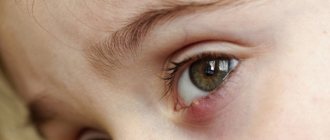
Symptoms of hemorrhage in the sclera are reduced to a visual defect in the form of a blood-red spot on a white background. The peculiarity of this hemorrhage is that over time it does not change its color like a bruise, but as it develops it simply becomes lighter until it disappears completely. Quite rarely, eye discomfort can be observed in the form of a feeling of a foreign body, slight itching, which are more likely to be of psychological origin.
Treatment of subconjunctival hemorrhage usually does not present any difficulties. In the vast majority of cases, reverse development occurs without the use of medications.
However, the following can help speed up resorption and limit the spread of hemorrhage:
- If you managed to catch the moment of hemorrhage formation under the conjunctiva and it increases “before your eyes,” vasoconstrictor eye drops (Visine, Naphthyzine, Octilya and others) are extremely effective; they will stop the outflow of blood from the vascular bed, which will stop the spread of hemorrhage;
- To accelerate the resorption of already formed hemorrhage, potassium iodide eye drops are effective.
A one-time hemorrhage in the sclera, which occurs even without an apparent reason and occurs without inflammation, decreased vision, “spots” and other symptoms, does not require examination or consultation with a doctor. In the case of frequent relapses or a complicated course, hyposphagma can signal serious diseases, both of the eye itself and of the body as a whole, which requires immediate contact with a medical institution to diagnose the pathology that caused it and prescribe treatment.
Video: about the causes of burst blood vessels in the eye
Diagnostics
To identify hyphema, a comprehensive diagnosis is necessary. Upon examination and questioning of the patient, it usually turns out that there is a history of trauma or that an ophthalmic operation has recently been undergone.
Before treatment you must:
- biomicroscopy using a three-mirror Goldmann lens. The doctor examines the conjunctiva, lens and vitreous body. The main object for study remains the anterior chamber. The procedure is painless for the patient; first, drops must be dropped into the eyes to dilate the pupil;
- gonioscopy – examination of the anterior chamber with visualization of inflammatory and degenerative changes. The procedure is non-invasive. Gonioscopy cannot be performed for conjunctivitis or keratitis. To avoid infection in the eye, the doctor must thoroughly clean the goniolens;
- ultrasound scanning. A highly informative and sensitive method that allows you to reproduce a two-dimensional image. Helps to identify the inflammatory focus and determine how extensive it is.
You also need to do visometry - a visual acuity test. It will help you understand how much visual acuity has decreased due to the hyphema; for this, it is enough to use a standard letter table. To monitor intraocular pressure, tonometry is performed using a contact or non-contact method.
Symptoms of the disease
Symptoms depend on the degree of damage to the eye. The first stage of hyphema can be virtually asymptomatic and is often diagnosed during a scheduled examination by a doctor. The second degree begins with deterioration of the patient’s vision and photophobia.
At this time, hemorrhage is detectable with the naked eye. At the next stage, the symptoms are pronounced, the patient notes a decrease in vision, feels pain and discomfort in the eye area, and external signs of hyphema are clearly visible. The fourth stage is characterized by significant pain and loss of vision (a person can only distinguish light).
General symptoms of hyphema include the following manifestations:
- The affected eye hurts
- Surrounding objects become foggy, visual ability gradually decreases
- Bright light is painful
- “Floaters” appear before the eyes
- Sick man suffering from dizziness
Treatment of hyphema
For grades 1 and 2 of disease severity, drug treatment is indicated. To do this, use eye drops containing corticosteroids and mydriatics - agents that dilate the pupil. Together, these substances not only effectively eliminate the problem, but also prevent relapses.
Read in a separate article: Hemangioma of the eye: what is it, treatment in adults and children
If hyphema is combined with increased intraocular pressure, anti-glaucoma drops are also used: Betaxolol, Pilocarpine, Travatan.
Surgical treatment is prescribed if medication does not have an effect, or the disease has progressed to stage 3 or 4. Trabectulectomy is used. It helps improve blood flow and normalizes intraocular pressure.
The operation is performed using anesthesia, the surgeon makes an incision on the conjunctiva, separates the fibrous plate and makes an incision on the tunica albuginea. Next, a path is formed for the outflow of intraocular fluid. To prevent the formation of a postoperative wound, antitumor antibiotics are used. Sutures are placed on the conjunctiva.
After the intervention, it is necessary to instill antibacterial drops and corticosteroids into the eyes for 4 weeks, and mydriatics will also be needed. It is necessary to keep the level of intraocular pressure under control; it is measured using non-contact tonometry. If the pressure rises, which is natural for the postoperative period, it is reduced with medication.
You need to remain in bed for several days after the operation, but in most cases a hospital stay is not required; the patient can undergo examinations on an outpatient basis.
A decisive examination of the anterior chamber of the eyeball, from which it can be concluded that the patient is healthy, can be carried out no earlier than 3 weeks after the operation.
Possible complications
All complications of hyphema can cause a decrease in visual acuity, therefore, if you suspect a pathological condition, you must immediately see a doctor or call an ambulance.
The prognosis for hyphema largely depends on its stage and the adequacy of treatment. Thus, the 1st and 2nd stages, with appropriate drug treatment, are usually subject to traceless involution and in the future only need to prevent the underlying disease that caused hemorrhage in the frontal chamber of the eye.
In patients with stages 3 and 4, in the presence of errors in drug treatment and untimely and delayed surgical treatment, the possibility of maintaining vision at the level of light perception, when a person can only distinguish day from night, is not excluded.
In many cases, subsequent loss of vision is associated not with the hyphema itself, but with concomitant damage to the iris, lens, cornea, retina, as well as with pathology caused by increased intraocular pressure - optic nerve dystrophy.
If signs of bleeding into the frontal chamber of the eye are detected, you should consult an ophthalmologist as soon as possible; self-treatment and the use of traditional medicine will inevitably lead to a drop in visual acuity, and possibly to absolute blindness.
A hematoma on the eye is fraught with the following complications:
- staining of the cornea with blood and, as a result, an increase in size;
- deterioration of vision, up to blindness;
- optic nerve atrophy;
- fusion of the cornea and iris;
- amblyopia, in which one eye does not participate in the visual process;
- secondary glaucoma.
What is hyphema
When the disease occurs, blood and clots collect in the anterior chamber of the eye. The larger the congestion there, the worse the visual visibility. When the chamber is completely filled, vision is completely lost. This appears due to injuries, rupture of recently formed vessels, or due to the presence of hematological diseases.
Hyphema caused by injury is most often observed in men. And other causes of the disease are noted in the same proportion. Patients over 40 years of age have an increased risk of developing hyphema. In infants, the disease can appear due to shaking or congenital blood diseases.
The pathology is associated with a complex course; without appropriate treatment there will be progression. Lack of therapeutic action will lead to complete loss of vision. In 35% of cases, a relapse occurs on day 3.
conclusions
Non-traumatic hyphemas can occur in patients with severe blood diseases or cancer. The main symptom of eye damage is hemorrhage in the anterior chamber, but additional symptoms may be present. Often only they can help detect hyphema.
In any case, the disease is a dangerous pathology that threatens reduction and even loss of vision and should be treated only under the supervision of an ophthalmologist.
Read more about strabismus in newborns and whether conjunctivitis is transmitted.
Hyphema or hemorrhage in the eye, or more precisely in the anterior chamber, can be either completely harmless or lead to serious complications. Therefore, in case of injury (or bruise), you should visit an ophthalmologist.
Prevention
There are no special preventive measures that can guarantee the prevention of hyphema, but the following recommendations should be taken into account:
- if the patient has blood diseases, he should undergo regular tests and keep hematological parameters under control;
- for diabetes mellitus type 1, 2 and arterial hypertension, it is necessary to be examined by an ophthalmologist 2 times a year;
- for malignant tumors of the eyeball, consultation with an ophthalmo-oncologist is necessary every 3 months.
Eyes should be protected from mechanical damage; when performing traumatic work, safety glasses should be worn.
With timely treatment of hyphema, the prognosis for life is favorable, the patient remains able to work.
If, due to the advanced state of the disease, visual function has been seriously affected, a disability may be issued by decision of a special commission.
Preventive measures
To avoid the formation of a hyphema, you should:
- observe safety precautions at enterprises;
- treat any pathologies of the visual organs or correct them.
People with blood pathologies should regularly monitor hematological parameters. Patients with diabetes and hypertension should visit an ophthalmologist twice a year.
A timely visit to an ophthalmologist will help avoid decreased vision and the development of further disability.
Treatment of the disease
The pathology is eliminated by using Atropine. In the early stages of the development of the disease, conservative therapy is used with the help of Prednisolone and Atropine. Corticosteroid drugs stop repeated relapses of hyphema. If a patient has been diagnosed with increased eye pressure, carbonic anhydrase inhibitors are prescribed. Treatment with surgical intervention is recommended at stages 3-4 of the disease. As a result of two parallel punctures, a crystalloid solution is injected into the eye cavity and the accumulated clots are washed out. Trabeculectomy improves fluid outflow and lowers IOP. The disease is treated on an outpatient basis. Only patients with high intraocular pressure, pathological diseases of the hematopoietic system, and children under 3 years of age are recommended to be hospitalized.
Alternative medicine
Traditional methods can be used as an auxiliary therapy. A decoction of chicory will help eliminate the manifestations of hemorrhage. To prepare, you need to take 3 large spoons of the plant root, add 0.5 liters of water, let it boil, and leave for 15 minutes. It is recommended to strain the resulting liquid through cheesecloth. Drink ½ cup 3 times a day, or make compresses based on the resulting decoction.
With microhyphema, there is no need to treat the disease; the blood in the eye cavity can resolve spontaneously. People who have been diagnosed with hyphema should be seen by an ophthalmologist.
How to treat
Treatment of hyphema depends entirely on the factors of pathological development, its severity, duration and development of complications. If the patient has previously taken anticoagulants, he should stop taking them for a while.
He is prescribed vaso-strengthening, hemostatic and absorbable agents. The collected blood is removed through surgery.
If there was only a small amount of it, it may resolve on its own in a few days.
During therapy, the doctor must monitor intraocular pressure. Sometimes blood cells can get into the drainage system and clog it.
During treatment, doctors recommend:
- avoid any physical stress;
- contact the clinic immediately if severe pain occurs;
- regularly use the prescribed drops;
- protect the eye from damage with a soft bandage;
- visit an ophthalmologist daily.
In almost 20% of patients with the disorder, hemorrhage increases in the first 3-5 days. To prevent this from happening, you must follow all the doctor’s recommendations.
Surgery
When blood enters the sclera in large quantities, there may be no effect from conservative resorption therapy. In this case, it is washed out surgically.
The operation is usually prescribed for:
- the appearance of bloody smears on the cornea;
- the occurrence of clots;
- complete filling of the anterior chamber;
- preservation of blood in the organ for more than 5 days;
- increase in pressure.
Basically, the operation is performed at the third and fourth stages. During it, two parallel holes are made. A balanced crystalloid solution is injected into one, and blood and clots are removed through the second.
If the complication appears due to injury, after its treatment it is necessary to undergo an annual examination by an ophthalmologist.
This will help diagnose glaucoma at an early stage. It is mainly formed due to damage to the drainage system of the eye chambers.
In the hospital
In most cases, treatment is carried out on an outpatient basis. Inpatient treatment is provided for patients with high intraocular pressure, a disease that cannot be corrected with medication, hematological pathologies, and children under 3 years of age.
In this case, bed rest with minimal physical activity is required. You will need to wear a protective bandage over the injured eye.
Treatment of the disease
The ophthalmologist prescribes treatment after determining the degree of hyphema. In the initial stage, conservative therapy is chosen, and if there is significant hemorrhage, the patient is operated on. Most procedures are performed in an outpatient clinic; children and patients with hemoglobinopathy or serious complications are treated in the hospital.
Recommendations for patients
The treatment prescribed varies, but there are general tips:
- Need to rest and stay in bed
- You need to sleep with a high headboard, raising the top of a hospital bed or placing several pillows at home (the angle should be about 50–60 ̊)
- You can’t bend over, you need to avoid physical activity for a while
- If you need pain medications, consult your doctor. Most of these medications alter blood clotting rates, so refrain from self-administering medications
- If your doctor has advised you to wear a bandage, you should not ignore his recommendation.
Therapy with medications
To treat hyphema, drugs of different effects are prescribed, they are widely used and produce a good effect:
- Medicines to strengthen the walls of blood vessels (Emoprox, Diclofenac, Actovegin, Taufon, Okovit)
- Painkillers (Tylenol, Acetaminophen, Aldolor, etc.)
- Mydriatics to prevent adhesions near the lens and iris (Atropine 1%)
- Medicines to stop bleeding (Purolase, Aminocaproic acid, etc.)
- Medicines with absorbable action (Glycerol, Mannitol, Mannitol, etc.)
- Means for normalizing IOP (Diacarb, Timol, etc.)
Corticosteroid drops
The basis of treatment is corticosteroids, which reduce the likelihood of recurrent hemorrhage (numerous studies indicate a decrease in rates from 12% to 5% of cases). With the help of drops with corticosteroids, inflammation and pain are eliminated.
| The optimal dose and duration of use of these drugs is determined by the doctor for each patient. |
Surgical intervention
The operation is envisaged for the third and fourth degrees of hyphema. The anterior chamber of the affected eye is carefully washed. To do this, the surgeon makes two incisions: a special solution is poured through one, and liquid with blood clots is pumped out of the second. If IOP is high, trabeculectomy is performed, with the help of which blood flow is activated and the pressure is normalized. Indications for surgery are:
- Staining the cornea red
- Formation of blood clots
- Increased IOP
- Lack of expected results after drug treatment
- Third and fourth degrees of pathology
Return to contents
Treatment prognosis
In mild cases, it takes several days to a week for the hyphema to completely resolve. But approximately 1/5 of the patients experience more intense bleeding within 3-5 days, so it is very important to consult a doctor and follow his recommendations exactly.
In general, the prognosis regarding the restoration of previous visual acuity may be different, since this is influenced by many factors, especially the volume of blood shed and the value of intraocular pressure. If the latter remains within normal limits and there is no other damage to the structures of the eye, vision is usually completely restored after a few weeks.
Provoking factors
The disease can be a concomitant illness with other ophthalmological pathologies. Causes of hyphema of the visual organs:
- injury to the organs of vision;
- surgical intervention;
- other diseases.
In addition to ophthalmological diseases, intraocular bleeding can be provoked by diseases such as diabetes, blood clots, hemophilia, neoplasms in the eye area, leukemia, severe anemia, and excessive addiction to alcoholic beverages.
Recovery forecasts
The disease can have a different and unpredictable course. 1/5 of all patients experience a sharp deterioration in their condition in the first few days of illness. With mild severity of the disease, vision is completely restored within a week, provided that therapy is started in a timely manner.
If bleeding does not stop in a timely manner and there are concomitant pathologies, this can lead to:
- staining of the cornea with blood (leads to impaired quality of vision);
- secondary glaucoma (intraocular pressure increases, which leads to complete blindness).
Most often, complications occur when the anterior chamber of the eye is completely filled with blood.