Yellow spots on the white membrane of the eyeball indicate pathological conditions or disturbances in the functioning of internal organs.
Atypical manifestations worry a person.
Orange spots at a young age are unlikely to pose a threat to health and visual perception, but it may be a symptom of a disease that leads to complete blindness. Therefore, it is important to know why it appears and how it is treated.
We recommend reading: Red spot on the white of the eyes
Causes
Yellow spots on the tunica albuginea are a manifestation of Gospel disease. This pathology develops due to a violation of bilirubin metabolism in the body.
Common reasons:
- malignant tumors in the liver;
- obesity;
- cirrhosis;
- anemia;
- cholelithiasis;
- inflammatory pathologies of the liver.
If the yellowish spot is a general cause, you need to contact a specialized specialist. This sign indicates serious disorders in the body that require immediate treatment.
The second group of reasons are specific pathological conditions that lead to the formation of orange spots on the tunica albuginea. The following pathologies cause this symptom:
- A pinguecula is a yellow formation that is often found on the conjunctiva in elderly patients. Appears on both sides of the cornea. This is a sign of aging of the conjunctiva. Manifested by irritation, dry eye syndrome, redness.
- Pterygium or pterygoid hymen is a dystrophic pathology characterized by the formation of a fold of the connective membrane. The hymen grows in the inner corner and looks like a yellow triangular spot. Pterygium is manifested by redness, itching, swelling, and a feeling of a foreign body.
- A connective membrane cyst is a formation that has formed in the area of the outer membrane. This is a thin-walled seal with exudate. It may be yellowish in color and appear as a raised spot.
- A nevus is a benign formation consisting of an accumulation of melanocytes. These are moles or birthmarks that are yellow or brown in color.
- Horner-Trantas spots appear due to allergic reactions. These are small grains formed on the albumen. The cause is conjunctivitis and allergic keratitis.
- Leukoma is a disease that appears as a white spot on the cornea, sometimes manifesting as a yellow formation. Formed due to cicatricial changes after inflammation or ulceration.
Homeopathic treatment
To eliminate unpleasant symptoms, moisturize the conjunctiva
, use the following drugs:
- Fagopyrum (Fagopyrum) drug reduces itching in the eyes
, eliminates inflammation, swelling and redness. - Heracleum sphondylium improves the condition of mucous membranes
and skin, relieves inflammation. - (Senega) is effective for swelling, neoplasms in the eye
, clouding of the cornea, photophobia. - (Euphrasia) relieves unpleasant symptoms in the eye area, eliminates the feeling of dust and sand under the eyelid
. - (Causticum) prevents the formation of nodules, compactions, and eliminates dry mucous membranes
.
This drug cannot be combined with the homeopathic remedy Phosphorus
. - Silica acts on foci of chronic inflammation, on connective tissue, mucous membranes, reduces pain and itching in the acute period of the disease
, reduces swelling, congestion, and improves the condition of patients with photophobia. - To prevent dryness
, fatigue and inflammation of the eyes, a complex homeopathic preparation has been developed - Similasan drops containing harmless mineral and herbal components.
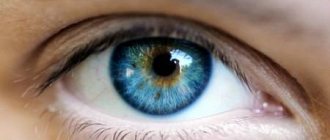
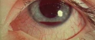
In a healthy person, the whites of the eyes have a snow-white tint.
Therefore, if yellowish spots appear on it, you should immediately contact an ophthalmologist, because this is the first symptom that the functioning of the internal organs is impaired. If such a pathology manifests itself in adulthood, this is a sign of the development of a pinguecula. The disease does not require treatment, but a consultation with an ophthalmologist will definitely not be superfluous. The front part of the eye, colored white, is called the sclera. In some cases, a thin network of blood vessels is visible on it. Yellowing of the eyeball is often a sign of the development of a certain type of disease.
At the same time, the spot does not cause discomfort and does not affect visual acuity. However, the absence of dangerous symptoms is not a reason to refuse a visit to the doctor. After all, often the disease does not manifest itself in any way in the initial stages.
Symptoms
A yellow formation near the pupil is almost never associated with pathologies of the optical system. But it is a sign of a progressive disease. Accordingly, the symptoms will be different, depending on the cause of its appearance.
Progressive eye pathologies cause:
- doubling of objects;
- pain syndrome;
- unbearable itching;
- mucous and purulent discharge;
- sensitivity to bright light.
In the case where the orange spot is a sign of problems with internal organs, the clinical picture of the disease is supplemented by an increase in body temperature, poor appetite, asthenopia, trembling and the occurrence of a gag reflex.
Table. Signs depending on the cause
| Name of pathology | Clinical picture |
| Cirrhosis | icteric discoloration of the skin and mucous membranes; skin and eye itching; abdominal pain on the right; weakness; swelling of the legs, arms and face (especially the eyes). |
| Anemia | pallor; sticking on the lips; asthenia; dry mucous membranes; taste disturbance. |
| Hepatitis | increased body temperature; yellowness with a greenish tint to the skin and tunica albuginea; dry conjunctiva; itching; lethargy; insomnia; sweetish odor from the mouth. |
| Gallstones | feeling of heaviness; nausea; sharp pain; light stool; change in color of the skin and sclera. |
| Leukoma | corneal clouding; deterioration in the quality of vision; complete blindness or partial loss of visual perception; increased intraocular pressure; disruption of fluid outflow. |
Photo
Why does it appear
There are many reasons that can cause a yellow spot to appear on the eyeball. It can be of different sizes and indicate various pathologies progressing in the body.
Nevus
Nevi are formations that can appear on any part of the skin or mucous membranes. Nevi can be congenital or appear during a person’s life. The cause of such formations is changes in hormone levels in the body during pregnancy and breastfeeding, as well as during menopause.
A nevus is a cluster of pigment cells that do not pose any danger to the organ of vision. Depending on the amount of pigment, the nevus can be yellow or dark in color.
Conjunctival cysts
In some cases, a yellow growth in the eye may be a cystic formation of the conjunctiva of a congenital or acquired nature. In childhood, a benign dermoid cyst is usually detected in the form of a capsule that contains fat cells.
After suffering inflammatory pathologies, injuries and eye surgeries, a person may develop acquired cystic formations on the proteins. They resemble nodules in which fluid accumulates.
A small cyst usually does not cause significant discomfort and does not affect the patient’s vision in any way. And if the formation reaches a large size, then it is better to remove it. A large cyst can cause damage to the tissues of the organ of vision and disrupt its normal functioning.
Leukoma
In medical practice, leukoma is called a corneal thorn. A fresh formation is usually milky in color, while an old leukoma takes on a yellow tint. Leukoma can appear as a result of scarring of eye tissue after an injury, including an inflammatory one.
The location of the formation may be the periphery of the corneal vision, and such a defect usually does not cause visual impairment. When the leukoma is located in the very center of the eyeball, vision may be impaired, and when the cornea is completely blocked, a person can only see silhouettes.
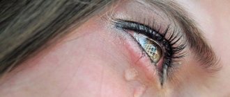
Pinguecula
Often yellow spots appear on the eyes of older people. Mainly the place of its localization is the inner corner of the organ of vision and such a formation is called penventiculum. It indicates that the choroid of the eye is aging.
Pingeventicula does not affect visual acuity in any way and does not cause unpleasant symptoms. The main reason for its formation on the eye is considered to be working in unfavorable conditions, prolonged exposure to a computer monitor and UV radiation from the sun.
The listed factors do not always cause pigmentation to appear in the eyes, but damage to the membrane of the eye increases the risk of its growth several times. Patients who use contact lenses need to be especially careful.
Horner-Trantas points
Small yellow spots may appear on the white of the eye around the cornea. Their appearance resembles small round grains. The main reason for the appearance of Horner-Trantas points is an allergic reaction that occurs with certain pathologies of the organ of vision. The impetus for the appearance of such formations in the eyes can be allergic keratitis or conjunctivitis.
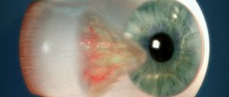
Pterygium
When the normal growth of the mucous membrane of the organ of vision is disrupted, a fold is formed on the sclera, which is called a pterygium. Mostly the place of its localization becomes the inner edge, on which formations appear in the form of a triangle. Subsequently, the pterygium grows towards the pupil and can cause decreased vision.
The provoking factor for the appearance of such a formation on the protein may be the syndrome of increased dryness of the eye and working in very dusty rooms. In addition, pterygium can form when exposed to sunlight for too long or under the influence of computer radiation.
Diagnostics
It is impossible to make a diagnosis after a visual examination; even an experienced ophthalmologist will not be able to immediately determine the cause of the macular spot. The patient will need to undergo a series of tests:
- First, the ophthalmologist collects the patient's medical history. Finds out what symptoms are bothering you, except for the detected yellow spot near the pupil.
- Performs palpation if there is a history of liver disease.
- Biomicroscopy makes it possible to study the structure of the eye, detect damage to the cornea, cataracts, inflammatory disease or degenerative changes in the optic nerve.
- Ultrasound and computed tomography allow you to examine the internal organs located in the abdominal cavity. Performed if hepatitis, cirrhosis, or stones in the gallbladder are suspected.
Laboratory tests are performed to confirm jaundice and anemia. The patient is given a referral to donate blood, urine and feces. With their help, the level of hemoglobin and bile pigment is determined.
Treatment of these diseases
After diagnostic measures have been carried out, when the exact cause of the disease has been established, treatment begins.
If it is jaundice or any other type of hepatitis, it is treated with interferon and other drugs used in such cases. A patient with jaundice is hospitalized. A course of intensive therapy is carried out, which in the case of viral hepatitis gives positive results within 30-40 days.
The patient is prescribed a special diet and should not engage in heavy physical work for 6 months. Such measures usually help with hepatitis types A and B. Almost all patients recover. If hepatitis B has entered the chronic stage (this occurs in 10-15% of patients), then recovery is extremely rare, but modern drugs make it possible to avoid the disease developing into cirrhosis.
If a patient is diagnosed with liver cancer, surgery may be performed when the disease is at an early stage of development. Typically, the survival rate of such patients is no more than 20% of the total number for 5 years after surgery.
Gallstones can be removed using special medications or surgical methods. To eliminate the symptoms of anemia, medications and traditional medicine recipes are used.
Eye diseases pterygium and pinguecula can be easily cured at the initial stage of their development with moisturizing gels or drops. Sometimes the patient, along with the above symptoms of these ailments, has redness around the eye and irritation. To remove them, doctors prescribe a course of special anti-inflammatory therapy.
But these diseases cannot always be cured with medication. If conservative treatment methods are found to be ineffective, surgery is prescribed. The same measures are taken if there is a threat of vision loss in the patient. Some people are concerned about the cosmetic appearance of these eye diseases. Therefore, they prefer to remove yellow spots through surgery.
Treatment
After determining the cause that caused the formation of a yellow spot on the tunica albuginea, treatment is prescribed. You can remove the yellowish halo after getting rid of the cause.
Methods of therapy:
- For jaundice, phototherapy is prescribed until bilirubin is completely destroyed. They take Karsil and Hofitol. In severe cases of the disease, physiotherapeutic procedures are prescribed. Treatment takes place in a hospital setting.
- In case of cirrhosis, it is important to follow a diet and take diuretics. Glucocorticoid hormones and antiviral treatment will help slow the course of the disease. Additionally, medications are prescribed that reduce pressure in the collar area and plasmapheresis (the procedure allows you to purify the blood and reduce jaundice).
- If the cause of the macula is anemia, therapy will be based on taking medications containing iron, folic acid and vitamin B12. A blood transfusion is prescribed.
- If there are stones in the gall bladder, they are dissolved with special preparations. Diuretics are prescribed to help small stones pass out quickly in the urine. In this case, extracorporeal shock wave lithotripsy is performed. If the stones are large, surgery is performed, then medications are prescribed for rapid recovery.
- Leukoma is treated with surgery. Keratoplasty and keratoprosthesis are mainly performed.
- Pinguecula is treated with medications that relieve discomfort. These are moisturizing drops. If you have pinguecula, it is not advisable to wear contact lenses until it is completely cured. Anti-inflammatory and antibacterial medications are prescribed. If the patient is concerned about aesthetic discomfort, surgery is performed to remove the stain.
- Pterygium is treated surgically. It is advisable to carry out the operation in the early stages of development. Treatment with traditional methods is ineffective and can aggravate the course of the pathology.
- The nevus is removed using surgical removal or a laser beam. The latter method is used more often because it is painless and the rehabilitation period is reduced.
- Conjunctival cysts are treated with conservative and surgical methods. Glucocorticosteroids are prescribed, then trichloroacetic acid is injected into the cavity, which has a sclerosing effect.
Diagnosis and treatment of yellow spots on the white of the eye
Yellow spots on the white of the eye can be eliminated only after identifying the root cause of their appearance and promptly prescribing the correct treatment. In each case it will be individual.
For example, pinguecula and pterygium in the initial stages are treated with drops and gels that moisturize the conjunctiva, or with anti-inflammatory drugs. Surgical intervention is used only in cases where long-term therapy has not helped. Drug therapy is also used to treat Horner-Trantas spots.
But advanced cases of conjunctival cysts are treated only surgically. Modern technologies make it possible to do this using a laser painlessly for the Patient.
Doctors at Dr. Belikova’s Eye Clinic will conduct a comprehensive examination of the visual organs and, if necessary, select the most effective treatment or give recommendations for visiting a specialized specialist.
Prevention
To prevent yellow spots from appearing on the sclera, you must initially protect your eyes. Preventive measures are as follows:
- an active lifestyle and proper nutrition (especially for patients suffering from liver diseases);
- combining long work with a little rest for the eyes;
- correct use of contact vision correction products (regular cleaning and changing of lenses in accordance with the wearing period);
- moisturizing mucous membranes when working in the sun, in a dusty room or in an air-conditioned room.
It is important to protect the visual organs from direct ultraviolet radiation, dust and wind using protective glasses. Taking vitamin complexes and healthy sleep for 8 hours will reduce the likelihood of certain diseases that lead to the formation of a yellow spot on the white of the eye.
Preventive measures
A proper daily routine and a balanced diet will help prevent the development of pathologies or their relapse. Following simple medical recommendations will help you avoid eye health problems:
- Sleep at least eight hours a day;
- Move more, walk in the fresh air;
- When working at a computer for a long time, use safety glasses;
- Take vitamin complexes;
- Use medications to moisturize the mucous membrane;
- Visit your eye doctor regularly.
| On sunny days, wear optical devices marked UV400; they protect your eyes 100% from harmful ultraviolet radiation. |