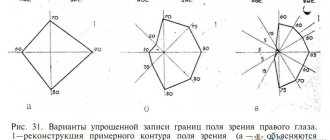
Refractometry is an analytical method based on the phenomenon of light refraction when rays pass from one medium to another, which is explained by changes in the speed of light distribution in different environments.
Today, this method of analysis is widely used in many fields: refractometry is often used in pharmaceutical and food analysis, as well as in the study of eyes.
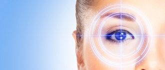
Refractometry in ophthalmology is one of the objective methods for studying the refractive power of the eye - refraction, which is carried out using specialized equipment - an eye refractometer. The refractometry method is used to identify eye diseases such as:
- nearsightedness (myopia);
- farsightedness (hypermetropia);
- astigmatism.
This research method allows doctors to quickly obtain accurate data about the patient’s eye health. The procedure is possible at any age: both children and adults - this is a definite advantage of the method.
Equipment and procedure
As already mentioned, refractometry is carried out using specialized ophthalmological equipment - refractometers, which come in several types:
Hartinger refractometer
Consists of the following parts:
- lighting system;
- optical system;
- measuring scale.
The procedure itself is as follows: a test symbol is introduced into the optical system, which is three vertical and two horizontal stripes. The light beam from the device is directed into the patient's eye under examination and projects onto the retina an image of test symbols, which are referred by the optical system of the eyes to the focal plane of the refractometer. The initial position of the device's optics is a measuring scale with zero values, which are associated with the far points of pure vision of the emmetropic eye. The doctor sees the test symbol through the eyepiece of the device.
With normal refraction of the eye, the two parts of the half-picture of vertical and horizontal stripes merge, but in the case of hypermetropia and myopia, on the contrary, they diverge. Horizontal displacement of the stripes and along the vertical axis indicates astigmatism.
By turning the device horizontally, the ophthalmologist minimizes the divergence of the bands by installing the device in one of the main meridians. In this way, refraction is measured in a specific meridian. The doctor, by rotating a special ring located near the eyepiece of the device, achieves the merging of the stripes, and the scale of the refractometric device indicates the type and size of the refractive abilities of the eye apparatus. The measurement limit for this type of equipment is from -20.0 to +20.0 diopters, but the accuracy is up to 0.25 diopters.
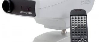
Computer type
The most commonly used today are automatic computerized refractometers. The essence of their work is also based on the emission of microscopic beams of infrared rays, which cross the pupil and the refractive medium, are reflected from the fundus of the eye and go in the opposite direction. The device’s sensor reads out the received information, and a special application analyzes the original and newly obtained data, through which the clinical refraction of the eyes is calculated. All results obtained are instantly transferred to the monitor and printed.
The procedure for measuring refraction occurs as follows:
- The patient sits in front of the device.
- His chin is fixed in a special socket, and his forehead is pressed against the top panel.
- The doctor fixes the patient's head in the required position so that it remains motionless during the examination.
- The patient is allowed to blink.
- Each eye is examined separately.
- The subject needs to focus his gaze on the fixation image, the sharpness of which will gradually change.
- More modern devices can use quite complex pictures that can arouse interest even in the smallest patient, which is important for the success of the procedure, since small children are not known for their perseverance.
- Next, using a joystick, the doctor places the refractometer in the very middle of the pupil and begins complex measurements in manual or automatic mode.
- In total, the procedure can last from one to two minutes.
What is refractometry?
So, based on the above information, the definition of refractometry as the process of measuring refraction becomes more clear. Clinical refraction is examined, since the ability to focus images on the retina is important. Moreover, both the static and dynamic components are studied.
Some time ago, refraction could only be measured manually. For this purpose, special eye diagrams and manual refraction measurement techniques were used. In terms of accuracy, they were in many ways inferior to modern instruments; in addition, the possibility of error could not be ruled out.
General concept of the method
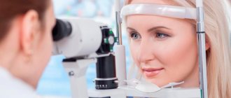
Today, refractometry is a high-tech procedure that takes no more than five minutes. For this diagnostic method, special devices are used - refractometers. The operating principle of this device is infrared radiation. The refractometer is located on the table, is about half a meter in height and has “outputs” on both sides - a screen with a control panel for the doctor and a special device where the patient looks. From a special lens, a beam of rays in the infrared spectrum is directed towards the pupils of the subject, which, penetrating through a hole in the iris, fall on the retina. There is a reflection from the bottom of the eye and return to the sensors of the device. The doctor only needs to direct the rays through the patient's pupil. The devices, in turn, record the received data, and the computer calculates the necessary indicators. The calculations are immediately displayed on the screen and can then be printed.
Refractometer
How to decipher the results
The finished printout contains all the information about the state of refraction of our eyes and their health. And naturally, the results for any patient are of considerable interest. However, not everyone can read a refractogram fluently. How are the indicators deciphered?
The finished printout consists of three columns:
- The first is called SPH - “sphere”. It contains information about the type of refraction found in the subject. Simply put, this column tells us whether there is a disease of myopia, or, conversely, the patient suffers from farsightedness.
- The next CYL column is “cylinder”. It contains information about lenses that are necessary for vision correction. If there is a need for such, of course.
- The last column of AXIS is “axis”. It contains data on the required lens angle.
- And finally, the printout, at the very bottom, contains another value - PD, which is used to indicate the interpupillary distance.
Refractometry values change throughout life. For example, farsightedness is most often detected in a newborn child, but by the age of 20 this anomaly remains in only a third. About 40% of young people have normal refraction, while the rest suffer from myopia. And with age, refraction worsens, which is caused by age-related changes in the lens, at which time patients begin to develop presbyopia. Therefore, it is extremely important to undergo periodic examinations in order to promptly prevent the development of eye diseases.
Theoretical part.
The refractive index (refractive index) is the ratio of the speed of light in a vacuum to the speed of light in the test substance (absolute refractive index). The refractive index depends on the temperature and wavelength of light at which the determination is made. In solutions, the refractive index also depends on the concentration of the substance and the nature of the solvent. In this case, in practice, the so-called relative refractive index (n) is determined, which is calculated as the ratio of the sine of the angle of incidence of the beam (α) to the sine of the angle of refraction (β) for two contacting media.
The refractive index is also equal to the ratio of the speeds of light propagation in these media:
In laboratory conditions, the so-called relative refractive index (RI) of a substance with respect to air is usually determined. PP is measured using refractometers of various systems. Previously, RI measurements were most often carried out using Abbe refractometers, which operate on the principle of total internal reflection when light passes through the interface between two media with different refractive indices. Currently, automatic refractometers ATAGO RX series can be increasingly found in the laboratory.
The range of measured RIs when measured in transmitted light using Abbe refractometers is 1.3000 – 1.7000. If it is necessary to push the boundaries of the ranges, special models with low or high ranges, as well as multi-wavelength Abbe refractometers, are used.
The range of measured PPs when measured on automatic refractometers of the RX series is 1.32500 – 1.70000. The accuracy of refractive index measurement must be no lower than ±2·10-4. The value of the refractive index depends on the nature of the substance, the wavelength of the light, the temperature at which the measurement is made, and the concentration of the substance in the solution. Typically, refractive index measurements are made at a light wavelength of 589.3 nm (line D of the sodium spectrum). But in some cases, different wavelengths are used in the range from 450nm to 1550nm. A very important condition for determining PP is compliance with the temperature regime. Typically, determination is performed at 20 degrees Celsius. At temperatures above 20 degrees, the PP value decreases; at temperatures below 20 degrees, the PP value increases.
Temperature correction is calculated using the formula: n1=n20+(20-T)*0.0002
The refractive index measured at 20°C and a light wavelength of 589.3 nm is designated n20. The refractive index can be used as a constant to determine the identity and purity of those drugs that are liquid in nature. The refractometric method is widely used in pharmaceutical analysis to quantify the concentration of substances in a solution, which is found from a graph of the refractive index of a solution versus concentration. On the graph, select the concentration range in which a linear relationship is observed between the refractive index and concentration. This method can be used in the practice of intrapharmacy control.
The dependence of the refractive index on the concentration of a substance in percent is expressed by the formula:
where n and n0 are the refractive indices of the solution and solvent; C is the concentration of the substance in solution; F is the refractive index factor.
The refractive index of a solution is the sum of the refractive index of the solvent and the refractive index of the dissolved substances. The values of refractive indices and factors for various concentrations of solutions of medicinal substances are given in refractometric tables, which are available in the manual for intrapharmacy control. Using tables greatly simplifies calculations.
Dependence of the refractive index of aqueous solutions of some substances on concentration:
Preparation
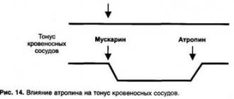
To obtain the most accurate results, before the procedure, the ophthalmologist prescribes a course of atropinization, which the patient undergoes for three days. This procedure consists of daily instillation of an atropine solution twice: in the morning and in the evening. The concentration of the drug is set in accordance with the age group of the subject, but can be changed due to individual factors.
- children under one year of age are prescribed the drug with a concentration of 0.1%;
- in the age group up to three years, the concentration of the drug should be 0.5%;
- Children after three years of age and adults are prescribed a one percent atropine solution.
It is strictly forbidden to start using drops on your own, as this can lead not only to false readings, but also worsen eye health. Another important factor in the success of the procedure is avoiding alcohol a few days before refractometry.
If an allergic reaction to atropine occurs, you must immediately notify the treating ophthalmologist and stop instilling the drug.
How is a refractometer used?
The procedure does not require special preparation of the patient, since it does not involve direct contact with the eye. For bacterial conjunctivitis, it is necessary to rinse the eyes from purulent discharge before the test, otherwise it may distort the data.
During the examination, the doctor can eliminate the accommodation of the pupil using special drugs such as Atropine. These methods allow the doctor not only to conduct refractometry, but also to examine the fundus.
2-3 days before the examination, the doctor prescribes a drug to the person being examined that eliminates the constriction of the pupil under the influence of bright light.
The test takes place in a sitting position, the patient places his chin on a special stopper of the device and fixes his gaze at one point without moving his head. A beam of light is directed into the eyeball and data is obtained, which is processed by a semi-automatic analyzer.
The procedure is not contraindicated even for people with highly sensitive eyes, since it takes no more than 2 minutes.
Indications and contraindications
This procedure is indicated in the following cases:
- blurred vision;
- diagnostics before surgery;
- check after surgery to assess the effectiveness of the intervention.
But, like any procedure, this method has a number of contraindications:
- It is prohibited to conduct examinations of persons with serious mental disorders;
- state of alcohol and drug intoxication;
- cataracts, cataracts and vitreous opacities.
The procedure does not cause complications.
Indications for the procedure
This test tells the doctor whether the patient needs glasses or prescription lenses, and which prescription lenses the patient will see best with.
The results of computer refractometry are used to diagnose the following conditions:
- astigmatism, a refractive problem with the eye related to the shape of the lens that causes blurred vision;
- farsightedness, which is also known as hyperopia;
- nearsightedness, which is also known as myopia;
- presbyopia, a condition associated with aging that causes the eye's lens to have trouble focusing.
The test results can help diagnose the following conditions:
- macular degeneration, a condition associated with aging that affects sharp central vision;
- retinal vessel occlusion, a condition that causes small blood vessels near the retina to become blocked;
- retinitis pigmentosa, a rare genetic condition that damages the retina;
- retinal detachment, when the retina separates from the rest of the eye.
Prevention of visual impairment
A set of preventive measures to restore vision and prevent its deterioration is truly effective if performed regularly. The main difficulty for people whose work involves the use of digital devices is the inability to take breaks, but it is precisely because of eye strain that the blood supply and oxygen supply to the organs of vision are disrupted, and a spasm of the ciliary muscle appears.
How to prevent visual impairment:
- take breaks from work, special programs that remind you of rest will help with this - if the planners’ signals were previously ignored, check in the settings to prohibit canceling the break; such programs darken the screen or cover it with a picture and can even turn on musical accompaniment for relaxation;
- acupressure eye massage, gymnastics - exercises for training accommodation, to relax the eyes are simple and do not take much time, sometimes it is enough to look away from the computer out the window and look at distant objects there;
- moisturizing eye drops - use moisturizing drops that restore the natural tear film, relieve eye fatigue, and protect the eyes from external factors.
Particular attention should be paid to diet. A balanced diet with a varied diet that includes leafy greens, vegetables, fruits, fish and lean meat, as well as taking vitamin supplements, all have a positive effect on the condition of the visual organs and prevent the development of many ophthalmic diseases.
What eye diseases can refractometry determine in ophthalmology?
Refractometry allows you to identify various refractive errors, even at an early stage, for example the following:
- Myopia (nearsightedness).
- Astigmatism.
- Hypermetropia (farsightedness).
If a patient is diagnosed with a disease such as myopia (myopia), this means that the light beam is focused not in the central part of the retina, but in front of it, as a result of which the picture before the eyes becomes blurry. In order to see it, a person has to bring the object closer to a closer distance.
Astigmatism is a condition characterized by an irregularly shaped cornea, causing the eye to lack focus. For normal functioning of the eyes, the surface of the cornea must be spherical and smooth, since in the presence of any deviations, vision will not be clear. This disease is characterized by the fact that the image is focused in front of the retina, and not on it (the contours of objects will be blurry, blurred). Even an ordinary point for such a person will be seen as an oval or a dash.
In people with farsightedness (hyperopia), the light beam is focused behind the central recess of the macula. A blurred image occurs as a result of the fact that the retina has to take on a beam of converging rays.
Myopia and hypermetropia in ophthalmology are included in one term - “ametropia”, which refers to refractive errors of the eyes. Some people may experience a phenomenon called anisotropy - this is a condition when the refraction of each eye is different. Astigmatism is also classified as ametropia.
The worse the patient’s vision condition, the less accurate the data obtained will be, therefore, in addition to this technique, other methods are also used that make it possible to most effectively determine the condition of the eyes. These methods include: touch-skiascopy and skiascopy (the duration of the procedure will be 10 minutes).
When conducting an examination using skiascopy, the doctor is at a distance of 1 meter from the patient. The eye is illuminated using a concave or flat mirror, which helps determine ametropia. The result is determined by moving the skiascope in the horizontal and vertical directions. The results are deciphered as follows:
- if the examination is carried out with a flat mirror, the pupil will move in exactly the same way (in the same direction as the mirror - hypermetropia). If a person’s vision is normal, or there is myopia, there will be a deviation of 1.0 diopters. The presence of pronounced myopia will be indicated by a deviation of more than 1.0 diopters - the pupil moves in the direction opposite to the mirror;
- examination with a concave mirror (the movement of the pupils occurs in the opposite direction), and if there is still no shadow, then this indicates that the patient has myopia of 1.0 diopters.