What is fundus pressure?
The human eye is a complex organ with a well-functioning self-regulation system. Its functioning largely depends on intraocular pressure (IOP), which is responsible for maintaining normal shape and size. This indicator has a constant value, which is measured in millimeters of mercury. This characterizes the force with which the fluids inside the organ of vision press on the eye wall. Even with minor deviations of this parameter from the norm, the functioning of the human optical system is disrupted, which leads to a decrease in the clarity of vision and other symptoms.
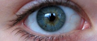
Maintaining normal metabolism in the eyeball is possible only if intraocular pressure is normal. Normally, this parameter varies between 10 and 20 mmHg. Art. Ophthalmologists say that on average it is 15.5 mm Hg. Art. In a healthy person, this indicator is constant and almost never changes. However, slight fluctuations in fundus pressure of up to 2.75 mmHg may occur throughout the day. Art., which is the norm. Immediately after waking up it is slightly higher than in the evening before going to bed. Experts attribute this to the predominance of the parasympathetic nervous system at night and prolonged stay of the body in a horizontal position.
Low eye pressure
Low fundus pressure is dangerous because it does not have pronounced symptoms. The patient experiences a gradual decrease in vision, so often the problem can be detected at an advanced stage. Symptoms such as dizziness, pain in the temples, as well as pain and burning in the eyes are absent.
With a long course of the disease, you can see a visual decrease in the size of the eyes - they lose their shine and become “dry”.
In the most severe cases, the eyeballs become sunken and emergency medical attention is required.
A decrease in eye pressure can be caused by a number of reasons, one of which is a decrease in blood pressure. This is due to the fact that with hypotension, the pressure in the capillaries of the eye drops significantly, which negatively affects the state of the visual system. The problem is also often associated with dehydration, purulent-inflammatory processes of the eyeball (uveitis, iritis, etc.) and infections. In this case, there is a sharp deterioration in vision.
Retinal detachment leads to a decrease in fundus pressure, since this disease is accompanied by a violation of the mechanism of formation of intraocular fluid. Serious injuries and foreign bodies in the eye can also cause this problem.
With severe mechanical damage to the organs of vision, a progressive decrease in IOP indicates the initial stage of atrophy of the eyeball. Less common causes of the disease include ketoacidosis, an acute condition that occurs in patients with diabetes, as well as severe liver disease.
Low eye pressure: symptoms
- Gradual deterioration of vision;
- Visual reduction in eye size with a long course of the disease;
- Recession of the eyeballs (in the most severe cases);
- A sharp deterioration in vision (at the initial stage of optic nerve atrophy caused by severe mechanical damage).
High eye pressure
There are transient and labile increases in intraocular pressure. At the same time, it increases for a short time (several hours) or longer periods (several days).
The most common causes of increased intraocular pressure include hypertension - a general increase in blood pressure, as well as excessive eye fatigue during prolonged work at the computer.
At the same time, the pressure in the capillaries, arteries and veins of the eyeball increases, and an increase in intracranial pressure is often observed.
During violent emotional reactions and stress, as well as when the nervous system is disrupted, a temporary increase in fundus pressure may occur. With some kidney diseases and heart failure, fluid retention occurs in the body, which can also cause an increase in intraocular pressure.
The problem can also be caused by various endocrine pathologies, for example, hypothyroidism (insufficient production of thyroid hormone) and Itsenko-Cushing syndrome (increased production of adrenal hormones).
An increase in intraocular pressure can occur due to drug poisoning, the presence of tumor processes, as well as inflammatory diseases in the body. In all of the above cases, the problem is intermittent (occurs periodically).
Common symptoms of high eye pressure:
- Rapid eye fatigue;
- Decreased vision clarity;
- Pain in temples;
- Severe dry keratitis (dry eye syndrome);
- Dizziness, nausea.
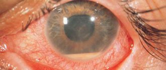
If a patient experiences a persistent increase in IOP, the ophthalmologist will often diagnose glaucoma. This is a serious disease that, if not treated in a timely manner, leads to the destruction of retinal cells, atrophy of the optic nerve, disruption of the flow of visual signals to the brain and complete blindness.
Increased intraocular pressure
Discomfort and vision problems in most cases are caused by increased intraocular pressure. This problem often occurs in older people, but also young men and women, and sometimes even children can suffer from illnesses with such symptoms. The definition of pathology is available only to a doctor. The patient may only notice symptoms that should prompt a visit to a specialist. This will help to cure the disease in a timely manner. How the doctor will reduce the indicators depends on the degree of the disease and its characteristics.
Increased eye pressure - causes
Before prescribing therapy for the pathology, the ophthalmologist must determine the causes of increased eye pressure. Modern medicine identifies several main factors by which IOP can increase:
- a functional disorder in the functioning of the body, as a result of which the secretion of fluid in the organs of vision is activated;
- disruptions in the functions of the cardiovascular system, which cause hypertension and increased ophthalmotonus;
- heavy physical or psychological stress;
- stressful situations;
- as a consequence of a previous illness;
- age-related changes;
- chemical poisoning;
- anatomical changes in the organs of vision: atherosclerosis, farsightedness.
How can you measure eye pressure?
Many people are interested in how to measure intraocular pressure. This procedure, as a rule, is mandatory when undergoing a comprehensive examination of the visual organs in the office of an ophthalmologist. Its principle is based on tracking the degree of deformation of the eyeball under various influences on the cornea. This type of ophthalmological examination is carried out using a special device - a tonometer. As a result, the doctor receives data in a generally accepted unit of measurement (millimeter of mercury).
Various techniques are used to perform eye tonometry, three of which are most actively used: digital, non-contact and contact.
The finger method involves determining the tension of the eyeball by feeling it with the fingertips of an ophthalmologist. It does not guarantee a reliable result and, as a rule, is carried out in the postoperative period, when the eyes cannot be subjected to instrumental influence. It is not suitable for those who need to accurately determine eye pressure.
The non-contact method is based on studying the speed and degree of deformation of the cornea in response to the influence of air flow. The data obtained as a result of the study is instantly processed on a computer, and within a couple of seconds the patient can see the result. Measuring eye pressure in this way is convenient because local anesthesia is not required and there is no risk of complications.
What research methods are there?
- Finger method (feeling the tension of the eyeball by an ophthalmologist);
- Non-contact method (study of the speed and degree of deformation of the cornea in response to air flow pressure);
- Contact method (contact of the tonometer with the surface of the organs of vision).
How to determine eye pressure using the contact method?
Since this method involves direct contact of the tonometer with the surface of the organs of vision, in order to avoid pain it is necessary to additionally use anesthetic drops. The results of studies conducted by this method are as accurate and reliable as possible. They are suitable for professional diagnostics and expert assessment. Contact tonometry has several subtypes. In each specific case, a different technique is used, corresponding to the individual characteristics of the patient’s eyes.
Types of contact tonometry:
- Impressive. Involves the use of a Schiotz or Icare tonometer. It is based on soft indentation of the cornea using a special rod (plunger). This type of tonometry is quite painless and quick, so it is often used when examining children.
- Applanation. For measurements, Maklakov weights or a Goldman tonometer are used. These methods have a common operating principle. A small weight moistened with paint is placed on the eye. Next, its imprint is made on paper and special measurements are taken. The higher the fundus pressure, the less ink will remain on the paper.
- Dynamic. This type of tonometry takes into account the individual characteristics of the blood supply to the patient’s visual system, but is somewhat inferior to previous methods in the accuracy of measurements.
Diagnostics
Let's figure out how to check eye pressure. There are several effective ways to measure intraocular pressure. Almost all of these techniques require consultation with a specialist.
A palpation-orientation technique is often used . The doctor approximately determines the level of pressure by the level of resistance of the eyeball when pressing on it with your fingers.
The patient lowers his gaze, the specialist fixes his eyes with one finger, and presses the apple with the other. Measures the density of the sclera, it should deform.
With hypotension (low blood pressure), the sclera is soft, normal, moderate. With increased IOP - stone.
This is the most gentle technique that can be done at home. However, you should first consult an ophthalmologist.
Tonometers
Measuring IOP with a tonometer is an important procedure performed by specialists. This is a necessary test when assessing the condition of a patient's eyes that are at risk for glaucoma.
The most common device is Maklakov tonometer . Refers to applanation apparatus. Anesthesia is carried out first, as the sensations will be unpleasant.
This is a cylindrical device with glass plates on which a dye is applied. Ocular pressure is measured using special weights.
Research algorithm:
- the areas of the device are wiped with alcohol, then with a sterile swab;
- special paint is applied;
- Dicaine solution is dripped into the eyes;
- the patient's gaze is fixed on the index finger, as the weight should fall on the corneal center;
- A colored plate is lowered onto the cornea, an unpainted spot appears on the plate;
- the cornea is flattened, the paint is washed off with a tear at the site of contact with the platform;
- a light circle remaining at the point of contact is imprinted.
The size of the spot depends on the degree of flattening of the cornea, indicating the amount of pressure. If the contact area is large, the eye is soft, the pressure is low. The measurement is repeated with the opposite plate. The readings are measured with a ruler on paper.
The Goldmann tonometer is a more modern method of applanation tonometry . The device is mounted on the slit lamp. A prism is installed on it.
Anesthesia is given, a fluorescein solution is instilled, and a prism is applied to the cornea. Its pressure on the cornea is carefully adjusted. It is flattened until the colored half rings meet at one point. IOP is determined using the instrument scale.
The operation of the impression tonometer is based on the Schiotz method . Pressure is measured by pressing a rod of a certain mass onto the cornea. The method also requires pain relief.
A piston with a weight is placed on the eye. The force of the IOP moves it, deflecting the needle on the scale. The readings are checked against the values of the calibration tables.
Dynamic contour tonometry is carried out with a Pascal tonometer . Measurements are taken without flattening the cornea.
The device is mounted on the optical axis of the slit lamp. The doctor sees the contact boundary between the cornea and the tip of the device, which determines the pressure.
A pressure sensor is built into the tip. It is designed to generate an electrical signal. The higher the beep, the higher the pressure. It is listened to for 5 seconds.
How to measure blood pressure at home?
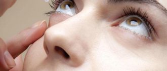
People often ask on forums how to measure fundus pressure at home. To obtain reliable data, modern high-precision equipment is required. However, you can approximately control the intraocular pressure reading yourself. The technique is quite simple. It is necessary to feel the eyeball through closed eyelids with a fingertip. To evaluate the result, you should carefully apply slight pressure.
With normal fundus pressure, you will tactilely feel the eye like an elastic ball, which is slightly pressed under external influence. If the organ of vision is excessively hard, like a stone, and is quite difficult to deform when pressed, which means there is a risk of increasing IOP. Conversely, if your finger easily “falls in” and you cannot feel the spherical shape of the eye, this may indicate a significant reduction in eye size.
It is only possible to accurately measure fundus pressure in a specialized ophthalmology clinic. You won't be able to do this at home.
If you use contact lenses, we recommend that you familiarize yourself with the wide range of products in the Ochkov.Net online store. Here you can purchase goods from world brands at competitive prices!
Portable blood pressure monitors
For people suffering from severe glaucoma, it is important to determine ophthalmotonus several times a day. How to measure eye pressure at home without visiting a doctor every time? There are special portable devices for this. They determine IOP by contact method.
The ICare device is often used to measure eye pressure at home. It has replaceable sensors that need to be momentarily applied to the cornea of the eye. Since the sensor is disposable, infection is practically excluded. A short contact time ensures that the procedure is painless and can be done independently or with the help of loved ones.
Important! The pressure inside the eye is constantly changing. It depends on breathing, heartbeat and other factors. Therefore, for more accurate readings, it is advisable to take several readings at a time, and take their average value as the true one.
The amount of intraocular pressure is an important indicator of the health of the visual organ. It allows us to judge the presence of a disease such as glaucoma, which constantly progresses and, if left untreated, leads to blindness. With regular violations of ophthalmotonus, it is important to determine their cause. Perhaps some chronic disease contributes to this. Then treatment of the underlying disease will help normalize the pressure inside the eye.