Before diagnosing any problem in the body, you should analyze its condition. In some cases, you need to undergo a series of tests and take other measures, but in the field of ophthalmology, primary diagnosis is much simpler.
Every person has encountered vision tests at least once during a medical examination. You can arrange such a check not only in the ophthalmologist’s office, but also in your own home.
Before you do any of the tests, remember what vision problems you have encountered. Do you suspect farsightedness, nearsightedness or astigmatism? Choose the appropriate test. And do not forget that the test should be carried out when you are feeling well.
Vision test. Eye test at home
Checking visual acuity at home
You are probably familiar with Sivtsev’s table, because it is the most relevant and widespread. Its main purpose is to test visual acuity. With its help, it is easy to calculate the capabilities of each eye.
The tablet has twelve lines. They contain several identical letters arranged in a chaotic order. Its standard format is two hundred and ninety-seven millimeters in width by four hundred and eighty millimeters in length.
This test is easy to use:
- Draw or print the sheet so that the format is identical to the required one.
- Attach the resulting test to a flat and preferably plain, discreet surface. The position of the middle of the table should coincide with eye level.
- Move five meters away from the leaf.
- Try to read the third line from the bottom.
- Close one eye at a time and read the indicated line again.
Sivtsev table for vision testing
Have you noticed that one eye sees much worse or you couldn’t see the line at all? You should consult a doctor!
It is not so easy to check children's vision using this table, since children may not know the letters. For this case, there is another test with small images. The essence of checking children's vision is simple - you should print the attached plate in the same format as the previous one and arrange it in the same way. Ask your child to describe the objects seen in the third line from the bottom.
Table for checking children's vision
In some situations, neither one nor the other test option for checking visual acuity is suitable. We are talking about determining improvements in vision during its correction. The problem is that letters and symbols are remembered. The solution is to create the tables yourself.
Below is an example of such a table. Change the size of the letters so that they are arranged from smallest to largest from top to bottom, print the test, stick it on the wall, determine for yourself a point where the bottom third of the chart will be difficult to see, and evaluate the results by taking the test weekly. If you memorize letters, change them.
Example of a vision test table
You can use not only letters, but also symbols and single digits. The main thing is that their color is the same (black) and their sizes correspond to the sizes of the letters in the initially created table.
Degree of disease by number of diopters
There are several degrees of the disease, characterized by a certain number of diopters that are necessary for complete restoration of vision:
- weak degree (I) - 1.5-2 diopters ;
- average (II) - 3.5-4 diopters;
- high (III) - above 4.0 diopters.
If a weak degree can be restored by correct refraction, then for a medium and high degree additional correction is needed.
A mild degree occurs in children under 18 years of age and does not require surgical intervention.
People over 45 often feel a lack of diopters, and to correct the optical system they use laser vision correction if recommended by a doctor.
Medium and high degrees of farsightedness are dangerous because they lead to complications such as strabismus , and in older people, cataracts or the initial stage of glaucoma . Moderate and high degrees of strabismus are not treated until vision is completely restored. With vision correction you can only reduce the number of diopters.
Dioptres is the optical power of a lens. Diopter is measured as one meter divided by focal length. A + or - indicates the strength of the scattering system. This calculation helps to understand the quality and degree of refraction.
Vision index 1, what does it mean?
It is generally accepted that eyes with an index of 1 can distinguish between two separate focal points with an angle between them of 1.6 degrees . This means that vision is 100 percent , and any deviation requires further diagnosis.
Calculating diopters using cycloplegia determines the extent of the disease and the possibility of restoring vision. The system helps compensate for defects of accommodation and its treatment. Diopters help in determining the exact focal length and actual visual field of a person.
How to recognize color blindness?
It is worth recalling that color blindness is the inability or poor ability to recognize tones and colors. The disease is classified as congenital and hereditary. Degrees of color blindness can vary. Some people are extremely poor at distinguishing color variations of objects, while others simply do not understand shades, considering them to be one color.
There are a great many tests for color blindness. Any of them is a picture consisting of roundness of different shades. The roundness of certain colors forms numbers that must be recognized.
Color vision chart for color blindness
Before checking, it is advisable to print the picture. Place the image in front of you and try to understand which numbers form the circles. At the same time, it is strictly forbidden to squint and lower your head.
In the case when all the numbers can be seen, there is no need to worry about the problem of color blindness. But if you saw only contrasting images or nothing at all, you have problems with color perception.
Definition
Color blindness (color vision test) - Also known as color blindness, this is a condition that affects the perception of color. This is due to the fact that the eye is missing one of the three pigments, the combination of which gives all the variety of colors. The test comes after a short section of theory. This can be verified using a very common graphic format - RGB, which stands for "Red, Green and Blue", that is, "red, green and blue". These same colors are found in our eyes. Typically, color blindness is caused by genetic changes that affect the ability to perceive individual colors and their shades. However, acquired color blindness is also common. It can develop due to diseases affecting the optic nerve or due to injury to the eye. Also, simply age-related changes contribute to the development of such a condition.
It is worth noting that color blindness may be a clear prohibition for certain types of work.
For example, until recently, it was forbidden to drive a vehicle if color vision was impaired. There is a ban on people flying an airplane. This is due to the fact that in the cockpit there are many multi-colored sensors that need to be clearly visible.
However, one hundred percent vision is required not only for pilots, but also for many other professions. For example, this applies to some types of production, as well as for electric train drivers. Particularly stringent requirements for health in general, and for vision in particular, are imposed in the metro, where color vision tests are carried out annually.
What is used for diagnosis
In order to check your color perception, you can get tested by an ophthalmologist. It is quite simple and does not take much time, but it allows you to accurately determine whether you have color weakness. To determine color blindness, Rabkin tables, sometimes called Ryabtsev tables, are used. Typically, this is a book that contains a set of pictures. They contain circles of different sizes and colors, which make up different numbers or geometric shapes.
Each picture has a specific combination of colors of the same brightness, which will accurately determine whether you have color deficiency. If perception is impaired, a person will see either a jumble of circles or incorrect symbols that are also encrypted there. This test also consists of two parts. The first allows you to understand whether there is a vision anomaly, and the second serves as an auxiliary one. In the second part of the test, images are selected that will indicate which color you have problems with perceiving. In addition, Yustova’s tables are no less popular. They allow us to conduct threshold studies of color vision and determine which shades are perceived with the greatest difficulty. This is necessary for a more accurate examination.
However, it is possible to undergo a similar examination on the Internet.
Now there are many sites on which the above tables are posted. Some of the resources offer a survey in the form of a test, where you need to choose one of several answer options. Others lay out tables under which the correct answers are indicated, with explanations.
However, it is worth noting that online testing is not always reliable. This is due to the fact that the monitor is not always correctly configured for color reproduction. In addition, there may be malfunctions in its matrix, which also distorts the perception of colors, and therefore makes the test unreliable. However, with the help of such tests, you can get an idea that something is wrong with your vision, which means it’s time to see a doctor.
Some people, in order to get a desired position, enter queries into a search engine, such as “test with answers” , in the hope of learning the correct answers to tests. However, with such scams, they expose themselves and others to danger. If you managed to get a physical copy of the tables, or cards, then you can take the tests yourself. To do this, you should use the following rules, which also apply in the ophthalmologist’s office:
- You should sit with your back to the window, and the tables should be opposite you.
- The distance to the tables should be one meter.
- Keep them at the same level as your eyes, vertically.
- It takes from five to seven seconds to view the image and answer.
It is worth remembering that you should take the test when you are in good health, as illness or fatigue can affect the clarity of your vision. Also, you need to relax before starting testing - nervousness is not the best assistant in this matter.
Test for macular degeneration
Macular degeneration refers to problems with the retina of the eye. You should consider this test if, when focusing your gaze, the remaining picture appears distorted, or if there are other vision problems.
Testing is carried out as follows:
- Print the attached table. The format does not play an important role, but it is desirable that it fills most of the A4 sheet.
- Attach the sheet to the wall so that the center point of the image is at eye level.
- Move back twenty-five to thirty centimeters.
- Look at the entire image first, and then try to focus your eyes on the small black circle in the center.
Test for macular degeneration
Now it's time to evaluate the results. Were all the square shapes the same size? Were all the lines straight? If there are no problems, the answer to both questions should be yes. But if the retina of the eye is damaged, distorted visibility of the image is possible.
If the case is neglected, gray spots may appear in the field of view, and square outlines will take on completely different shapes when focusing on the circle. In this situation, you should visit an ophthalmologist as soon as possible.
Last problem
And finally, the last stage of our attentiveness test is a humorous problem. Find 8 differences in this picture.
Answer
In one picture we see a spider with legs, in the other we see a black ball (or a potential spider carcass without legs). As you know, a spider has 8 legs, hence the 8 differences.
[collapse]
We hope that our test in pictures left you with a pleasant impression and that you completed all the tasks. If they caused you difficulties, read our article “How to become more attentive?”
Finally, we invite you to watch one well-known social advertisement, which also contains an attentiveness task.
Checking the contrast of vision
Golovin’s tablet is very similar to Sivtsev’s table. The difference is that the first test helps to identify not only the acuity, but also the contrast of vision.
Golovin's table is twelve rows of roundness with a gap. Your goal when testing is to see breaks in the shapes in a certain row. People with normal contrast vision will be able to easily see the gaps, and with low contrast, the outlines of small figures will appear continuous.
Golovin table for testing visual contrast
The instructions for using the test are simple:
- We print the plate in a format of two hundred and ninety-seven millimeters in width by four hundred and eighty millimeters in length.
- We attach the table at eye level.
- We move away from the test five meters.
- Let's recognize breaks in the figures from the third row from the bottom. First, we look at the image with both eyes, and then cover each one in turn.
Couldn't see the location of the breaks? This means that the visual contrast is below normal.
Landing
- The organs of vision, which are located in a certain depression, are called deep-set. They can be visually brought forward if you use warm shades of shadows on the upper eyelid, and dark shades a little higher.
- Eyeballs, which have clearly visible spherical contours, are convex. Sometimes it seems that a person is surprised or is looking closely at you, but in fact, for those with bulging eyes, this is a normal facial expression. Their owners should use dark and medium shades of eyeshadow on both eyelids.
- If a person does not show signs of deep-set and protruding eyes, then he has normal eyes.
Is it easy to detect myopia or farsightedness?
Myopia is difficulty recognizing objects that are far away, while farsightedness is difficulty recognizing what is close to the eyes.
With myopia, it is possible to focus the gaze on everything that is close, but with distant objects, focusing is difficult or impossible, since the picture appears blurry.
With farsightedness, it is possible to focus your gaze on both distant and close objects, but what is close will have blurry outlines.
Table for testing vision for nearsightedness and farsightedness
Have you ever noticed something like this in yourself? Check it out, because the test is very simple. You just need to print the attached picture in the maximum format on an A4 sheet of paper on a color printer. Hang it on a plain wall and try to determine which color the letters appear brightest on.
The brightness of the letters on a scarlet background indicates farsightedness. Did the letters seem clearer in bright green? You are probably nearsighted. In the case where there are no deviations, the brightness of the letters will be identical on each of the color canvases.
Indications
The eyes quickly get tired and watery.
Basically, the disease manifests itself from school age at the time of active growth of the body, and begins to progress from 19-25 years. By the age of 27, the process remains more or less stable and becomes active again after reaching the age of 42-45. There are general reasons for testing for myopia, these include:
- headache, especially in the frontal and temporal parts;
- squinting when looking at a distant image;
- fatigue in the eyes when reading, writing, working at the computer;
- blurry picture at a distance.
What are the tests for astigmatism?
Astigmatism is a refractive error of the eye. The consequence of pathology is complete or partial distortion of the image. It consists of blurred outlines and duplication. This vision defect is often accompanied by other problems (myopia, reduced visual acuity, etc.).
Astigmatism test. Table 1
Attention: before testing for astigmatism, you should be very careful about how you feel. Their specificity can indicate the presence of astigmatism in a person without vision problems, if he is tired, stared at the monitor screen for a long time, or is thirsty for sleep.
Attached below are two test images. The check should be carried out as follows:
- Print out the tests on A4 sheets of paper. You can choose another format, but not smaller.
- Hang them on the wall at eye level. You need to move a short distance.
- Cover one eye at a time, and then look at the picture with both eyes.
Astigmatism test. table 2
The result that a person without astigmatism should get is immobilized, identical, straight lines. There should be no blurriness, no movement, no different sizes. For any irregularities and similar deviations, it is advisable to visit an ophthalmologist, especially if you have been diagnosed with other vision defects.
Myopia and causes
The inability to focus the picture or seeing it in a blurry, fuzzy state suggests that the patient has myopia. In this case, a defect occurs with the image protruding in front of the retina. Vision problems occur at any age and for various reasons. Eye diseases are divided into 2 main types:
- Congenital: Genetic predisposition. The disease is inherited and its manifestations are possible both from infancy and into adulthood.
- Injuries, bruises. Due to injury to the mucous membrane of the eye, pupil or eyelid, an immediate or gradual (from 1 week to 6 months) decrease in vision occurs.
You need to undergo diagnostics once every 6-12 months. This will help to identify the disease at an early stage and avoid the consequences of a severe course.
Wide-set eyes
People with such eyes are attracted to the big picture, and are not interested in small details and nuances. Excessive fuss, sudden movements or presentation of an ultimatum are not their style. You have to try very hard to piss off such a person. Regardless of age, he tends to always remain a child. He derives a sense of reliability and security from his family. Having such support behind him, the “wide-eyed” takes risks without looking back, and is ready for any adventure. These types of eyes are characteristic of cheerful and optimistic natures. They know how to enjoy life and enjoy the feeling of freedom.
Structure of the eye
2
Where do eyes get a certain color? The eyeball consists of a nucleus and surrounding membranes - outer, middle and inner. The middle membrane is divided into 3 components: the anterior, iris and pupil, the middle - the eyelash area and the posterior - vascular. It is the iris that has a certain color. It depends on the presence of pigment and on the level of light reflection. If there is little pigment, then the eyes are blue, if there is a lot, then brown. Residents of the northern regions often have light eyes, while residents of the middle zone and south often have dark eyes. Only the Yakuts and Nenets are exceptions. They have dark eyes and hair. This happened because the eyes have to protect themselves from the brightest light reflected from the ice. That's why their eyes have a lot of pigment. In children, eye color is established by 2-3 years. They are born blue-eyed.
Binocular vision test
In the absence of ocular anomalies, a person sees not only the boundaries of surrounding objects, he evaluates their size, color, depth, distance or proximity. The world around us would not be bright and voluminous if people did not have binocular vision.
There are different types of binocular vision tests.
- Sokolov's experience. To perform this, you need a “spyglass” - any object rolled up in the shape of a tube. Such a tube is brought to the eye, and the palm of the second hand covers the other eye. With binocular vision, a hole appears in the palm through which surrounding objects will be visible.
- Kalf's experience. Two pencils are placed one vertically, the other horizontally. The test taker is asked to hit a horizontal pencil with a vertical pencil. With binocular vision, such an experience will pass without difficulty; the person evaluates the size of the pencil and the degree of their distance from each other.
- Reading experience. You will need a piece of paper with text and a pencil, which is placed in front of the eyes and removed 2-3 cm from the tip of the nose. When reading, you cannot move the text, move your head or hand. With binocular vision, the pencil will not interfere; the images from both eyes will merge into one.
5 / 5 ( 8 votes)
Asian eye type
Slanting eye shape is most often found in people of the Mongoloid race. They have such a structure of their skull that there is no clearly defined frontal bone. Because of this, there is no fold in the upper eyelid. However, it would be wrong to say that this shape is unique to Asians, since you can find a large number of Europeans with the same cut.
According to psychologists, the distinctive features of people with this eye shape are tolerance, kindness, sentimentality and the desire to care for others. They are so sensitive that even seemingly frivolous events touch them.
This type is considered the most common. Psychologists talk about the determination and success of the owners of such eyes. However, despite all their strength of character, these people do not like to burden themselves with responsibility. If there is a need to make a decision that will entail significant changes in life, then it will be very difficult to make. Therefore, they never realized many great goals.
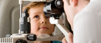
Effective methods that will help you independently identify heterotropia in children
You can detect strabismus in a child at home using two tools: a flashlight and a camera. What are they needed for? You need to shine a flashlight into the baby’s eyes and watch the reflection. If the reflection in the pupils of both organs of vision is the same, you can relax - this indicates the absence of strabismus. A different reflection indicates the presence of a vision defect.
An equally effective way to detect heterotropia is photography taken with a flash. To determine from the photo whether the baby has this pathology, you should carefully look at the glare from the flash - when the baby has no vision problems, the glare in both eyes is the same.
The development of strabismus in young children is indicated by the inability to focus on certain objects. While watching the baby, parents may notice how he periodically tilts his head, trying to look at this or that object, and rubs his eyes at the same time. These symptoms should not be ignored, as they may signal the onset of the development of the disease!
Determining strabismus in young children is not always easy.
Fact: A newborn may have slight strabismus in the first six months of life for the simple reason that the child is not yet able to physically control both eyes at the same time. After this period, the effect of the deviation goes away, but at the beginning it is insignificant.
If strabismus does not go away in a baby even at one year of age, or it is initially pronounced, you need to contact a pediatrician, who will refer you to an ophthalmologist.
The most difficult thing to identify in a child is hidden strabismus, especially in the first 2 years of life.