When the corneal tissue becomes bent or clouded, the light rays become distorted. The image of objects on the retina is distorted, visual acuity decreases up to absolute blindness. Pathological processes are irreversible; correcting vision with lenses and glasses is ineffective. In many cases, only transplantation can eliminate corneal problems.
Cornea transplantation is a microsurgical intervention aimed at removing diseased tissue, eliminating various pathologies and restoring vision. Through surgery, the altered corneal areas are replaced with healthy donor tissues; the flap can be placed only on the superficial layers or on the entire thickness of the corneal tissue. Penetrating keratoplasty allows you to restore visual acuity, improve your appearance, eliminate opacities and other pathologies. Every year tens of thousands of such transplantations are performed around the world.
In Moscow today, ophthalmological interventions of varying levels of complexity are carried out. Russian surgeons successfully transplant endothelium, perform lamellar keratoplasty, and successfully correct congenital and acquired eye pathologies.
End-to-end corneal transplant - 50,000 - 90,000 rubles.
Use of a biological product for cornea transplantation—RUB 120,000.
(duration of operation)
Indications
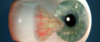
- Progressive keratoconus and keratoglobus, when the cornea is very thin and cone-shaped.
- Injuries and other defects of corneal tissue.
- Congenital abnormalities of eye tissue.
- Post-traumatic scars of the cornea.
- Various corneal dystrophies, as a result of inflammation, operations, etc.
- Fuchs' dystrophy, when the endothelium of the cornea is destroyed.
- The appearance of a cataract (clouding) of the cornea.
- Infectious lesions and ulcers of corneal tissue of a fungal, viral, bacterial or parasitic nature.
- Bullous keratopathy, manifested by corneal edema.
Contraindications
- Acute inflammatory diseases of the eye (blepharitis).
- Eversion or inversion of the eyelid.
- Acute infectious diseases.
Types of keratoplasty
- end-to-end corneal transplant;
- anterior and posterior layer-by-layer transplantation of corneal tissue;
- replacement of the inner layer of the cornea;
- keratoprosthesis.
End-to-end corneal tissue transplantation is the most common type of surgery. Performed for keratoconus, various injuries, burns and degenerative changes. A round trephine is used to excise the entire thickness of the damaged tissue and replace it with a donor graft. Healthy corneal material is cut out in the same way. Stitches can be removed after 6 months.
Treatment and restoration of the cornea of the eye
How is the cornea treated if a foreign object gets into it? First of all, treatment should only be carried out by a doctor. Turning to traditional medicine is possible in consultation with your doctor. A huge number of complications, including loss of vision, are suffered by people who self-medicate and wash their eyes with inappropriate means. As a result of a foreign object entering the eye, the integrity of the cornea is disrupted and erosion occurs. The depth and shape of the wound depend on what object caused it.
Damage to the corneal epithelium is a very common eye injury, especially in children. Erosion is often complicated by the addition of an infection, in which case an inflammatory process with suppuration develops. The sooner you see a doctor, the fewer consequences this eye damage will have.
small items
Foreign bodies can be located deep and on the surface of the cornea. The doctor must remove foreign objects in order not to cause even more harm to the victim. If grains of dust, metal or other substances remain in the cornea for a long time, an inflammatory process begins and traumatic keratitis occurs. Getting metal shavings into your eyes is especially dangerous because metals such as copper, lead and iron oxidize easily. Suppuration occurs around the site of penetration. Naturally, the eye is deprived of foreign objects, pushing them to the surface of the cornea. The victim of such an injury feels:
- sharp pain in the eye;
- feeling of dryness and unevenness of the eyeball;
- burning and itching;
- increased tearfulness.
The eye turns red and the eyelids swell. Due to the large mass, small dispersion of glass, fragments of stone or gunpowder may not rise to the surface layers of the cornea on their own. The reaction to them may not be so bright, but the consequences in any case will be very dangerous. For diagnosis, the ophthalmologist uses:
- visual inspection to determine the nature of the damage;
- focal lighting;
- instillation of a 1% fluorescein solution, which allows you to see the edges of the wound.
Often victims cannot look at the light, close their eyelids and experience intense pain. For anesthesia the following are used:
- 0.5% dicaine solution;
- 4% lidocaine solution;
- 0.4% solution of oxybuprocaine.
The doctor then removes any accessible foreign bodies using a cotton pad or swab. If possible, deeply located particles are removed as they rise into the upper layers of the cornea. If particles of a substance pose a hazard to the eye. For example, if it is metal dust, then removal must be carried out promptly. If metal is attracted to a magnet, a magnetic field is used to lift the particles. If foreign objects cannot be influenced by a magnet, layer-by-layer extraction is carried out under local anesthesia using a needle or a special spear. To prevent infection from getting into the wound, antibacterial agents are instilled:
- 0.25% solution of chloramphenicol;
- 20% sodium sulfacyl solution.
If it is possible to remove all foreign particles, the patient is treated at home; if this fails, hospitalization is carried out to monitor his condition. The main danger of corneal erosion is that without proper help, an inflammatory process easily develops, festering ulcers appear on the epithelium, and necrosis begins. In order to stimulate the regeneration process, ophthalmologists use:
- 1% solution of emoxipine;
- 5% ointment Korneregel;
- 20% solcoseryl gel;
- 20% Actovegin gel.
If an inflammatory process has begun, the attending physician may additionally use antibiotics, for example, a 4% solution of gentamicin sulfate or a 30% solution of lincomycin hydrochloride. The dosage and frequency of administration of all drugs are selected individually. Self-treatment of the cornea of the eye can pose a threat to the victim’s health. Recovery time depends on the severity of the damage and the presence of complications; in mild cases it is 1 week, in severe cases it is several months.
large objects
For injuries caused by large objects, restoration of the cornea may be necessary. More dangerous are penetrating injuries, in which a foreign body remains in the eye. Without immediate help, purulent inflammation may develop, which, together with prolapse of the membranes of the eye, represents a severe case. When providing first aid if large objects get into the eye, blood clots should not be removed, as the membranes of the eye may be mistakenly touched and fall out. Treatment of severe corneal wounds takes place only in a hospital.
For relatively small wounds, conservative therapy is possible. To prevent infection, the wound is temporarily covered with a lens. If the wound is large in area, an operation with suturing is performed. Sutures can bring the edges of the wound together on the surface of the cornea or penetrate through its thickness for a tighter grip. If the iris falls out during a wound, then until a purulent infiltrate has formed, you can set it back by pre-treating it with a broad-spectrum antibiotic. The operation is called iridoplasty, and sutures are used to reattach the iris. To prevent complications, many anti-inflammatory drugs are prescribed in the form of:
- eye drops;
- eye ointments and gels;
- tablets;
- injections;
- rectal suppositories.
The dosage and name of medications are selected by the doctor taking into account the clinical picture. Properly organized and timely treatment of the cornea of the eye in most cases allows the victim to preserve his vision.
However, despite all the achievements of ophthalmology, there is a possibility of complications, especially if the foreign body that got into the eye was metal.
Complications after injury
The addition of a bacterial or fungal infection leads to the development of a purulent inflammatory process in the eye; without immediate help, tissue death and spread of inflammation to neighboring areas are possible. The eyes are located very close to the brain, and there is a direct connection between them. The infection can cause meningitis and death in a short time. In the inpatient department, the inflammatory process can be quickly stopped. Other common complications after a foreign body enters the eye:
- decreased vision;
- post-traumatic glaucoma;
- traumatic cataract;
- retinal detachment;
- endophthalmitis;
- vitreous prolapse;
- panophthalmitis;
- fibrinous plastic iridocyclitis.
Treatment of these complications can be lengthy and painful; successful results do not occur in all cases. Glaucoma and cataracts often occur in old age, when quite a lot of time has passed since the injury.
First aid
If foreign objects get into the eye, rinse it with water, removing only the particles that are on the mucous membrane. You cannot remove foreign objects yourself using needles, toothpicks, nails or other scratching objects. If bleeding occurs as a result of the injury, you should treat only the outside of the eyelid, do not remove blood clots from the eye, and do nothing until the ambulance arrives. For pain relief, you can only use an ice compress, which is applied through the tissue in the area near the eye, but not directly on the eye. There should be no pressure on the eyeball, no rubbing of the eyes, and no close closure.
Treatment of the cornea of the eye should be carried out only under the supervision of an ophthalmologist; it is unacceptable to practice traditional medicine methods, especially on children. After washing your eyes, you should visit an ophthalmologist, even if the pain and burning have gone away. In some cases, inflammation does not develop instantly, but within 1-2 days. During the inflammatory process, the eyelid swells, redness appears in the eye, blurred vision and discomfort when blinking are possible. The sooner the victim goes to a medical facility, the greater the chance of a successful outcome.
Keratoplasty technique
In Moscow, a microsurgical operation takes place in a one-day hospitalization regime. The patient is positioned on the operating table, and a special eyelid dilator is placed on the eyes to prevent involuntary blinking. Depending on the scope of the proposed work, the age and individual characteristics of the person, the ophthalmologist prescribes general anesthesia or decides that local anesthesia is sufficient. Regardless of the chosen method, the patient does not feel pain during the manipulations.
During the intervention, a microsurgical instrument and an operating microscope are used. The surgeon determines the diameter of the damaged corneal flaps and separates the fragments using a surgical trephine and other instruments. Healthy donor tissues are placed on the prepared site. The flaps are sutured to the patient's cornea using thin suture material.
Keratoplasty ends with revision of the established graft. The ophthalmologist evaluates the uniformity of the location of the fragment, the evenness of the corneal surface, etc. If necessary, manipulations are performed to align the shape of the cornea. To prevent inflammatory processes, glucocorticosteroids are injected under the conjunctiva of the eye. To protect against damage, a bandage or protective lens is placed on the operated eye.
Symptoms of Corneal Transplant Rejection
The following ophthalmological symptoms appear:
- new precipitates are visible on the posterior surface of the cornea;
- a line of precipitates appears on the endothelium (Khodadoust line, or endothelial rejection line);
- there is swelling of the stroma;
- subepithelial infiltrates appear;
- an unevenly elevated epithelial line or local neovascularization is visualized in combination with an infiltrate in the stroma.
Other signs of rejection are also noted:
- conjunctival pericorneal injection;
- symptoms of inflammation in the anterior chamber;
- graft suture rupture;
- neovascularization that extends toward or grows into the graft;
- lacrimation.
If corneal graft detachment is suspected, there is a need for differential diagnosis with the following diseases:
- Suture abscess or infectious process in the cornea of the eye. There may be a corneal infiltrate, purulent discharge, or hypopyon. In this case, you should remove the suture and take smears and cultures, including cultures from the suture. Slowly reduce the frequency of steroid use. Intensive local therapy is carried out with fluoroquinolones or enhanced antibiotics.
- Uveitis. With this disease, cells are found in the moisture of the anterior chamber. There is turbidity of the moisture and the appearance of precipitates on the endothelium. Even if there is a history of previous uveitis, this condition is treated in the same way as graft rejection.
- Intraocular hypertension. If intraocular pressure increases significantly, swelling of the corneal epithelium may develop. There will be no other signs of graft rejection. After reducing intraocular pressure, the swelling disappears without a trace.
Failures during keratoplasty can occur for other reasons:
- recurrence of the disease in the graft (herpetic keratitis or corneal dystrophy);
- decompensation of the endothelium in the graft.
If corneal graft rejection is suspected, the patient is examined:
- Anamnesis collection. They clarify when the corneal transplant was performed, what medications the patient receives as local therapy, whether the regimen of corticosteroids has changed recently, what were the indications for corneal transplantation.
- Slit lamp examination. The ophthalmologist looks for endothelial rejection lines, subepithelial infiltrates, and precipitates on the endothelium.
Reviews of doctors providing the service – Keratoplasty
When illness strikes, it is very important to find a highly qualified, caring doctor.
Tatyana Igorevna is exactly such a doctor! Behind each of her recommendations is enormous knowledge and experience, as well as respect for the patient’s personality. This is what gives hope for the best! THANK YOU. Read full review Svetlana
06.09.2019
Vladimir Yuryevich Makhmutov is a doctor of rare talent. Out of this world! Talent, kindness, courtesy, attentiveness. Skillful fingers. Many thanks and low bow. Shchedrova E. Read full review
Shchedrova Ekaterina
25.03.2019