Briefly about the procedure
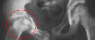
Biometrics is a procedure that helps a specialist study the structure of the eye in more detail and detect the presence of possible vision pathologies. This diagnostic method allows doctors to measure the thickness of the cornea and lens, find out the length of the axis of the eye, and the size of its anterior chamber. Using the procedure, you can monitor how the eye recovers after surgery, as well as determine the refraction of the cornea. Biometrics allows us to study in more detail the structure of the iris.
This is a modern diagnostic method that can replace several other studies at once, including keratometry, pachymetry and pupillometry. The procedure does not cause discomfort or pain to the patient, since the equipment with which it is performed does not irritate the optical media of the eye. It can be done for both adults and children.
Biometric results are much more accurate than alternative research options, which helps achieve better results from treatment or surgery. The procedure can also be carried out for preventive purposes in order to detect the development of vision pathology in time. There are two variations of this study: ultrasound and optical.
Optical and ultrasound biometrics
Biometry is the study of the parameters of the eyeball: the axial length of the eye, the thickness of the cornea, the depth of the anterior chamber, the thickness of the lens, etc.
This examination method is used before cataract surgery, to calculate the strength of an artificial lens, before excimer laser vision correction and the selection of contact lenses and glasses. Biometrics is quite simple to perform, painless and provides the doctor with the necessary information to choose methods of vision correction. We offer two types of biometrics - optical and ultrasound.
How to prepare
No specific preparation is required for biometrics. It is not recommended to use alcohol, tobacco products or drugs before the study. Women should avoid wearing makeup around the eyes. You should not wear contact lenses on the day of the procedure.
Indications and contraindications
Although biometrics are also carried out for preventive purposes, doctors often prescribe it if the patient has any pathology. So, you should undergo it if there is a sudden deterioration in vision, age-related macular degeneration, or diabetes. Biometrics must be carried out in case of foreign objects getting into the eyes or various injuries to this organ.
With its help, you can find out if a patient has glaucoma and identify the progression of myopia. The study helps in diagnosing cancer. It is indicated for suspected retinal detachment. Biometrics must be performed before surgical interventions, including laser correction of ametropia.
There are practically no contraindications for this type of diagnosis. The procedure can be performed on young children, pregnant women, and the elderly. The only exception is the presence of purulent inflammation in the area of study.
How is an ultrasound examination of the eyes performed?
The procedure is carried out in a sitting or lying position. It does not require any special preparation and its duration on average ranges from 15 to 30 minutes.
Diagnostics in A-mode is carried out by contact method. The patient is with his eyes open. The sterile probe of the ultrasound machine comes into contact with the eye and is slowly moved across the surface. Tear fluid acts as a natural contact medium. To reduce discomfort and lacrimation due to direct contact of the sensor with the eye, an anesthetic drug is instilled into the patient before the procedure.
Diagnosis in B-mode is carried out with eyes closed. An easily washable water-soluble gel is applied to the eyelids, acting as a contact medium. The specialist moves the sensor across the eyelid, periodically indicating what actions the patient needs to perform with his eyes. The procedure does not involve the use of an anesthetic.
How to do it
Biometrics is a simple procedure that can be completed in 10-15 minutes. The patient does not require anesthesia or pain medication. The only inconvenience is immobility, since the person will have to sit or lie down during the entire study. The procedure for conducting biometrics will depend on its type.
For children and adults
Carrying out biometrics in children is practically no different from a similar procedure in adults. It is also prescribed to newborns if the disease is suspected. It is recommended that premature babies undergo ultrasound eye biometry twice – when they are 6 months and 1 year old. For children under 5 years of age, it is prescribed when congenital lesions and opaque media in the eyes are detected. For minor patients, the examination should be performed by a pediatric ophthalmologist. However, age is not a contraindication for biometrics.
Ultrasonic and optical
The progress of the study will depend on the type of diagnosis (ultrasound or optical).
Ultrasound biometry is considered a cheaper, but outdated research method. When performing ultrasound biometry, the doctor places the patient on a couch, closes his eyes, and then applies a thick layer of a special drug to his eyelids, which is used to perform an ultrasound. The specialist places a sensor on the eye and runs it over the skin. He may also ask the patient to hold their breath for a few seconds. Echobiometry of the eye is not much different from ultrasound of other areas of the body and lasts about 10-15 minutes. After its completion, the gel is washed off from the eyelids.
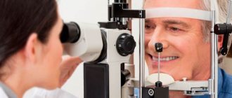
Optical biometry is considered a modern and automated diagnostic method. In this case, the procedure is carried out using a special device. It is used for non-contact eye scanning. The entire procedure is fully automated. First, the device determines which eye it is studying: right or left. Then it scans each of them in turn and then displays the results.
The advantage of this type of diagnosis is the speed and safety of the procedure, the absence of contraindications, and the accuracy of the research. Since all measurements are taken by the device, the risk of medical error is reduced to zero.
Optical biometrics
Optical biometry is considered more accurate than ultrasound, and helps to calculate the required strength of the artificial lens and all parameters of the eyeball in a non-contact manner.
How does optical biometrics work?
Optical biometrics does not use a gel or sensor: the procedure is non-contact, which further increases patient comfort and completely eliminates the risk of infection.
Our clinic uses the AL-Scan optical biometer from NIDEK (Japan), which within 10 seconds allows you to evaluate all parameters of the eyeball, even with a dense lens, and accurately calculate the power of the intraocular lens (IOL) using the latest generation formulas.
Biometrics is necessary for patients who are preparing for lens replacement surgery: in the treatment of cataracts or correction of high degrees of myopia and farsightedness. Modern devices select the desired artificial lens with high precision in just a minute. Optical biometrics is also suitable for examining the condition of the eyes of children.
You can undergo optical and ultrasound biometry in Moscow at the Eye Clinic of Dr. Belikova. We use only high-quality modern equipment and carefully approach each patient.
Decoding the results: normal and pathological
Based on the results, the specialist determines whether the patient has myopia, farsightedness or other pathologies. So, the normal length of the eye axis is 23 mm. With farsightedness, this figure decreases to 19 mm. If the patient has myopia, the length will be increased. The more pronounced it is, the higher this number will be. Using the data obtained, individual lenses or glasses are selected, treatment is prescribed, or surgery is recommended.
If the results are not accurate enough, the doctor may recommend repeat biometric testing. Since this procedure is completely safe for the patient, it can be done even after a short period of time.
A-Scan Plus Accutome by Keeler (USA)
The new Accutome A-Scan Plus ultrasonic ophthalmological scanner will provide accurate and fast diagnostics and calculations using the latest formulas!
The medical terms “A-scan of the eye” and “echobiometry” are used to refer to a diagnostic method aimed at measuring the depth of the anterior eye chamber, the length of the eyeball and the thickness of the lens. These measurements not only have diagnostic value in determining myopia and other disorders, but also, along with data on the parameters of corneal curvature, make it possible to determine the strength of the IOL before surgery. MC Healer conducts comprehensive research using modern equipment, allowing us to obtain accurate information, thanks to which the results of any treatment will be better.
The essence of the method
An A-scan is an example of a one-dimensional scan. During its implementation, the following is measured:
- depth of the eye chamber (exclusively anterior);
- lens thickness;
- eye length - this indicator helps to clearly establish the degree of myopia.
The received information is reflected on the monitor in the form of a graph with two axes – vertical and horizontal. The obtained indicators of echobiometry of the eye are used to analyze all structures of the eye, which allows us to obtain a comprehensive picture.
Diagnostic device
Echobiometry lasts on average from 15 minutes to half an hour. Eyes must be open throughout this time. The procedure does not require the use of painkillers, so it is recommended for both adults and children.
Indications and contraindications for eye tonometry
The manipulation is definitely not performed on patients who have an allergic reaction to anesthetic drops. Without them, it is impossible to carry out the procedure. The exception is the non-contact tonometer.
Also, persons with: viral or bacterial eye diseases should refrain from tonometry of the eyeballs; injury or disruption of the integrity of the retina; high myopia; diseases of the transparent outer membrane of the eyes.
Doctors also prohibit performing such a procedure on patients who are under the influence of alcohol, drugs, or in an unbalanced state (disturbed mental system).
Tonometry of the eyeball is performed in patients with:
- chronic increased intraocular pressure;
- detachment of the inner membrane of the eyeball, which is responsible for the perception of the color palette;
- pathological processes affecting the cardiovascular system;
- neurological diseases;
- diseases of the endocrine system;
- developmental disorders of the eyeball;
- complications after the operation.
Ophthalmologists advise patients over 40 years of age to undergo this diagnostic procedure once a year.
BIOMETRICS
BIOMETRICS
(Greek, bios life + metreo measure) - a set of methods and techniques for mathematical processing of quantitative data in biology and medicine.
The term “biometrics” was proposed by the English scientist Francis Galton (F. Galton) in the book “Natural Inheritance” (1889); the beginning of its application to biological problems dates back to 1901, when the special journal Biometrika was founded.
The first attempt at a quantitative interpretation of the variability of physical characteristics and human behavior was made by the Belgian scientist L. Quetelet in his book “The Experience of Social Physics” (1835). And in the middle of the 19th century. Many outstanding biologists have already noted the importance of mathematics in biology. B. reached a special development in the 20th century. in connection with progress in the field of probability theory and mathematical statistics, the emergence of cybernetics. Mathematical statistics is widely used in studying the variability of signs of the structure and functioning of the human body depending on living conditions and age. Recently, not only the morphological characteristics of a person have been subjected to biometric study (see Anthropometry), but also physiological and biochemical characteristics. The variability of the latter is characterized by right-sided asymmetry, which is often evidenced by
about the presence in the studied material of individuals with certain pathologically altered properties. Physiological, biochemical and mental indicators and their age-related dynamics and variations are especially important in pediatrics and gerontology. Biometric studies of species and intraspecific variability of pathogenic organisms and viruses make it possible to establish differences between pathogenic and non-pathogenic forms. The statistical method in biology has great prospects for resolving issues of assigning individual individuals or groups to certain systematic categories (subspecies, races, species, etc.). The method of discriminant functions, proposed by R. Fisher in 1936, was improved by A. A. Lyubishchev (1962) and applied in anthropological research when deciding whether individual skulls belong to a particular group and some issues of medical diagnostics.
To determine the dependence of certain biological phenomena on environmental factors, for example, the heart rate of cold-blooded animals on temperature, the survival of lower organisms at different temperatures, etc., they use the method of plotting experimental data on graphs limited to two coordinates. Curves are drawn through the observation points, or first, empirical formulas for the dependence are found using the least squares method (see Least squares method) and empirical curves are constructed using the formulas. In some cases, with three variables related to each other, diagrams with three coordinates are constructed. Then the relationship between phenomena is expressed by surface area, not by curve. A larger number of independent variables is difficult to depict graphically, but can be studied mathematically using appropriate methods. Empirical dependencies can be rectilinear, parabolic, exponential, logarithmic, etc. To quickly find the constants of empirical formulas, it is advisable to convert curvilinear dependencies into rectilinear ones by changing the coordinates. For this purpose, there are special stencils - grids with coordinate axes (semi-logarithmic, in which the divisions of one coordinate axis are plotted logarithmically, double logarithmic, etc.). In pharmacology, the alignment of experimental data on the effect of various doses of medicinal substances on certain functions of the body is also widely used: the S-shaped relationship between dose and effect turns into a linear one. For this purpose they use the so-called. the method of analyzing mortality curves, or the probit method, which allows you to objectively compare the activity of drugs and toxic substances. This method is used in microbiology, radiobiology, toxicology, for biological standardization of substances, chemistry. the nature of which has not yet been clarified and which cannot be standardized by the amount of certain chemically precisely determined components.
Modern therapeutic statistics arose on the basis of methods of mathematical statistics developed in relation to field agronomic and zootechnical experiments. The introduction of a new therapeutic or surgical method can only be recommended after statistically based tests that provide high confidence that the effect obtained is not random. Testing a new preventive or therapeutic method takes place in three stages:
1) create a test design or research plan; 2) select experimental and control groups of subjects; 3) make a statistical assessment of the results obtained.
The next stage in the mathematization of biology and medicine was mathematical modeling. However, its use is still limited to certain areas of biology and medicine, chap. arr. when solving theoretical issues. In general physiology, and general biophysics, in particular, it is used in the study of the physiology of processes occurring in the nervous system and sensory organs. The method of mathematical analysis is as follows: the researcher creates a working hypothesis about the connection of certain phenomena with each other in mathematical expression - the so-called. mathematical model.
The results obtained from mathematical modeling can be further tested experimentally, and if the conclusions are confirmed, the initial hypothesis becomes a scientific theory. The founders of the mathematical direction in biophysics can be considered G. Helmholtz, W. Nernst and P. P. Lazarev. Mathematical analysis is also used in biology when studying the dynamics of populations of various organisms, including pathogenic ones. The founder of these studies is the English epidemiologist R. Ross, who used mathematics to study the relationship between the size of the human population affected by malarial plasmodium and the number of mosquitoes that transmit the infection.
Biometrics in cybernetics is the main quantitative method for collecting and processing information characterizing the morphology and functioning of biological objects at various structural levels, as well as for assessing pathological changes in the body and developing decisions or control commands when taking measures to normalize severe conditions. B.'s means are used when clarifying anamnesis and during preventive examinations, as well as in diagnosing conditions (in automatic control systems for active intervention devices and artificial organs).
Rice. 1. Enlarged block diagram of an open-loop measuring registration and information system (explanations in the text). Rice. 2. Enlarged block diagram of a closed registration, information and control system (explanations in the text). Rice. 3. Enlarged block diagram of an open-loop measuring and recording system for mass preventive examinations (explanations in the text).
The collection and processing of the necessary information is carried out by primary information sensors (PIS), which convert the measured value (for example, body temperature, blood pressure, etc.) into an electrical signal, amplification devices, information transmission lines and means for its processing. In addition, biometric systems usually have recorders (ink or thermal recorders, magnetic recorders, etc.) - block P2 (Fig. 1-3) and means of displaying information (oscilloscopes, digital cells, etc.) - block Reject (Fig. 1-3). When measuring electrophysiological parameters, the role of DPI is performed by electrodes that remove biopotentials from a certain area of the body.
According to their structure, biometric systems are divided into open-loop (measuring registration and information systems) and closed (registration, information and control systems). Figures 1 and 2 show enlarged block diagrams of both types of systems.
Depending on the frequency characteristics, biological processes are divided into rapidly changing (RIC) - signal frequency within 1-100 Hz and slowly changing (SIC) - frequency less than 1 Hz. MIPs have a numerical expression and can be represented either by the magnitude of electrical voltage, or after amplification (in the US block) and conversion (in the Prb block) in a digital display or in the corresponding code. An automatic state analyzer (AAS) can be introduced into the biometric system, designed for complex processing of information about MIP and BIP and issuing a generalized decision about the state of the controlled organism. The main indicators characterizing MIP include: heart rate, central and peripheral pulse, respiratory rate, inhalation and exhalation volume, minute volume of respiration, air flow rate during inhalation and exhalation, temperature (body surface, rectal, esophageal), arterial and venous pressure (maximum, minimum and average), percentage of oxygen and carbon dioxide in the blood, partial pressure of hydrogen, oxygen, carbon dioxide, content of sodium and potassium ions, volumetric blood flow velocity, etc.
BIPs are recorded in the form of voltage curves and do not have a direct numerical expression. In this regard, in order to use information about the BIP in automated open-loop, as well as in closed-loop, control diagnostic complexes, preliminary processing of signals is necessary in order to identify informative features. This operation and coding of features are carried out in a special computing device - an informative feature extraction unit (ICU). The useful information identified in this way from the BIP signal enters the automatic state analyzer.
The most widespread recorded BIPs include: electrical activity of the heart (electrocardiogram), electrical activity of the brain (electroencephalogram), electrical activity of motor muscles (electromyogram), mechanical activity of the heart (ballistocardiogram, mechanocardiogram, seismocardiogram, valvulocardiogram, etc.), intracavitary acoustic field of the heart. (phonocardiogram), changes in electrical characteristics under the influence of blood circulation dynamics (rheogram), galvanic skin response (GSR), changes in blood filling of blood vessels (plethysmogram), eye motor activity (oculogram), changes in arterial and venous pressure, volumetric blood flow velocity, etc.
Depending on the purpose of the measuring registration and information complexes, the amount of information from the MIP and BIP can vary within fairly wide limits.
In continuous monitoring systems, especially those serving resuscitation and surgical complexes, as well as in physiological laboratories, the so-called acute measurement methods, in which the DPI is placed on various parts of the body with a violation of the skin. During preventive examinations, as well as during continuous monitoring of the activities of healthy people (for example, operators servicing control systems), acute measurement methods are usually not used.
Closed systems are fundamentally different from open systems by the presence of a control part, which includes a control unit for active intervention devices (BUPAV), active intervention devices (ASD) and a system for monitoring the operation of active intervention devices (SAM Control).
BUPAV is a computer system that implements a rigid or adaptive program for controlling surfactant modes. In this case, the control algorithm provides for the use of information from AAS and Control. Surfactant for selecting and optimizing the operating mode of the surfactant.
In accordance with the functions they perform, surfactants are divided into the following groups: 1) artificial organs (artificial heart-lung machine, heart-lung machine, artificial kidneys, artificial respiration devices, etc.); 2) stimulants (electrical pacemakers, pharmacological stimulants, etc.); 3) systems for controlling environmental parameters (temperature, humidity, illumination, content of gas components in the air, sources of electromagnetic and radiation fields, etc.).
The main requirement to ensure sufficient reliability of the information obtained using biometric systems is a minimum of interference. From this point of view, microminiature sensors and wireless biotelemetry methods are promising (see).
One of the varieties of open-loop measuring and recording systems are automated systems (Fig. 3). These systems are designed for systematic mass preventive examination. They can also play the role of a subsystem in automated control systems for large industrial enterprises.
In these systems, the parameters of the body are measured using the DPI, Prb blocks. and BVIP are entered into the comparison block (BC). It also contains data characterizing the normal state of the subject, stored in the corresponding block of random access memory (RAM). If at least one of the measured parameters deviates from the norm, an alarm signal is sent from the comparison unit. The results of examinations can be automatically entered into the system's long-term memory unit (LMB), which is an automated archive, from which, if necessary, relevant statistical information can be extracted.
In recent years, in microscopic studies, the method of optical-structural machine analysis proposed by K. M. Bogdanov has found application (see Medical Morphometry). The method is based on statistical analysis of a scanogram (signal obtained by sequential photometry of a sample) and allows one to obtain quantitative characteristics of the distribution of structural components of cells and tissues in health and disease, and to calculate the orderliness and information entropy of the structure. To implement the method, an automated microscope analyzer “Protva” was developed.
See also Mathematical methods (in medicine).
Bibliography:
Alpatov V.V. Norm and pathology in the variability of human blood pressure, Vopr, anthropopol., v. 23, p. 66, 1966, bibliogr.; aka, The law of the system’s response to external influences in relation to biological and demographic phenomena, ibid., c. 34, p. 148, 1970, bibliogr.; Bailey N. Mathematics in biology and medicine, por. from English, M., 1970; Bogdanov K. M. Method of quantitative analysis of morphological structures based on their statistical characteristics, in the book: Machine analysis of microscopic objects, ed. G. M. Frank, p. 21, M., 1968; Plokhinsky N.A. Biometrics, M., 1970, bibliogr.; Application of mathematical methods in biology, ed. P. V. Terentyeva, collection. 1 - 4, JI., 1960-1969; Rokitsky P. F. Biological statistics, Minsk, 1967; Sepetliev D. A. Statistical methods in scientific medical research, trans. from Bulgarian, M., 1968, bibliogr.; Urbakh V. Yu. Biometric methods, M., 1964; Arm it age P. Statistical methods in medical research, Oxford - Edinburgh, 1971, bibliogr.; Bliss G h. I. Statistics in biology, v. 1, NY, 1967; Bourke GJ a. McGil-v g a y J. Interpretation and uses of medical statistics, Oxford - Edinburgh, 1969; Campbell RC Statistics for biologists, Cambridge, 1967; With ava 1 1 i -Sforza L. Biometrie, Jena, 1969, Bibliogr.; Finney DJ Probit analysis, a statistical treatment of sygmoid response curve, Cambridge, 1952; aka, Statistical method in biological assay, L., 1952; Fisher RA a. Y ates F. Statistical tables for biological, agricultural and medical research, L.—Edinburgh, 1953; Herdan G. Statistics of therapeutic trials, Amsterdam, 1955; H i 1 1 AB Principles of medical statistics, L., 1966; S o ka 1 RR a. R o h 1 f FJ Biometry, San Francisco, 1969, bibliogr.
B. in cybernetics
— Akhutin V.M. On the principles of constructing complexes for continuous monitoring of the human body and automatic normalization of its states, in the book: Bioelectric. control, human and automatic systems, ed. V. A. Trapeznikova, p. 519, M., 1970, bibliogr.; Akhutin V. M., Neroslavsky I. A. and Stein JI. B. Specialized computing devices for automatic control of the functional state of the human operator’s body, in the book: Automation, organization, diagnostics, ed. V.V. Parina et al., part 2, p. 691, M., 1971, bibliogr.; Akhutin V.M. et al. Automatic control of the physiological functions of the body during surgery, Med. technology, no. 2, p. 5, 1968, bibliogr.; Wiener N. Cybernetics or control and communication in animals and machines, trans. from English, M., 1968; Parin V.V. and Baevsky R.M. Introduction to medical cybernetics, p. 154, M., 1966; Utyamyshev R.I. Radioelectronic equipment for the study of physiological processes, M., 1969, bibliogr.
V. A. Alpatov, V. M. Akhutin.
Types of ocular tonometry
Today there are four types of tonometry. The principle of the procedure is almost the same; different devices are used.
Applanation tonometer
This type of tonometer allows you to influence the cornea as little as possible. In addition to the tonometer, the ophthalmologist will additionally use a microscope and a slit lamp to completely examine your eye. The applanation tonometer is considered the most accurate and is used after the patient has passed a standard vision test.
Electronic tonometer
To measure intraocular pressure in the eyeball, this type of tonometer is most often used. It also stands out for its accuracy, but its readings can differ significantly from the applanation one. A special electronic tonometer sensor is placed on the outer mucous membrane of your eye, which slowly begins to read pressure readings. All readings are recorded on a small computer panel.
Non-contact tonometer
It is also called a pneumotonometer. During the procedure, nothing is attached to your eye. How then is pressure measured? The fact is that the measurement is carried out by supplying air. Unfortunately, the contactless procedure cannot be called accurate. But it is still used in ophthalmological practice, namely when measuring intraocular pressure in children. Doctors may also prescribe the procedure to patients who have just undergone eye surgery, and any contact can cause serious complications. During the procedure, the patient does not need anesthetic drops.
Schiotz impression tonometer
To measure pressure, doctors use a special rod that gently presses on the outer shell of the eye. The ophthalmologist then places small weights on the second part of the machine. Conclusions are obtained by calculating the weight that is necessary to return the mucous membrane to its previous position. It is used very rarely, since many scientific studies have not proven the reliability of its results. It can also be found among doctors who come on house calls.