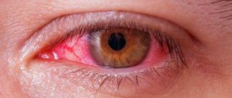
A diagnosis with code H10 includes 8 clarifying diagnoses (ICD-10 subheadings): Chain in classification:
The diagnosis does not include: – keratoconjunctivitis (H16.2)
mkb10.su - International classification of diseases, 10th revision. Online version of 2020 with search for diseases by code and decoding.
| Pharm. groups | Active substance | Trade names |
| H1-antihistamines | Astemizole* | Gistalong ® |
| Terfenadine* | Bronal | |
| Alpha adrenergic agonists | Tetrizoline* | Berberyl ® N |
| Aminoglycosides | Gentamicin* | Garamycin |
| Gentamicin | ||
| Tobramycin* | Tobrex 2X | |
| Tobrex ® | ||
| Antiseptics and disinfectants | Picloxidine* | Vitabakt ® |
| Chlorhexidine* | Plivasept | |
| Dietary supplements vitamin and mineral complexes | Arthromax | |
| Vitamins and vitamin-like products | Retinol* | Retinol acetate solution in oil |
| Retinol acetate injection solution in oil | ||
| Riboflavin* | Riboflavin | |
| Glucocorticosteroids | Hydrocortisone* | Hydrocortisone |
| Dexamethasone* | Dexamethasone | |
| Maxidex | ||
| Desonide* | Prenatsid | |
| Glucocorticosteroids in combinations | Gramicidin C + Dexamethasone + Framycetin* | Sofradex ® |
| Dexamethasone + Neomycin + Polymyxin B* | Maxitrol | |
| Dexamethasone + Tobramycin* | Tobradex ® | |
| Homeopathic remedies | Mucosa compositum ® | |
| Okulochel ® | ||
| Other antibiotics | Fusidic acid* | Fucithalmic ® |
| Other antimicrobial, antiparasitic and anthelmintic agents in combinations | Colistimethate sodium + Rolytetracycline + Chloramphenicol* | Colbiocin |
| Colistimethate sodium + Tetracycline + Chloramphenicol* | Colbiocin | |
| Other synthetic antibacterial agents | Nitrofural* | Furacilin |
| FURACILIN AVEXIMA | ||
| Furacilina tablets for external use 0.02 g | ||
| Furazidin | Furagin tablets 0.05 g | |
| m-Anticholinergics | Tropicamide* | Midroom |
| Macrolides and azalides | Erythromycin* | Grunamycin syrup |
| Ilozon | ||
| Ermiced | ||
| NSAIDs Acetic acid derivatives and related compounds | Diclofenac* | Naklof |
| General tonics and adaptogens | Aloe arborescens leaves | Aloe extract liquid for injection |
| Aloe extract liquid for injection in ampoules of 1 ml | ||
| Ophthalmic products | Benzyldimethylmyristoylaminopropylammonium | Okomistin ® |
| Ofloxacin* | Uniflox | |
| Chloramphenicol* | Levomycetin | |
| Ciprofloxacin* | Ciprofloxacin-AKOS | |
| Ophthalmic agents in combinations | Oftalmo-Septonex | |
| Gentamicin + Dexamethasone* | Dexa-Gentamicin | |
| Dexamethasone + Tobramycin* | Tobrazon | |
| Antiviral (except HIV) agents in combinations | Polyadenylic acid + Polyuridylic acid | Poludan ® |
| Sulfonamides | Sulfacetamide* | Sulfacyl sodium |
| Tetracyclines | Oxytetracycline* | Innolir |
| Quinolones/fluoroquinolones | Lomefloxacin* | Okatsin |
| Moxifloxacin* | Vigamox ® | |
| Norfloxacin* | Chibroxin | |
| Ofloxacin* | Zanotsin ® | |
| Ofloxacin | ||
| Phloxal ® | ||
| Ciprofloxacin* | Zindolin 250 | |
| Liproquin | ||
| Ciloxane | ||
| Tsiprobay ® | ||
| Tsiprosan | ||
| Ciprofloxacin hydrochloride | ||
| Cifloxinal ® | ||
| Cephalosporins | Cefazolin* | Totacef™ |
| Cefezol ® | ||
| Cefapirin* | Cefatrexil | |
| Ceftazidime* | Ceftidine |
Official website of the RLS ® company. Home Encyclopedia of medicines and pharmaceutical assortment of goods of the Russian Internet. The directory of medicines Rlsnet.ru provides users with access to instructions, prices and descriptions of medicines, dietary supplements, medical products, medical devices and other goods. The pharmacological reference book includes information on the composition and form of release, pharmacological action, indications for use, contraindications, side effects, drug interactions, method of use of drugs, pharmaceutical companies. The medicinal reference book contains prices for medicines and pharmaceutical market products in Moscow and other cities of Russia.
Classification
Conjunctivitis is divided into infectious and non-infectious. The first develops under the influence of bacteria, fungi, viruses and other pathogenic microorganisms. The second is provoked by external irritating factors (allergens, small objects - dust, sand, etc. that get on the mucous membrane of the eye). In some cases, the cornea or eyelids may become inflamed along with the mucous membrane, which implies keratoconjunctivitis or blepharoconjunctivitis.
In this case, acute conjunctivitis is divided into the following types (depending on the causes):
- Bacterial - an infectious lesion of the mucous membrane of the eye caused by various types of bacteria (gram-positive and gram-negative). The greatest danger is chlamydial conjunctivitis.
- Viral - inflammation of the conjunctiva associated with the entry into the body or activation (for example, in the case of weakened immunity) of existing viral infections - adenoviruses, herpes viruses, etc. Very often this form of the disease is accompanied by cold symptoms (or develops against the background of acute respiratory infections).
- Allergic - the inflammatory process of the mucous membrane serves as a response to contact with a particular allergen. This form is most often diagnosed at a young age. The danger of allergic conjunctivitis is that in the absence of adequate or timely treatment, any infection can join the allergy (since the mucous membrane of the eye is inflamed and therefore sensitive to the effects of foreign microorganisms).
Allergic conjunctivitis
Allergic eye diseases are an important problem in practical ophthalmology and allergology, and according to foreign epidemiological studies, their prevalence among the population of Western countries is about 15–20% (Current ocular therapy (2000).
An analysis of the recent NHANES III study (Third National Health and Nutrition Examination Survey) conducted in the United States showed that symptoms such as “episodes of watery, itchy eyes during the last 12 months” bother 40% of the adult population, and the prevalence of such symptoms with age is significantly haven't changed. Clinical symptoms of AK are caused by hypersensitivity reactions triggered by immunological mechanisms. In most patients, AK develops by an IgE-dependent mechanism. The interaction of a specific allergen with IgE - antibodies fixed on high-affinity receptors of this immunoglobulin (Fcε receptors type I - FCεRI), presented on mast cells and basophils, leads to activation and release of allergy mediators (histamine, serotonin, platelet activating factor) from target cells , leukotrienes, prostaglandins, chemotactic factors, proteoglycans, enzymes, etc.), responsible for the development of symptoms of the disease.
Allergic conjunctivitis is a disease characterized by allergic inflammation of the conjunctiva of the eyes, caused by an etiologically significant allergen.
Classification according to ICD-10
- H10 – conjunctivitis,
- H10.1 – acute atopic conjunctivitis,
- H10.2 – other acute conjunctivitis,
- H10.3 – acute conjunctivitis, unspecified,
- H10.4 – chronic conjunctivitis,
- H10.9 – unspecified conjunctivitis.
Prevention
Primary prevention is aimed at preventing the development of allergic conjunctivitis, which primarily includes the formation in patients of a competent attitude towards their health, familiarization with the causes and mechanisms of the development of the disease based on broad information about the features of the development of AK, possible etiological and provoking factors, the need for elimination measures and mandatory compliance with doctor's orders. Primary prevention should be carried out starting during pregnancy and from the first days of life. During pregnancy, it is necessary to provide proper and varied nutrition, corresponding in volume and ratio of food ingredients to age, body weight, concomitant diseases and energy costs, strict exclusion of active and passive smoking is required, etc. It is necessary to fight and maintain motivation for breastfeeding children. For infants and children at high risk of developing asthma and allergies (this includes children from families where at least one parent or sibling has an allergy), multiple interventions are offered to reduce early life exposure to house dust mites, such as bed covers and special coverings for parent and child beds, washing bedding and soft toys at temperatures exceeding 60 ºС, the use of acaricides, smooth floors without carpets, etc.
Secondary prevention - prevention of exacerbation of allergic conjunctivitis in those people who suffer from allergies: - carefully collect and analyze allergic, pharmacological and nutritional history; – limit contact with the causative allergen as much as possible; – do not prescribe medications manufactured or containing plant materials to patients with seasonal allergic conjunctivitis caused by sensitization to plant pollen; – for patients with allergic conjunctivitis caused by sensitization to medications, do not prescribe these drugs and those similar in chemical structure, and clarify the synonyms of the drugs, since the drug produced by different companies may have different trade names; – do not use cosmetics containing plant materials in patients with seasonal allergic conjunctivitis caused by sensitization to plant pollen;
– do not consume food products of plant origin that have cross-reactions with causally significant pollen or fungal allergens.
Tertiary prevention is important for people who have suffered severe complicated manifestations of allergic conjunctivitis, and includes the development of measures for long-term control over the symptoms of the disease: - constant monitoring by an allergist-immunologist; – the patient has a written treatment plan;
– education and training of patients, including in allergy schools.
Classification
There is no unified classification of allergic conjunctivitis. Allergic conjunctivitis is classified by form, by mechanisms of development, by severity and stage of progression.
Classification of AK by shape:
Develops with sensitization to pollen (pollen from trees, cereals, weeds, etc.) and fungal (spores of Cladosporium, Penicillium, Alternaria, etc.) allergens. It is characterized by seasonality of clinical manifestations, coinciding with the dusting period of causally significant allergens.
Develops with sensitization to house dust allergens, house dust mites, library dust, wool, dander, animal saliva, bird down and feathers, mold fungi, food allergens, insect, occupational and other allergens. It is characterized by the absence of seasonality and year-round flow.
Classification of AK according to development mechanisms
According to development mechanisms, there are:
– IgE-mediated allergic conjunctivitis , which includes acute allergic conjunctivitis, seasonal allergic conjunctivitis and perennial allergic conjunctivitis.
– Mixed – IgE and cell (Th2) caused by AK . These include giant papillary conjunctivitis (GPC), vernal keratoconjunctivitis (VKK), and atopic keratoconjunctivitis (AKK).
– Non-IgE-related – dermatoconjunctivitis/allergic contact conjunctivitis.
Classification of AK by severity:
- – mild degree;
- – moderate degree;
- – severe.
Classification of AK by stage of flow:
Diagnostics
The diagnosis of AK is based on the results of a comprehensive examination, including the following data:
- – medical history;
- – physical data (clinical manifestations);
- – ophthalmoscopy;
- – allergy history;
- – results of clinical and laboratory examination;
- – results of allergological examination.
Separately, atopic keratoconjunctivitis is distinguished. ICD code H10.1 – acute atopic conjunctivitis. There are two known forms of atopic keratoconjunctivitis: childhood and adult. The childhood form develops in children under 5 years of age. In adults, it most often develops at the age of 35–40 years.
The causes and mechanisms of development of atopic keratoconjunctivitis are the same as AK. There is a close relationship with allergen exposure and an elimination effect is noted. The eye damage is bilateral.
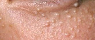
In contrast to AK, ophthalmoscopy reveals pallor of the conjunctiva and the presence of yellowish-white dots in the limbus (Trantas dots or grains, Horner spots, which are pinpoint lesions of degeneratively changed eosinophils).
Clinical, laboratory and allergological indicators for atopic keratoconjunctivitis are the same as for AK.
Differential diagnosis
It is necessary to exclude non-allergic forms of conjunctivitis and keratoconjunctivitis:
- – viral, bacterial, chlamydial conjunctivitis and keratoconjunctivitis;
- – irritant, drug-induced conjunctivitis;
- – “red eye” syndrome;
- – dry eye syndrome, keratoconjunctivitis sicca;
- – glaucoma;
- – blepharoconjunctivitis, uveitis, corneal lesions;
- – conjunctivitis in systemic diseases, autoimmune diseases, etc.
Treatment
Indications for hospitalization
As a rule, treatment of AK is carried out in an outpatient setting. Hospitalization is indicated only for severe and/or complicated AK that threatens visual impairment. Hospitalization is also indicated if it is necessary to carry out ASIT (allergen-specific immunotherapy) using an accelerated method.
Non-drug treatment
Elimination measures . Eliminating contact with the allergen (for example, stopping contact with pets and creating a hypoallergenic lifestyle for household and epidermal allergies, elimination diets for food allergies, eliminating professional contact with the causative allergen, etc.).
Educational programs (allergy schools) for patients.
Drug treatment
Treatment of seasonal conjunctivitis
2–3 weeks before the onset of an expected exacerbation of AK, preventive therapy is prescribed (cromoglycic acid preparations in the form of eye drops, non-sedating antihistamines of the 2nd generation.
Treatment of exacerbation of AK
Preparations for topical use - preparations of cromoglycic acid in the form of eye drops, at a dose of 1-2 drops 4-6 times a day.
Antihistamines and combination drugs in the form of eye drops: – azelastine, at a dose of 1 drop in each conjunctival sac 2 times a day; – olopatadine hydrochloride, in the form of eye drops, in a dose of 1 drop 2 times a day into the conjunctival sac. Shake the bottle before use.
– ketotifen, eye drops, adults and children, 1 drop in each conjunctival sac 2 times a day; – diphenhydramine, in a dose of 1 drop of 0.2% and 0.5% solution in each conjunctival sac 2–5 times a day.
Drops containing diphenhydramine: polynadim (diphenhydramine - 1 mg, naphazoline - 0.25 mg) in a dose of 1 drop every 3 hours into the conjunctival sac until swelling and irritation of the eye decrease, then 1 drop 2-3 times a day until clinical symptoms disappear symptoms. Do not use polynadim for more than 5 days without consulting your doctor.
- betadrine (diphenhydramine hydrochloride 1 mg, naphazoline nitrate 330 µg) 1-2 drops into the lower conjunctival sac, no more than every 6-8 hours. Duration of use - 3-5 days; – okumetil – a combined drug diphenhydramine + naphazoline + zinc sulfate in the form of eye drops, at a dose of 1 drop in each conjunctival sac 2-3 times a day (D).
When a secondary infection occurs, complex drugs are prescribed, including antibacterial and GCS components (steroid).
For moderate and severe AK, the following drugs are used:
– dexamethasone in the form of eye drops, at a dose of 1–2 drops of a 0.1% solution 4–5 times a day for two days, then 3–4 times a day, but not longer than 3–6 weeks;
– hydrocortisone in the form of eye ointment, 2-3 times a day, for 2-3 weeks. GCS for topical use are contraindicated in conjunctivitis of viral origin.
Systemic drugs
H1 receptor blockers - antihistamines (AGP):
– AGPs: loratadine, desloratadine, cetirizine, fexofenadine, quifenadine, sehifenadine, ebastine, levocetirizine, rupatadine. In case of AK, preference is given to second-generation antihypertensive drugs (non-sedating). Antihypertensive drugs are prescribed in accordance with the instructions for use of the drugs.
– If parenteral administration of antihypertensive drugs is necessary, 1st generation histamine H1 receptor blockers: clemastine, IM, at a dose of 1 mg 2–3 times a day, chloropyramine, at a dose of 25 mg 2–3 times a day. In severe forms of AK: clemastine, administered intramuscularly, at a dose of 2 mg 1–2 times a day, chloropyramine, at a dose of 40 mg 1–2 times a day.
Treatment of year-round conjunctivitis
The following are prescribed as basic therapy:
Systemic drugs
H1 receptor blockers
– AGPs: loratadine, desloratadine, cetirizine, fexofenadine, quifenadine, sehifenadine, ebastine, levocetirizine, rupatadine. In case of AK, preference is given to second-generation antihypertensive drugs (non-sedating). Antihypertensive drugs are prescribed in accordance with the instructions for use of the drugs;
– AGP with a stabilizing effect on mast cell membranes: ketotifen at a dose of 1 mg 2 times a day for 3 months;
– preparations of cromoglycic acid (B), in the form of eye drops.
Treatment of exacerbation of year-round AK is carried out according to a scheme similar to seasonal AK. The main pathogenetic method of treating AK is ASIT. ASIT is prescribed and performed by an allergist-immunologist.
What not to do
Prescribe GCS for conjunctivitis of viral origin. Prescribe eye drops and eye ointments containing antibiotics, antifungals and antivirals for uncomplicated forms of AK. It is impossible to prescribe planned surgical interventions with gases to patients with seasonal AK during the dusting season of etiologically significant allergens.
A patient with AK is subject to observation by an allergist-immunologist and an ophthalmologist:
- – prescribing ASIT outside the allergen dusting season;
- – clinical examination (screening): 2–3 weeks before the plant dusting season, correction of therapy for year-round course of AK, monitoring the adequacy of therapy for concomitant allergic diseases;
- – training at an allergy school.
Source:
Allergology. Federal clinical guidelines. Chief editors: acad. RAS R.M. Khaitov, prof. N.I. Ilyina - M.: Farmarus Print Media, 2014. 126 p.
allergies causes of allergies conjunctivitis redness of the eyes lacrimation
Prevention
To prevent allergic reactions, you should resort to the following rules:
- sensitization of the human body by allergens in small doses in the autumn-winter period;
- if a person develops an allergy for a short period of time, he can go to another place until the flowering of plants ends;
- annual preventive examination by an ophthalmologist, allergist;
- During the period when the patient is about to develop an allergy, he should begin therapy with medications in advance to reduce the degree of clinical manifestations.
Allergic rhinoconjunctivitis is a serious condition for patients, worsening their well-being. There are specific methods of prevention and treatment in which the effect of the allergen is reduced. Requires you to see an allergist, who will conduct a full diagnosis of the body’s condition and prescribe medications.
Allergic rhinitis (AR) has become the most common problem of “runny nose” in children in recent years! AR is an allergic disease of the nasal mucosa (the tissue that lines the inside of the nose). This disease can be seasonal, caused by pollen, or year-round, most often caused by house dust mite and mold allergens. Allergic rhinitis is often combined with bronchial asthma or allergic conjunctivitis.
The main symptoms of allergic rhinitis are:
- Rhinorrhea - watery discharge from the nose, leading to hyperemia, irritation of the skin of the wings of the nose and upper lip, sometimes so abundant that patients change handkerchiefs every 30-60 minutes
- Episodes of sneezing that may develop suddenly; Most often, sneezing occurs in attacks of 10 to 30 sneezes in a row
- Nasal congestion, caused by allergic swelling of the mucous membrane, leads to a narrowing of the lumen of the airways, difficulty in nasal breathing, up to its complete absence. Swelling of the nasal mucosa leads to decreased hearing, sense of smell, and headaches due to the development of negative pressure during the passage of air from the maxillary sinus to the middle ear;
- Itching in the nose, which can appear spontaneously or precede sneezing and is characterized by varying degrees of intensity
Allergic rhinitis is often accompanied by the presence of so-called “non-nasal” symptoms (itching of the palate, eyes, lacrimation, disturbance of voice timbre and distorted pronunciation of speech sounds - nasal sound, headache, increased fatigue, dry mouth)
Depending on the severity of the manifestations of allergic rhinitis during the year, there are two different variants of the disease: seasonal and year-round.
Seasonal allergic rhinitis
Seasonal allergic rhinitis (hay fever) is characterized by manifestations only during the flowering period of plants. Itching in the nose and other symptoms of hay fever are most intense in the mornings during the period of maximum concentration of pollen in the air. At the height of the dusting season of causative plants, patients may also experience general symptoms: irritability, easy fatigue, lack of appetite, depression, weight loss, etc.
After pollination of the causative plants is completed, the symptoms of pollen AR usually disappear. Sometimes the symptoms of allergic rhinitis can persist for 1-3 weeks after the cessation of plant pollination, which may be due to the presence of nonspecific hyperreactivity of the nasal mucosa in patients with hay fever, especially in environmentally unfavorable regions. In addition, consuming a number of foods that have cross properties with pollen can lead to symptoms of rhinitis even when the plant is not blooming.
Year-round allergic rhinitis
Perennial allergic rhinitis bothers the patient all year round. Clinical symptoms are similar to seasonal allergic rhinitis, but are usually less severe. Sometimes the only symptom of year-round allergic rhinitis may be nasal congestion. A characteristic feature of year-round allergic rhinitis is sneezing when waking up in the morning or at night.
The cause of year-round allergic rhinitis is most often sensitization to house dust mite allergens. In allergic rhinitis caused by animal allergens, symptoms occur after contact with an animal to which the patient is sensitive.
How to use intranasal sprays?
Before use, shake the bottle carefully, take it by placing your index and middle fingers on either side of the tip, and your thumb under the bottom. When using the drug for the first time or a break in its use for more than 1 week, you should check the serviceability of the sprayer (pointing the tip away from you, make several presses until a small cloud appears from the tip).
Next you need:
clear your nose (blow your nose lightly);
close one nostril and insert the tip into the other nostril;
tilt your head slightly forward, continuing to hold the aerosol bottle vertically;
start inhaling through your nose and, continuing to inhale, press once with your fingers to spray the drug;
exhale through the mouth
The following groups of drugs are used in the treatment of allergic rhinitis:
Antihistamines:
(orally or in sprays) - are very effective against itching in the nose, sneezing, and effectively reduce the volume of nasal discharge, and their effect develops within the first hour after use. However, they practically do not relieve nasal congestion
Intranasal corticosteroids:
They have a pronounced anti-inflammatory effect, and with long-term use (a week or more) they not only lead to a decrease in the severity of symptoms, including nasal congestion, but also prevent the recurrence of symptoms
Antileukotriene drugs - not only eliminate the symptoms of allergic rhinitis, including congestion, but also prevent the “lowering” of allergic inflammation into the bronchi, and therefore can prevent bronchial asthma
An allergist-immunologist should select the medications necessary for your child.
However, it must be remembered that the best and most modern drugs will not be effective enough if you do not follow a hypoallergenic diet and a hypoallergenic lifestyle!
Allergic conjunctivitis
Allergic conjunctivitis is an allergic eye disease (inflammation of the conjunctiva) caused by contact with a causal allergen.
This disease can be seasonal, for example, in spring or autumn (with allergies to pollen),
Sometimes allergic conjunctivitis can bother the patient throughout the year (in case of allergies to house dust mite allergens, mold or animal allergens).
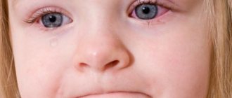
It is extremely rare that allergic conjunctivitis occurs without symptoms of allergic rhinitis. If your child's eyes become red and itchy and there is nasal congestion or sneezing, then it is necessary to be tested for allergies!
The main manifestations of allergic conjunctivitis:
itching of the conjunctiva of the eyes
lacrimation
redness of the conjunctiva
burning sensation in the eyes
The conjunctiva of both eyes is always affected
The causative allergens for conjunctivitis are usually the same as for rhinitis.
Treatment most often uses new generation antihistamines for oral administration and local agents in the form of eye drops containing antihistamines or cromones. For extremely severe manifestations, the doctor may prescribe corticosteroids in eye drops - but that’s it. As a rule, for a short time, followed by a transition to the above remedies.
Causes
The causes of acute conjunctivitis can be different (depending on the type of disease - bacterial, viral, allergic). Thus, pathology occurs under the influence of the following factors:
- Bacterial conjunctivitis - develops under the influence of pathogenic bacteria (staphylococci, streptococci, gonococci, pneumococci, chlamydia, diphtheria bacillus, etc.), which are introduced into the conjunctival cavity along with unwashed hands, dust or dirty water. Another cause of the bacterial form of the disease is improper care of contact lenses.
- Viral - the disease is transmitted through household contact or airborne droplets from carriers of a viral infection (from work colleagues, family members, casual acquaintances, etc.). The infection can also be caused by untreated ophthalmic instruments in medical institutions. The most common pathogens are adenoviruses, herpesviruses and enteroviruses.
- Allergic - occurs under the influence of various types of allergens (medicines, pollen of flowering plants, pet hair, house dust, cosmetics, household chemicals, etc.) that enter the mucous membrane and cause inflammation.
How not to harm the procedure
Is it possible to massage the prostate on your own without consulting a specialist? The answer to this question is always negative, since stimulation will only benefit patients who have no contraindications to this procedure. Even if a person has perfectly mastered the massage technique and performed therapeutic palpation many times, before the next course of massage he must make sure that the following diseases are absent.
Numerous inflammations, prolonged abstinence, and improper use of sulfonamide drugs can lead to the formation of stones in the tissues of the gland. In this case, massaging causes significant harm to the prostate. Reviews from doctors say that such manipulations cause pathological scarring, as a result of which the organ is deformed and cannot perform its functions fully. A man needs to pay special attention if the gland, soft to the touch, hurts unbearably when touched with a finger, as well as during sex, defecation and urination.
- Prostatitis or urinary tract infections
If the disease is bacterial in nature, when massaging the prostate, microorganisms can spread to neighboring organs. Even more harm to the body will be caused when pathogenic microbes enter the blood. Then any human organ may be under attack. In addition, the liver is often harmed, since its function is to filter the blood flow. Experts in reviews say that after treating the acute phase of the disease, prostate massage is extremely useful, as it allows you to get rid of the remnants of pus and unhealthy prostate secretions.
Prostate massage is not performed for hemorrhoids, proctitis, paraproctitis, the presence of wounds and cracks in the walls of the rectum. In this case, the harm from the procedure far outweighs the benefits of massaging. Thus, blood often stagnates in hemorrhoidal cones and blood clots form. If a blood clot breaks away from the vessel wall, it can migrate through the circulatory system and cause dangerous diseases such as myocardial infarction, ischemic stroke, and pulmonary embolism. Doctors in their reviews advise patients to get rid of ailments in the rectum before undergoing a massage course.
- Benign and malignant tumors
One of the reasons for conducting a thorough diagnostic examination before massage sessions is to exclude the presence of any neoplasms in the male genitourinary system. If a cancerous tumor is detected, massage is not only harmful, it acts as a catalyst for the metastasis of carcinoma. With benign tissue growth, the harm of massaging lies in the activation of the disease, due to which neighboring organs are displaced and the urethra is pathologically narrowed. In their reviews, urologists warn that with frequent stimulation of the diseased gland, the adenoma may degenerate into a malignant form.
In order not to harm the body, it is necessary not only to be sure that there are no contraindications, but also to master the technique of performing gland stimulation. So, correct body position during massage is of great importance. A person can lie on his side with his knees bent, stand on the floor bending over the tabletop, or squat with his knees apart.
Why do you need prostate massage? What are the benefits of massage? And what harm can the procedure cause? The doctor will give detailed answers to these questions during the consultation after evaluating the patient’s diagnostic examination. The need for manual stimulation of the prostate gland, the time and number of sessions are calculated individually for each man, taking into account his age and health status.
What is prostate massage and what is it for? “Prostate massage” is a medical manipulation performed by mechanical action on the organ. The procedure is used to ensure the outflow of prostate juice from the organ.
In practice, urologists use two methods of exposure:
- Through the wall of the rectum.
- Bougie massage.
IMPORTANT! Half an hour before the procedure, you need to give an enema and drink 1 liter of liquid (for maximum adherence of the prostate to the wall of the rectum).
Transrectal manipulation is carried out:
- in the knee-elbow position;
- in a “standing” position (bent over, with emphasis);
- lying down (on your side, with your knees pressed to your stomach).
The doctor places a finger (with a sterile glove, lubricated with Vaseline) into the anus. Anatomically, the edges of the prostate gland can be felt at a distance of 4-5 cm from the anal outlet.
Before the procedure, the doctor determines the shape and size of the gland, the symmetry of the lobules, consistency, and palpates the presence of painful and sensitive areas.
In the case of a professionally performed massage using a finger, there is no pain. From the first sessions there is a gradual increase in the intensity of the palpation effect on the gland (starting with light stroking).
MORE ABOUT: Massage after a broken arm - YouTube
The duration of medical therapeutic massage is from 1 to 5 minutes. The standard course of massage therapy is 10 sessions (breaks for 2-3 days).
If there is a need to collect a secretion, it is placed on a glass slide (or in a test tube). In cases where the secretion could not be obtained in the usual way, rinsing urine is studied.
If a prostate massage is performed on a bougie, the patient lies on his side, facing the urologist. A bougie (a medical instrument in the form of a rod) is inserted through the urethral canal and a massage is performed. Unprofessional implementation of this procedure can lead to swelling and delayed urine outflow.
Does a man need a prostate massage? Read below.
Symptoms
Depending on the type of disease, symptoms may vary slightly. Therefore, it is worth paying attention to the following characteristic signs:
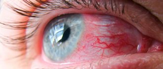
- Bacterial conjunctivitis - accompanied by severe pain in the eye, burning, itching, a feeling of “sand” or a foreign object, profuse lacrimation and yellowish-white purulent discharge. In this case, the mucous membrane turns red, photophobia occurs, and hemorrhages and the formation of follicles can be detected on the mucous membrane of the eyeball. Sometimes a person experiences headaches and insomnia. The duration of this form of the disease is 2 weeks. At first, one eye is always affected, and only after some time does the infection spread to the second.
- Viral - the disease is characterized by increased lacrimation, photophobia, redness of the mucous membrane and discomfort in the eye. But unlike the bacterial form, there is no purulent discharge. Along with this, cold symptoms appear (most often this is where it all begins): runny nose, headache, fever, etc. The infection first affects one eye and after a few days moves to the second. Recovery usually occurs within 4-10 days. In advanced cases, the disease can last for months and, in the absence of adequate therapy, becomes chronic.
- Allergic - the mucous membrane of the eye turns red, the person experiences severe itching and burning, photophobia and uncontrollable lacrimation occur (the liquid can be clear or white, which thickens over time), surrounding objects look blurry. Distinctive feature: the disease affects both eyes at the same time. When the irritant is eliminated, recovery occurs within a few days.
What it is?
Allergic conjunctivitis is an inflammation of the mucous membrane of the eye as a response to exposure to an allergen. To trigger this mechanism, a person must have increased sensitivity to a specific allergen. An immediate immune response begins when the allergen interacts with immunoglobulins - mast cells degranulate, and the active production of inflammatory mediators begins. Such immediate reactions develop half an hour after the allergen enters the mucous membrane. There are also delayed reactions that appear over several days. The severity of the inflammatory process depends on the concentration of the irritant and the sensitivity of the body.
Expert opinion Ermolaeva Tatyana Borisovna Ophthalmologist of the highest category, Candidate of Medical Sciences According to statistics, ophthalmological allergies of varying degrees of severity are observed in 20% of the population. As for all allergic eye lesions, conjunctivitis accounts for 90%.
The image shows Dermatitis
Quite often, the pathology is observed in people with a history of other allergic pathologies - dermatitis, rhinitis, pharyngitis, bronchial asthma. If it is severe, damage to the cornea is possible; this pathology is called allergic keratoconjunctivitis. In this case, vision may be significantly impaired.
Diagnostics
The diagnosis is made based on a combination of conjunctivitis with catarrh of the upper respiratory tract and regional adenopathy, as well as data from cytological, serological and virological studies.
When conducting a diagnosis, the doctor finds out the duration and strength of the symptoms, as well as their nature. Moreover, not only the signs of eye disease are important, but also the deterioration of the general condition of the body. The medical history also includes information about the patient’s suspected contact with adenovirus-infected people.
Particular attention is paid to bacteriological research:
- Conjunctival smear;
- Bacteriological culture;
- Cytological scraping.
Diagnosis of adenoviral conjunctivitis
Such tests make it possible to differentiate from bacterial lesions. A mandatory point is an examination using a slit lamp, which allows you to assess the extent of damage to the eye tissues. To identify a viral infection, the most informative technique is PCR (polymerase chain reaction).
If a patient suspects adenoviral conjunctivitis, an ophthalmologist determines whether there has been contact with an infected patient. It also reveals symptoms of conjunctivitis and catarrhal changes in the upper respiratory tract.
Laboratory virological, cytological, and serological methods are used to detect adenovirus. Early diagnosis of the disease, which consists of a bacteriological examination of a smear from the conjunctiva, makes it possible to isolate viral antigens. Also, during diagnosis, PCR scraping data is checked, and ELISA examination allows us to detect the presence of specific antibodies in the blood serum.
Diagnostic methods
The examination begins with an external examination , during which the ophthalmologist can detect a foreign body, wound, hematoma, bleeding, or swelling. In this case, the doctor always everts the upper eyelid to exclude or detect foreign bodies.
Visual acuity must , since in many injuries it decreases due to impaired transparency of the optical media of the eye (opacity of the vitreous body or lens). Intraocular pressure is measured, which can be either high or low.
Instrumental and hardware methods used in the diagnosis of eye injuries:
- Gonioscopy - examination of the iris and anterior chamber angle.
Photo 1. The process of performing gonioscopy. The iris and anterior chamber angle are examined for damage.
- X-ray of the skull in the orbital area in 2 projections to identify fractures and intraorbital foreign bodies.
- Ultrasound and CT of the eye allows you to assess the condition of the tissues.
- Perimetry is an assessment of the sensitivity of the cornea, which decreases with burns and some injuries.
- Ophthalmoscopy (direct, indirect and with a Goldmann lens) - detects retinal detachment or contusion, foreign bodies.
- Biomicroscopy - examination of the cornea with fluorescein drops.
The patient also takes a blood test (general, biochemistry, for syphilis and HIV) and urine (for leukocytes, sugar, etc.).
Treatment
Eye drops, ointments, antibiotics, etc. are used as therapy. Thus, depending on the type of disease, the following drugs are indicated:
- Bacterial conjunctivitis - the patient is prescribed drops with bactericidal and bacteriostatic action (Tobrex, Sulfacyl sodium, Levomycetin, Floxal, etc.), ointments (erythromycin, tetracycline, Oftocipro, etc.), as well as antibiotics in tablet form (Tetracycline, Tsiprolet , Amoxicillin, Ofloxacin, etc.).
- Viral - drops with antiviral action (Actipol, Oftalmoferon, etc.) and ointments (Zovirax, Virolex, Acyclovir, etc.) are recommended. A small amount of ointment is placed into the conjunctival sac, slightly retracting the lower eyelid.
- Allergic - use antihistamines for oral administration (Claritin, Zyrtec, Cetrin, etc.) and eye drops (Allergodil, Opatanol, Lecrolin, etc.).
Symptoms and treatment
Symptoms and treatment of eye injuries depend on the type of injury.
Superficial
With non-penetrating injuries, the integrity of the outer shell of the visual organ is not completely damaged. The foreign body may be absent, but if it is present, it most often injures the cornea. This is the most common type of eye injury.
Symptoms
Characteristic features:
- pain or tingling in the eye;
- redness of the white;
- sensation of a foreign body under the eyelid or closer to the pupil;
- photosensitivity and lacrimation.
The eyelids may be swollen and vision reduced.
Treatment
A foreign body from the surface of the conjunctiva or cornea is removed by washing. Pure water or any of the following solutions are suitable for this: table salt, furatsilin or potassium permanganate (very weak).
If a foreign body has penetrated into the tissue, it is carefully removed with a sterile injection needle after anesthetizing the eye with Lidocaine (2% solution) or Alcaine (0.5% solution). In case of deep penetration, the object is removed in a hospital setting.
Photo 2. Packaging of the drug Lidocaine in the form of an injection solution with a dosage of 20 mg/ml, 10 ampoules. .
After this, drops with sulfonamides and antibiotics are instilled into the eye 4-7 times a day . At night, ointments are placed under the eyelid. The sterile dressing is worn until complete healing, changing it regularly.
Penetrating
Such injuries are always associated with a violation of the integrity of the external capsule of the eye, regardless of whether the internal membranes are affected or not.
Symptoms
A sign of a penetrating injury is the presence of a through hole in the cornea, iris or sclera. A foreign object, prolapse of the eye membranes or vitreous may be detected. Sometimes the shape of the pupil changes, a hematoma occurs, and the iris comes off.
Photo 3. Penetrating eye injury. A nail got into the eyeball, in such a case surgical intervention is necessary.
Treatment
algorithm for penetrating eye injuries:
- instillation of antimicrobial drops ( Floxal or others);
- applying a binocular sterile dressing ;
- general or local anesthesia ;
- administration of antitetanus serum ;
- injections a broad-spectrum antibiotic
Attention! For penetrating eye injuries, surgical restoration of the integrity of the eyeball is always indicated, so the person must be taken to the emergency room as soon as possible.
Mechanical damage or concussion, their consequences
Concussion occurs:
- direct - direct impact on the eyeball;
- indirect - as a result of a concussion or injury to the spine or face;
- with a tear or complete rupture of the membranes of the visual organ.
Peculiarities
Any contusion is accompanied by hemorrhage, which is localized in the iris, albumen, anterior chamber, eyelids and other tissues. The victim feels sick, dizziness and pain. “Fog” appears before the eyes, vision is reduced. The head, neck, and facial bones may hurt.
Photo 4. Eye contusion. There is a large amount of hemorrhage in the white of the eye, and there is slight redness of the eyelids.
First aid
In case of an eye contusion, the first thing you need to do is disinfect the tissues of the eyelid with a weak solution of potassium permanganate or Furacilin, peroxide or other antiseptic , drip disinfectant drops and immediately apply something cold to the bruise. If there is bleeding, it is stopped by applying a bandage or using special means (biological swab, hemostatic sponge). If the pain is severe, you need to give the person an analgesic .
Treatment
Grade 1-2 contusion is usually treated outside the hospital after visiting an ophthalmologist, who prescribes:
- hemostatic agents in the form of tablets or injections ( Dicynon, Etamzilat );
- means for improving tissue trophism ( Emoxipin );
- iodine and calcium preparations ;
- antibiotics locally and, if necessary, orally ( Tetracycline, Amoxicillin , etc.);
- diuretics to relieve swelling;
- non-steroidal anti-inflammatory drugs ( Diclofenac, Indomethacin ) or hormonal drugs ( Dexamethasone, Prednisolone );
- physiotherapy for resorption of hematomas, improvement of blood supply and nutrition of the eye ( magnetic therapy, UHF ).
Consequences
Severe mechanical trauma can affect all structures of the eye and lead to their atrophy and, as a consequence, to a decrease or loss of vision. Complications of concussion:
- damage to the optic nerve leading to blurred vision;
- destruction of a cylindrical body;
- curvature of the fundus;
- loss of function of the ciliary and vitreous body;
- injuries of the iris, conjunctiva.
Is it necessary to massage the prostate to prevent prostatitis?
Prostate massage for preventive purposes at home will only be useful if done correctly.
Of course, it is advisable if it is carried out by a qualified specialist in the field of urology.
But not every man will voluntarily agree to visit a doctor’s office for this manipulation without having a problem.
It is important to remember that prostate massage should be performed with extreme caution. Rough and unprofessional movements can lead to cracks and tears, which can only aggravate the situation.
If a prostate massage was prescribed by a specialist solely for preventive purposes, then performing the procedure two or four times a week is sufficient.
In the case where specialized devices are used to carry out this procedure, the multiplicity can be increased several times.
Consequences and complications
Depending on the type of acute conjunctivitis, the following complications may occur:
- With the bacterial form of the disease, in advanced cases, blepharoconjunctivitis and keratoconjunctivitis develop, and visual acuity may decrease (even to the point of blindness).
- With viral conjunctivitis, the development of dry eye syndrome, myopia, farsightedness is possible; in severe cases, there is a high probability of cataracts and glaucoma.
- In the allergic type, a bacterial infection may join the underlying disease, which will cause deterioration and decreased vision.
Therefore, it is important to treat the disease when the first symptoms appear.
Treatment methods for acute conjunctivitis
IMPORTANT! As a rule, in the acute form of conjunctivitis, both eyes are affected. First, infection occurs on one visual organ, and then spreads to another. Therefore, it is important to take treatment measures as early as possible.
The first thing to do when treating acute conjunctivitis is to isolate the patient, because the disease is mainly infectious. Of course, this rule does not apply to the allergic form. It is imperative to prevent infection of family members, so the patient should be provided with a separate towel and other personal hygiene products. Treatment methods are based on neutralizing the pathogen and reducing the severity of symptoms. Treatment must be carried out immediately! This is the most important requirement to obtain the desired effect. The basic rules include the following:
- Treatment of the affected eye with rinsing, especially if purulent discharge is observed. For this purpose, special solutions and decoctions of medicinal herbs are used. It is also important to rinse your eyes before going to bed. This will prevent dry crusts from forming. Among the solutions you can use “Furacilin” and “Boric acid”, but you must observe the percentage of solutions. Therefore, a doctor's consultation is required. Remember that for rinsing you need to use new cotton pads and disinfected pipettes for each eye.
- Next, the doctor will prescribe eye drops or ointments. These can be antiviral drugs, antibacterial, antiallergic and even antibiotics. Among ointments, “tetracycline ointment” is considered the most popular. It can be used for any type of acute conjunctivitis.
- For any type of pathology, Albucid or Levomycetin eye drops are prescribed.
- Acute viral conjunctivitis involves the use of the drug "Interferon" in the form of drops or ointments. The following products are produced on its basis: “Oxolin”, “Zovirax”, “Florenal”, “Bonaftone”, “Tebrofen” or “Virolex”.
- For the allergic type of conjunctivitis in acute form, antiallergic drugs are used. For example, “Pure tear”, “Vizin”, “Artificial tear”, “Lycontin”. And also “Lecrolin”, “Allergodil”, “Opatanol”, or “Cromohexal”.
Definition of disease
For the adult population, viral eye damage in 80% of cases means an adenoviral infection, and if pus later appears in the eyes, this means that, through the fault of the patient himself, bacteria have joined the disease.
In childhood, viral and bacterial infections occur with approximately the same frequency, however, even here the adenovirus predominates among the viruses.
Most often, outbreaks of adenovirus infection occur in children's groups, where they spread very quickly in the spring and autumn. This is due to a temporary decrease in immunity due to adaptation to climatic conditions. However, there are also sporadic (random) spread of infection, again in children's groups.
In the adult population, infection most often affects older people and, as a rule, is single.
ICD 10 code for conjunctivitis: H10.
Treatment of the disease
Treatment of each type of conjunctivitis has its own characteristics. If an allergic or medicinal type manifests itself, it is necessary to limit contact with the allergen, which will lead to a weakening of the symptoms, and subsequently complete relief from them. If it is impossible to interrupt contact, antihistamine blockers are used.
Important! Despite the fact that conjunctivitis, code H10 according to the ICD, is a common disease, you should never self-medicate. If any symptoms occur, be sure to see a doctor.
Viral types are treated with special drops, but in addition, do not forget that, as a rule, in this case, conjunctivitis has concomitant diseases, which should also be gotten rid of. In the case of bacterial diseases, the doctor must prescribe antibiotics that attack the source of the problem, and ointments are used to relieve symptoms. Fluoroquinol drugs are also used.
