You should worry if
The baby was born before 34–35 weeks of gestational age (the time from the mother's last menstrual period until birth) weighing less than two kilograms.
This disease does not occur in children born after 36 weeks of pregnancy. Stages 3 and 4 of the disease are dangerous. With a successful outcome of treatment, the disease ends by the fiftieth to fifty-fifth week of total gestational age (gestational age + time from birth). Thus, the time at which retinopathy of prematurity ends directly depends on the period at which the child was born. For very premature babies (22-24 weeks of pregnancy), this may be the fourth or fifth month of life. Those born at 32 weeks have retinal problems as early as one and a half to two months. At the first stage of retinopathy of prematurity, a demarcation line is formed at the border of the vascular and avascular zones in the eye. This line demarcates areas of the retina with and without vessels. In the second stage, the line of demarcation begins to thicken because immature vessels form within it. A ridge or shaft is formed. With proper treatment, uncomplicated first and even second stages can be cured without a trace. At the third stage, the growth of newly formed vessels and fibrous tissue on the retina begins (extraretinal proliferation). At the fourth stage, fibers and effusions (free fluid that the walls of immature vessels have passed through) are organized into cords (exits of fibrous tissue). Partial retinal detachment occurs. The fourth stage is divided into 4a - when the process of formation of strands occurs along the edges of the retina, and 4b - when strands form in the center - in the central macular zone of the retina. If the disease progresses further, the fifth stage occurs with complete retinal detachment. In this case, the retina takes on a funnel-shaped shape. is distinguished as a separate, particularly severe form of the disease . With this course, the disease begins to develop very early, proceeds quickly, sometimes bypassing the first and second stages.
Symptoms
At the 1st stage of the disease, there are no obvious manifestations; diagnosis can only be made based on the results of an examination by an ophthalmologist. At the border of stages 2-3, as vascular disorders develop, a person will experience:
- decreased vision;
- floating spots before the eyes (scotomas);
- hemophthalmos (blood entering the vitreous body);
In a person suffering from diabetes, the disease will begin with farsightedness (visual acuity is impaired when examining nearby objects), the appearance of a veil before the eyes and vision. With any form of the disease, it is also possible that there is a violation of color perception, a decrease in the contrast of the picture before the eyes, and the appearance of flashes of light or sparks in the field of vision.
Primary retinopathy
The clinical picture of the disease, which does not have severe chronic pathologies among its prerequisites, is characterized by mild symptoms at the initial stage and slow progression in the external exudative form. As the disease progresses, each type of retinopathy acquires specific manifestations. Central serous is characterized by:
- narrowing of the viewing angle;
- decreased vision;
- scotomas (defects in the visual field, blind spots);
- micropsia (perception of the size of visible objects being reduced).
Upon examination, a person with the central serous form will notice gray or yellow precipitates (pigment epithelium particles on the posterior surface of the cornea), oval dark edema in the macular area. In acute posterior multifocal form, the following are added to the general symptoms:
- blurred vision;
- scotomas;
- episcleritis (inflammation of the outer layer of the sclera);
- swelling in blood vessels (peripheral);
- swelling of the optic nerve.
The external exudative form differs from other primary types of retinopathy in its erased symptoms: it is determined during diagnostic procedures when venous shunts and microaneurysms are detected on the periphery of the fundus. Due to the slow progression and absence of obvious symptoms, this form is noticed when retinal detachment and glaucoma develop.
Secondary
In the traumatic form of the disease, a person experiences hemorrhage, hypoxia with the release of exudate, blurred vision, and swelling of the retinal layers. Postthrombotic is similar to it: here, too, oxygen starvation of the retinal tissue and hemorrhage occurs, but occlusion (impaired patency) of blood vessels is added. The remaining forms should be considered according to their degrees of development. Hypertension can occur in 4 stages:
- Angiopathy (reversible processes in arterioles and veins).
- Angiosclerosis (decreased transparency in blood vessels, increased density, leading to organic damage).
- Active retinopathy (pathological lesions on the retina, scotomas, decreased vision).
- Neuroretinopathy (nerve swelling, exudation, retinal detachment).
Atherosclerotic disease proceeds similarly, only here a change in the color of the optic nerve, deposition of exudate on the veins and hemorrhage on the capillaries are added. In diabetics, as in hypertensive patients, the clinical picture depends on the stage of development of the disease:
- Background: expansion of veins, formation of microaneurysms on the walls of capillaries (they look like red dots). There are no clearly defined symptoms.
- Preproliferative: small hemorrhages, deposits of lipid exudate around the macula, tendency to edema. The phenomena are reversible.
- Proliferative (proliferating): an increase in glial cells, a dark veil before the eyes, the growth of new vessels with aneurysms into the vitreous body, its deformation, tractional retinal detachment.
Why does the disease develop?
The retina of a premature baby, due to the lack of blood vessels, experiences a deficiency of oxygen, which should bring blood to the tissue cells. To eliminate the problem, the body launches the rapid formation of blood vessels, but the vessels are too thin, their walls allow fluid to pass through, and exudate forms inside the eye - effusion, free fluid that should not be there. Hemorrhages may occur.
At the site of fluid accumulations, cords are formed - fibrous tissue outlets attached to the retina. Normally, the retina is not attached to anything and rests freely on the vitreous body, a special liquid that covers the lens. In retinopathy, cords attach the retina to the vessels below the vitreous and stretch it, like the ties of an apron.
Then the retina stretches and delaminates, the vitreous body is poured between its layers. In the most severe cases, the retina becomes detached and the child goes blind.
Can retinopathy of prematurity be prevented?
Can. To prevent ROP during primary resuscitation, a premature baby is given oxygen in a special dosage. This should prevent oxygen starvation of the brain and retina. In the future, in the incubator or in the ventilator, the oxygen level must be adjusted in a certain way.
The child is also given surfactants - special drugs that prevent the lungs from sticking together to avoid oxygen starvation. All of these are standard procedures for neonatal resuscitation.
But sometimes, even if all these procedures are carried out, the disease still develops - unfortunately, medicine is not able to control absolutely all processes in the body, and then special treatment is needed.
Where to give birth so that the child can be delivered
Since 2006, under the national project “Health”, more than fifty state perinatal centers have been built in Russian regions - in Bryansk, Saratov, Krasnodar, etc. - specializing in nursing children with various pathologies, including premature babies.
Premature birth is usually a predictable situation. If such a risk exists, expectant parents should be observed in a perinatal center.
In Moscow and St. Petersburg, some hospitals that have neonatal pathology departments specialize in nursing premature babies. In this case, the maternity hospital to which the woman is assigned plans in advance that the baby will be transported to one of these departments.
If it is not possible to transport the child to a specialized center, he should be delivered in a regular maternity hospital - the protocols for caring for premature babies are uniform and known.
In this case, you need to discuss in advance whether there is a specialist in the maternity hospital with experience in caring for such children.
Proper nursing can prevent the development of the disease, but this, alas, is not a guarantee, so
It is especially important that a child at risk for developing ROP is examined by an ophthalmologist in the fourth week of life. Further examinations by an ophthalmologist should take place every two weeks.
If a premature baby is transported to a perinatal center after birth, he will be examined by an ophthalmologist at the center. If the child has already been discharged home by this time, the parents should bring him to an ophthalmologist for examination. The optimal option for outpatient observation is a special follow-up room at a hospital where premature babies are cared for; The referral there is given by a pediatrician from the district clinic.
If the child was not transported to the perinatal center, but was cared for in the maternity hospital, the chief physician of the maternity hospital must call an ophthalmologist from the perinatal center (or organize transportation of the child), since the maternity hospital does not have its own ophthalmologist on staff. It is very important that the child comes to the first examination by an ophthalmologist on time - in the fourth week of life and then regularly - so as not to miss the onset of the disease, because it will be more difficult to treat.
Navoyan Maranush Meruzhanovna
Pediatric ophthalmologist.
Graduated from Moscow State Medical and Dental University. A.I. Evdakimova, 2014 Specializes in examination and assessment of visual functions at the initial appointment (skiascopy, direct and indirect ophthalmoscopy, biomicroscopy, tonometry, refractometry, determination of the angle of strabismus, study of binocular vision), performing p/b and s/c injections, examination with a Goldmann lens, selection of spectacle correction.
This might be interesting
- Pediatric ophthalmologist
- Treatment of dacryocystitis in newborns
- Strabismus in children
- Vision testing in children
- Farsightedness in children
The disease has been detected: what to do
If retinopathy develops, examinations by an ophthalmologist should be carried out every week until recovery. When diagnosing posterior aggressive retinopathy of prematurity (PARP), examinations are carried out every three days.
Procedures
In the early stages of ROP, the use of corticosteroids and antioxidants is indicated. Under the influence of treatment at these stages, the disease goes away without any consequences.
At the third stage of ROP, laser coagulation of the retina is indicated - cauterization of the avascular zone of the retina with a laser to prevent the development of newly formed vessels. In some cases, instead of laser coagulation, cryocoagulation - freezing the retina with a special cold effect.
If the disease has progressed to the fourth stage, laser coagulation is pointless. In this case, vicrectomy - removal of cords and part of the vitreous body. As a result, the detached areas of the retina are reattached to the eye and vision is restored.
All these procedures are carried out in specialized ophthalmological centers.
Another method of treatment, starting from the second+ stage, is the introduction of inhibitors of vascular endothelial growth factors ; in case of RARN, this method becomes the main one.
The introduction of inhibitors for ROP is a procedure when the drug is administered intravitreally - into the eye cavity.
Among the inhibitors of vascular growth factors, the drug Ranibizumab (Lucentis) is used in Russia (since July 2020, it has been included in the list of drugs allocated under compulsory medical insurance). Aflibercept (Elea) is currently undergoing clinical trials.
Combined procedures are also practiced, when laser coagulation is first indicated, and then the introduction of inhibitors. Unfortunately, there are a few cases when a child’s eyes do not respond to treatment and blindness occurs.
The main procedures for retinopathy of prematurity are carried out at the age of two to three months of the child’s life.
Do cases of ROP occur in children older than six months? Sergei Lesovoy , head of the ophthalmology department of the Children's City Clinical Hospital named after. Z.A. Bashlyaeva: “Relapses of retinopathy of prematurity after six months occur in children who received intravitreal administration of drugs, and where complete recovery was not achieved. The oldest child I have seen with a relapse of retinopathy of prematurity was 1 year and 7 months old. We did laser photocoagulation of his retina. The disease has subsided. But this is an isolated case."
Features of routing for RP in Russia
Laser coagulation in Russia is performed only in large ophthalmological centers. For example, in Bashkiria, to undergo this procedure, they will be sent to Ufa, where there are only five beds in the ophthalmology department of the Republican Children's Clinical Hospital.
Since the introduction of inhibitors in the regions of Russia is still not done, sometimes instead of this procedure, children were sent directly to vicrectomy. It is not right .
But in order to administer inhibitors (they are administered once), the child must be sent to Moscow.
To do this, the head of the ophthalmology department where the child is located (in the region) must contact the head of the department at the Moscow Children's City Clinical Hospital named after. BEHIND. Bashlyaeva (Tushinskaya Hospital), or Scientific and Practical Center for Specialized Care for Children named after. V.F. Voino-Yasenetsky (center in Solntsevo).
The Providence Foundation will provide parents with doctor's coordinates. Muscovites can also be given inhibitors for ROP at the Morozov Children's Hospital.
For hospitalization, you will need the child's birth certificate, referral and medical insurance. All procedures for the treatment of RN are carried out free of charge under compulsory medical insurance.
The main thing is to be on time, that is, as early as possible, in the first two months of the baby’s life.
But some children are still on mechanical ventilation or in pre-nursing at the time when the introduction of inhibitors is indicated for them. They will have to be transported to Moscow in a special vehicle, accompanied by a doctor, or by air ambulance.
Such services are not included in compulsory medical insurance. In any case, the child must arrive for the procedure as soon as doctors allow transportation.
If treatment is carried out correctly and on time, the child’s vision will be restored in most cases. Moreover, there are cases where children who have undergone laser coagulation have 100% vision by the age of seven or eight. Although, if a child has suffered stage 4–5 ROP, vision, as a rule, decreases.
On the other hand, myopia, astigmatism, strabismus, and damage to the visual pathways are more common in premature infants than in children born at term.
But this is not associated with retinopathy of prematurity. The main myth of retinopathy of prematurity: “Because the drops didn’t drop” Irina Astasheva , ophthalmologist, candidate of medical sciences, associate professor of the department of ophthalmology of the Russian National Research Medical University named after N.I. Pirogova: “There are no special drops for retinopathy of prematurity that would have a preventive or therapeutic effect. A drug whose effects are similar to laser coagulation is currently being introduced into practice, and clinical trials are being completed for another drug. But these drugs are not yet on the Russian market. After any ophthalmic surgery, antibacterial drops are prescribed. If they are not used, the eye may become infected. After laser coagulation, antibacterial drugs are dripped, usually for three days. After vicrectomy - ten days, this is determined by the doctor who performed the operation. As a rule, the child is still in the hospital all the time when the use of such drugs is necessary.”
Consequences and complications
The progression of retinopathy in premature infants does not go unnoticed. In the absence of adequate treatment, the following consequences are possible:
- loss of vision;
- retinal disinsertion;
- myopia;
- cataract;
- reduction in the volume of the eyeball;
- amblyopia - visual impairment that cannot be corrected with optical devices;
- optic nerve hypoplasia;
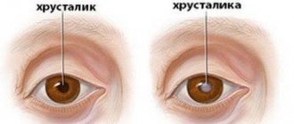
- change in the curvature of the lens - astigmatism;
- strabismus.
The fund will help
The Providence Foundation for helping premature babies and their families has been saving the sight of children for more than two years and recently published a book for parents “Look at the World”: Experts on Retinopathy of Prematurity,” written in simple, accessible language, and also launched an online course for parents from perinatal psychologists “Difficult childbirth: accepting and surviving.”
You can get detailed advice on what to do if a child is diagnosed with retinopathy by calling the fund:
+7;
+7.
Illustrations: Oksana Romanova
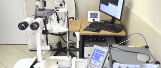
Diagnostics
The primary method of examining a patient with suspected retinopathy is ophthalmoscopy, used for children and adults: examination of the fundus of the eye with a special instrument using an ophthalmoscope. Afterwards, if necessary, several more diagnostic procedures are prescribed:
- tonometry (checking intraocular pressure);
- perimetry (study of the boundaries of visual fields);
- laser scanning of the retina;
- angiography (contrast x-ray examination of blood vessels);
- biomicroscopy of the eye;
- Ultrasound of the eye;
- differential diagnosis (especially for the hypertensive form, similar to occlusion of the central retinal vein).