Angle-closure glaucoma (ACG) is a condition of acutely elevated intraocular pressure (IOP) associated with a closed anterior chamber angle.
It can be chronic or, in about 10% of cases, acute.
PAH is a cause of severe visual impairment worldwide, with particularly high incidence in some Asian populations.
In a normal organ, a clear, jelly-like fluid is produced by the ciliated body behind the thin, moving diaphragm of the eye and flows through the apple of the eye into the reticular junction that lies around the circumference of the angle between the iris and the transparent outer membrane. This connection at the periphery of the anterior chamber is the anterior chamber angle.
Sometimes the iris becomes attached to the trabecular meshwork and thus blocks aqueous drainage. This results in increased IOP, which causes a range of symptoms and signs, depending on the type of angle closure.
Causes of angle-closure glaucoma
During an acute attack of pathology, a sudden blockage of the drainage of transparent jelly-like moisture occurs.
Fluid continues to be produced and IOP increases rapidly. This can cause damage to the optic nerve at the back of the eye and decreased vision.
Some patients are more prone to this form of the disease due to the structure of the visual analyzer. The iris may be thinner and more flexible, making it more likely to cause a reticular block.
The muscles of the iris control the diameter of the pupil. For those who are prone to the acute form of the disease, a dilated pupil may mean that the lens is sticking to the back of the iris. This process leads to blockage of the outflow of clear fluid.
Moisture collects behind the colored part of the eye shell and causes it to bulge forward, blocking the reticular junction. IOP increases sharply.
We recommend reading: Closed-angle and open-angle glaucoma
The following conditions can cause PAH:
- watching TV in dim light, which leads to expansion of the pupil, or a stressful situation;
- Some medications, such as tricyclic antidepressants or SSRIs, can cause an attack;
- overwork;
- hypertensive crisis;
- genetic predisposition.
Farsightedness, malignant tumors or swelling as a result of inflammation of the ciliary body are also considered causes of PAOG.
general information
Glaucoma is an insidious disease that can be asymptomatic for a long time. The danger of this pathology is that it can ultimately lead to permanent loss of vision.
The disease is characterized by damage to the optic nerve, decreased quality of vision, and a narrowing of the visual field. Most often, this all happens against the background of ocular hypertension, or increased IOP (intraocular pressure). The pressure inside the eye depends on the ratio of production and removal of aqueous humor. Ocular hypertension occurs when there are obstructions that interfere with the normal outflow of ocular secretions. The pressure rises to critical levels, which the visual organ is unable to cope with.
An acute attack of glaucoma, as a rule, occurs against the background of complete well-being. Pain in the eyeball gradually increases. In parallel with this, a headache occurs, most often in one part of the head. Most often, blood pressure readings are also elevated. A person may experience nausea, vomiting, and abdominal pain.
On a note! The development of this pathological process is based on a violation of the circulation of aqueous humor. Liquid accumulates, which is why pressure readings begin to rise. The structures of the eye experience stress that they simply cannot cope with. This leads to disruption of the blood supply to the visual organ. As a result, atrophic changes develop in the optic nerve. Signals from the eye stop reaching the brain.
The quality of vision deteriorates greatly. Patients see all objects as if through a fog. A black spot or halo may appear in front of the eyes when looking at a light source.
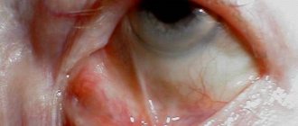
On visual examination, hyperemia (redness) of the eyeball is noticeable. Superficial vessels dilate and twist. The light iris takes on a greenish tint.
The trigger mechanism in the development of a glaucomatous attack can be a circulatory disorder, dystrophic changes in the optic fibers, or compression of the nerves. Provoking factors also include hypothermia, overwork, stress, and prolonged stay with your head down.
The initial stages of the disease may not manifest themselves at all. Often, the appearance of clinical symptoms indicates the development of irreversible processes in the optic nerve.
As the disease progresses, the narrowing of the field of vision becomes so severe that the person sees an incomplete picture. The so-called tunnel vision effect occurs. When trying to examine objects, defects appear in the form of circles, rings, and glows.
If you experience symptoms of an acute attack of glaucoma, you should immediately consult a doctor, otherwise you may lose your vision
There are two forms of the disease - open-angle and closed-angle. An acute attack is more typical in the second type of glaucoma, when the outflow of aqueous humor is suddenly blocked. The open-angle type is diagnosed in ninety percent of cases. In this case, the functions of the drainage system are disrupted, while natural access to it is open.
The increase in IOP in open-angle glaucoma occurs gradually. The pathological process can develop over several years. Sometimes a person discovers, absolutely by accident, that he has vision in only one eye.
Angle-closure glaucoma is characterized by overlap of the anterior chamber angle by the iris. Lack of access to the natural drainage system of the eye causes an acute attack.
IMPORTANT! Most often, an acute attack of glaucoma occurs in elderly people. As a rule, only one visual organ is affected.
Classification of angle-closure glaucoma
It can be primary and secondary. The primary form is a consequence of the anatomical features of the anterior chamber of the visual analyzer, the secondary form is provoked by another pathological condition.
Doctors distinguish between stabilized and unstabilized forms of the disease. Depending on the level of IOP, the pathology is divided into normotensive and hypertensive variants.
PAH has 4 stages of development:
- latent;
- in the second stage, a decrease in visual fields appears;
- then pain appears, and the quality of visual perception drops significantly;
- the last stage is damage to the second pair of cranial nerves and complete blindness.
Acute glaucoma: essence and mechanism of development
The eyeball is divided into two chambers. Between them is the lens with the iris. The cameras are equipped with special channels. The contents of the pockets circulate freely inside.
A certain pressure is automatically maintained in the healthy eye. The normal value is from 10 to 22 mmHg.
This is ensured by the balance of fluid inflow and outflow. The proper functioning of the eyes depends on the stability of pressure.
Sometimes the location of the lens changes. It moves forward, adjacent to the iris.
The cameras are isolated from each other. The pathological condition of the eyes disrupts the physiological circulation of moisture.
As the liquid substance accumulates, the pressure increases. The optic nerve and auxiliary anatomical formations experience excessive stress.
The blood supply to the eye structures is disrupted. As a result, the nerve atrophies.
The signals do not reach the brain, the person begins to see poorly and becomes blind.
Photo 1. Location of the lens in the eye
An acute attack of glaucoma is most often caused by its angle-closure form. Elderly people, after 60-70 years, are susceptible to the disease. But sometimes it is registered in younger people. There is also a congenital anomaly.
Symptoms of angle-closure glaucoma
Signs of the acute form begin suddenly. Clinical picture:
- sudden, severe pain in and around one eye;
- hyperemia;
- blurred vision, with rainbow-colored circles around bright light sources;
- pain extends to the head;
- patients experience nausea and vomiting;
- the visual analyzer is hard to the touch;
- the transparent surface of the eyeball appears hazy.
Symptoms begin in dim lighting, sudden agitation, after taking certain medications, or after general anesthesia. Signs worsen if timely treatment is not carried out.
Symptoms disappear when intraocular pressure falls, but recur when IOP rises. They are also typical for simple PACG.
What is an acute attack of glaucoma?
The cause of problems with the visual apparatus lies in a sharp increase in intraocular pressure.
Often an acute attack of glaucoma occurs against a background of chronic pathology due to a stressful situation, severe physical exertion or hypothermia. Sometimes an anomaly appears for no apparent reason. The exact factors influencing sudden changes in intraocular pressure in glaucoma have not yet been elucidated. Most often, IOP jumps to fifty millimeters of mercury as a result of strong excitement. For example, news of the death of a loved one or financial troubles can trigger an acute attack. Also, the cause of the development of pathology is sometimes hidden as a result of taking medications containing atropine. Therefore, such drugs are administered to elderly people only after measuring intraocular pressure.
Most often, glaucoma develops in elderly patients. In exceptional cases, it is diagnosed in people under fifty years of age.
Diagnosis of angle-closure glaucoma
The diagnosis is made based on the clinical picture and external signs. A presumptive diagnosis is made by a general practitioner, ophthalmologist or optician. Confirmed by an examination carried out in an ophthalmology clinic by an experienced specialist.
Upon examination, redness of the eyes is visible, expressed along the periphery of the cornea. The pupil is dilated. On palpation, the doctor notes hardness.
The slit lamp is used to detect swelling of the corneal epithelium and determine the degree of damage to the optic nerve.
If the acute attack is provoked by a secondary cause, examination may reveal peripheral anterior synechiae associated with uveitis.
Symptoms and signs are quite general. It is important to conduct a differential diagnosis to distinguish PAOG from optic neuritis, anterior uveitis, scleritis, endophthalmitis, pigmentary glaucoma, keratoconjunctivitis, keratitis, trauma, viral or infectious disease.
High intraocular pressure is the main evidence of PAOG. Therefore, tonometry is performed. Additionally, gonioscopy, biomicroscopy, and visometry are used.
What factors can trigger an attack?
The following factors can provoke an acute attack of glaucoma:
- increased physical activity;
- long work requiring head tilt;
- strong negative emotions, anxiety, stress;
- increase in blood pressure;
- eye injuries;
- the use of certain medications - certain types of antidepressants, eye drops to dilate the pupil, nasal drops that have a vasoconstrictor effect, etc.;
- drinking alcohol, salty or pickled foods;
- one-time consumption of large amounts of liquid.
However, an acute attack of glaucoma can begin for no apparent reason; the specific triggers for this condition have not been fully studied to date.
Treatment of angle-closure glaucoma
The first method of treatment is the use of local medications to reduce IOP. There are different types of medications and eye drops prescribed in different variations. The following medications are prescribed:
- Eye drops that block beta-adrenergic receptors when injected into the human body (to reduce fluid production in the eye) and steroids (to relieve the inflammatory response) - Timolol.
- Administration of the drug Acetazolamide.
- Carbachol reduces the diameter of the pupil and helps move the iris away from the trabecular meshwork. This helps improve the outflow of jelly-like fluid.
Painkillers and antiseptics may be prescribed if necessary.
When IOP has decreased, further therapy is necessary to prevent it from increasing. Follow-up therapy relies on using laser treatment or surgery to create a small hole in the iris. It allows aqueous humor to flow freely.
Laser therapy is called peripheral iridotomy. Two small holes are made in the iris of the visual analyzer. They are almost invisible to other people. Laser treatment is performed under local anesthesia on an outpatient basis.
A surgical treatment called iridectomy is another option. The surgeon makes a small triangular hole in the iris of the eye. Subsequently, it is noticeable, but insignificant, like a small black triangle on the edge of the iris.
First aid
Having discovered symptoms of an acute attack of glaucoma, it is important to urgently take emergency care measures and urgent consultation with an ophthalmologist.
If an attack occurs suddenly and unexpectedly, you should provide first aid yourself. This will reduce the risk of developing irreversible consequences. Activities that include emergency care are aimed at pain relief and reducing eye pressure. Necessary:
- Apply drops to the eyes, the effect of which is aimed at narrowing the pupil. The most commonly used is pilocarbine. Moreover, during the first hour this should be done approximately every 15 minutes;
- For severe pain, inject promedol subcutaneously and give a sedative.
Further treatment should be prescribed by an ophthalmologist after diagnosis. To make a diagnosis, eye pressure must be measured, as well as an examination of the eye and fundus. It is also important that the patient voice the symptoms of the disease and the time of the first signs. On examination it is usually noted:
- Significant redness of the affected eye;
- Swelling of the cornea;
- A significantly dilated pupil and a change in its shade to a greenish-gray tone.
The danger of an attack of glaucoma is that if timely assistance is not provided, there is a high probability of irreversible death of the optic nerve and the onset of complete blindness.
Delay in glaucoma treatment threatens complete blindness, and also provokes cardiovascular diseases and neurological pathologies. Therefore, if you notice the slightest signs of an acute attack, you should urgently call a doctor or an ambulance.
Before providing qualified care to a patient in a hospital, rest and horizontal position must be ensured.
In the hospital, first of all, it is necessary to normalize the intraocular pressure and blood flow of the eye. These measures are intended to restore the optic nerve and retinal function.
Usually, Promedol is prescribed subcutaneously to reduce pain. The eye drug Pilocarpine is instilled in 2 drops every 15 minutes during the first hour, then at intervals of half an hour.
Diuretics Diacarb or Furosemide are used for hypotensive results. Sedatives and painkillers are also prescribed.
Saline laxatives and hot foot baths will help improve eye blood circulation and help drain excess fluid.
These procedures can be performed by a paramedic or nurse in an outpatient clinic, but if no effect is observed, hospitalization in the ophthalmology department is required, where specialists will determine the further course of treatment.
If a person has the above symptoms and external changes are visible when diagnosing the eye, then this may be an attack of glaucoma. In this case, you should immediately call an ambulance.
Prevention
The best prevention of any disease is regular examinations with a doctor. Even in the absence of symptoms, it is recommended to visit an ophthalmologist every six months.
Preventive measures prevent further problems with the optical system.
Some people have an increased risk of developing acute glaucoma because they have a shallow anterior chamber or a narrow drainage angle. Sometimes this is noticed during a routine eye examination. The doctor should talk about this and advise you to be careful with certain medications and eye drops.
If the risk of developing acute glaucoma is very high, preventative treatment such as laser iridotomy may be recommended.
How to diagnose?
If you suspect an attack, you should urgently visit the ophthalmology department of the nearest hospital. If possible, visit a specialized clinic. Because if there is no effect from drug therapy, urgent surgical intervention will be required.
First of all, the doctor measures intraocular pressure, analyzes the patient’s complaints and the condition of the organ of vision. In some people, the pathology is accompanied by atypical symptoms. This especially applies to those with dark skin. Their attack manifests itself in minor pain, the pressure practically does not increase, but visual acuity rapidly decreases.
Biomicroscopy is mandatory. During the diagnostic process, the doctor may detect the following abnormalities:
- Swelling of the cornea.
- Cloudiness.
- Enlargement of the pupil oval.
- Stromal compaction.
- The appearance of epithelial blisters in the cornea.
- Reducing the size of the anterior chamber.
- Lack of reaction to light.
- Increased intraocular pressure.
| After removing the edema from the cornea, gonioscopy is performed. It is carried out during antihypertensive treatment using glycerol or hypertonic saline ointment. |
Gonioscopy of both eyes will help make an accurate diagnosis. The examination makes it possible to detect a hidden blockade of the anterior chamber of the eye.
Stages
There are several stages in the development of pathology. The stage of the disease is determined taking into account the level of damage to the optic nerve. The anomaly manifests itself in the form of narrowing of the visual fields. Ophthalmologists diagnose the following stages of the disorder:
- First degree - the visual fields narrow, but in each meridian they are more than 45 degrees;
- Second degree - narrowing occurs in each meridian. In at least one of them it is present within 15-45 degrees;
- Third degree - the visual field narrows in each meridian. At least in one of them it is from 0 to 15 degrees;
- Fourth degree – this stage is characterized by complete loss of vision. Sometimes there is residual vision, but it only allows you to recognize light or shadow.
Manifestations of open-angle glaucoma
In the early stages, open-angle glaucoma does not show characteristic symptoms: normal vision remains, pain and other changes in well-being are absent. Sometimes patients may complain of temporary rainbow circles before the eyes and asthenopia.
An undetected disease for a long time leads to deterioration of peripheral vision, when a person clearly sees objects located directly in front of him, but does not notice those located to the side or at an angle. At first, the narrowing of the visual fields mainly occurs from the side of the nose, but then it gradually begins to cover the peripheral parts concentrically, which ends in its complete loss. In addition, patients with open-angle glaucoma note the appearance of transparent or translucent spots in their field of vision. Dark adaptation or color perception may be impaired. Sometimes there is an uncorrectable decrease in vision, which indicates an advanced stage of open-angle glaucoma, with gradual atrophy of the optic nerve. The most severe manifestation of open-angle glaucoma is an acute attack of the disease.