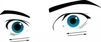
Anisocoria is a condition in which the pupils have different sizes. In this case, the reaction to light is different: one pupil remains motionless, while the second narrows and dilates. This pathology may be a consequence of ophthalmological or neurological disorders. Normally, the difference in pupil diameter should not exceed one millimeter.
Anisocoria is not an independent disease, but only a manifestation of other pathologies. A change in pupil size does not always mean the development of some disease. In this article we will talk in more detail about why the pupils are different sizes and find out how this condition can be dealt with. But first, let's find out what it means when the pupils are different sizes.
Types of anisocoria
Sometimes a person may have one pupil smaller in size compared to the other. Experts distinguish physiological and congenital anisocoria. In the first case, the difference in pupil size is no more than one millimeter, and during the examination the doctor does not detect any ophthalmological disorders. This feature can occur in absolutely healthy people.
The congenital form is formed due to defects of the visual apparatus. Moreover, visual acuity is different in each eye. Congenital pathology can also be a consequence of damage to the nervous system of the eyes. This type of anisocoria appears from birth. At the same time, the child does not have a lag in physical and mental development. In some cases, this feature can go away on its own by the age of five, while in others it remains for life.
Acquired anisocoria in adults can be a consequence of injury or ophthalmological diseases. In some cases, this condition may be caused by exposure to inorganic substances, such as belladonna or atropine.
Depending on the degree of damage, the pathology can be unilateral or bilateral. Affecting both eyes is quite rare.
If one pupil is larger than the other, you should consult an ophthalmologist
Normal and pathological functioning of the pupils
The organ of vision helps to receive visual information from the surrounding world. The pupil of the eye (the round hole in the center of the iris) performs the most important function of forming a beam of light waves entering the retina: the pupillary reflex ensures the narrowing and expansion of the hole in response to changes in the light flux.
Muscular structures of the iris
The pupil is a round hole in the center of the iris.
The anterior choroid of the eyeball is a round disc with a hole in the center. The iris acts as a diaphragm, expanding and narrowing the central area. The reflex action of the pupils is ensured by the contraction of 2 types of muscle fibers:
- ring-shaped (constricting sphincter);
- radial (expanding dilator).
The sphincter muscle fibers surround the edge of the opening, and the dilator muscles are located peripherally and radially. Contraction of a specific muscle in response to external influences in the form of an increase or decrease in the amount of light leads to a change in the size of the central disk of the iris.
Normal operation
The work of each muscle is regulated by nerves (oculomotor and sympathetic). The pupillary reflex is a chain of the following sequential events:
- Bright light irritates the retinal receptors that send information to the brain structures;
- The signal from the brain reaches the muscles of the iris through the nerves;
- Contraction of the sphincter leads to a sharp narrowing of the opening.
With a pronounced decrease in illumination, the brain forces the dilator to work: muscle contraction of the radial fibers leads to dilation of the pupils and an increase in light intake. All these events occur very quickly - the reflex should protect the retinal photoreceptors from being burned by bright rays and ensure the visibility of objects at dusk.
Standard options
The size of the central opening of the iris changes with age: wide pupils will be typical for a child, and eyes with a small opening diameter will be typical for an elderly person. Physiological options for changing the pupillary reflex include:
- emotional reaction;
- response to pain;
- strong fear;
- alcohol intoxication;
- dream;
- severe eye fatigue.
Each situation must be approached individually: during the examination, the doctor must take into account the age and psychological characteristics of the person.
Diagnostic methods
In addition to an external examination of the organ of vision, the doctor will use the following procedures during the initial examination:
- examination under lateral lighting with pupillometry (measurement of the width of the central opening);
- determining the reaction of the eyes to direct rays of light;
- checking the friendly reaction of the organ of vision (closing one eye of the patient, the doctor evaluates the reaction of the second);
- reaction to convergence and accommodation (change in the size of the pupillary opening when quickly moving the gaze from a distant point to a nearby object).
Standard diagnostic procedures will help identify reflex changes indicating various types of pathology.
Variants of diseases
Anisocoria is an asynchronous change in the size of the pupils.
All problems associated with the pupillary reflex arise against the background of impaired muscle contraction. There are 4 main types of pathology:
- mydriasis (constant enlargement of the hole);
- miosis (severe constriction of the pupils);
- anisocoria (asynchronous change in size on the right and left);
- hippus (attack of rhythmic contractions and expansions).
Pathological changes in the size of the pupillary opening are caused by the following types of diseases and conditions:
- traumatic head injuries;
- benign and malignant brain tumors;
- infectious and inflammatory diseases of the central nervous system;
- acute cardiovascular pathology (stroke, heart attack, thromboembolism);
- endocrinopathies (thyroid diseases);
- pathology of the organ of vision (glaucoma, iridocyclitis, cataract);
- poisoning by poisons, drugs or excess alcoholic beverages;
- the influence of drugs when used locally or generally.
Testing the pupillary reflex is one of the important diagnostic procedures for any acute and life-threatening conditions: an experienced doctor, based on the reaction of the eyes to a light source, can with a high probability guess the cause of loss of consciousness and the prognosis for life.
Causes
The causes of anisocoria can be very different. Different pupil sizes may be a consequence of ophthalmological factors, namely:
- uveitis;
- iritis;
- iridocyclitis;
- lens implantation;
- surgery on the eye.
Pupils of different sizes can also occur due to other reasons:
Two pupils in one eye
- cerebral aneurysms;
- traumatic brain injury;
- pathologies of the oculomotor nerve;
- migraine;
- hemorrhages;
- infectious processes;
- genetic predisposition;
- cervical osteochondrosis;
- syphilis;
- epidemic encephalitis;
- stroke;
- tumor process;
- glaucoma;
- taking certain medications;
- cerebrovascular accidents;
- herpes zoster.
Causes
The main factors for the development of anisocoria are the following:
- Genetics. If close relatives were diagnosed with this disease, and the child inherited it, then such a hereditary predisposition does not pose a threat. Usually no treatment is required.
- Muscle damage. The muscles responsible for the constriction of the pupil are damaged by infection and inflammation. Accordingly, they cannot sufficiently perform their functions.
- Some medications and inhalers for asthma lead to asymmetry. Also, different pupil sizes are found after taking drugs.
- The disease may be a consequence of damage to the optic nerve or tract, as well as other plexuses of the optical system running along the brain stem to the center of the hypothalamus.
When should you see a doctor?
If one pupil has become wider and this condition does not go away, and it cannot be explained, be sure to consult an ophthalmologist. You should be especially wary of the following symptoms:
- blurred vision;
- fever;
- photosensitivity;
- double vision;
- Pain in the eyes;
- headache;
- disturbance of consciousness;
- temperature increase;
- attack of nausea and vomiting.
If you notice that your pupils are different sizes, do not hesitate to consult a doctor.
Reasons for different pupils
In young children, the pupils are usually enlarged, but evenly. The condition in which their diameters differ is called anisocoria. If the difference is less than 1 mm and there are no pathological manifestations, this is considered normal.
Physiological anisocoria is observed in 20% of people from birth and is usually hereditary. By 5-6 years it can disappear without a trace.
Pathological anisocoria occurs due to an imbalance in the functioning of the eye muscles. Why is this happening? The most common cause is the use of eye drops or accidental exposure of the conjunctiva to certain medications. In addition, taking medications with narcotic effects can lead to uneven dilation of the pupils. The diameters of the holes in the iris become the same after stopping the use and removal of drugs from the body.
The remaining causes of anisocoria can be divided into ophthalmological and those related to the functioning of the central nervous system. Main ophthalmological factors:
- congenital deficiency of the eye muscles, which may be accompanied by strabismus or decreased visual acuity;
- injuries accompanied by damage to the iris, muscles and nerve fibers;
- iridocyclitis – inflammation of the ciliary body and iris;
- glaucoma – increased pressure inside the eye (extremely rare in children);
- herpetic eye lesions.
Neurological causes of anisocoria in infants:
- damage to the cervical spine during childbirth;
- a rapidly growing tumor in the brain;
- aneurysm;
- cerebral hemorrhage;
- meningitis;
- neurosyphilis;
- encephalitis;
- traumatic brain injuries;
- tuberculosis;
- thrombosis of the carotid artery.
Disturbance in the functioning of the pupils in these pathologies occurs due to compression of the nerve responsible for eye movement, or damage to the visual areas of the cerebral cortex. These conditions are always accompanied by other symptoms of trouble, if detected, you should immediately seek medical help. Possible manifestations:
- increased body temperature;
- vomit;
- restless behavior and sharp cry due to pain;
- neck muscle tension;
- weakness, apathy, drowsiness;
- photophobia;
- decreased vision and so on.
Anisocoria may be one of the signs of Horner's syndrome. In infancy, this disease is most often congenital or develops due to trauma to the cervical spine during childbirth. Its symptoms result from compression of the sympathetic nerve and damage to the eye muscles. Main signs (appear on one side of the face):
- anisocoria with delayed dilation of one of the pupils;
- drooping eyelid (ptosis);
- sunken eyeball;
- different colors of the irises (not always observed);
- lack of sweating on the face.
Diseases that cause changes in the pupil
Let's talk about pathologies, one of the symptoms of which is anisocoria. First, let's discuss paresis of the oculomotor nerve.
Oculomotor nerve palsy
The slightest changes in the functioning of the oculomotor nerve affect a person’s quality of life. Children suffer from this disease quite rarely. It is almost impossible to recognize the disease in the initial stages, since it does not manifest itself in any way.
The following reasons can cause nerve paresis:
- cervical osteochondrosis;
- diabetes;
- hypertension;
- vasculitis;
- carotid artery aneurysm;
- tumor process;
- heart attack;
- stroke;
- syphilis, diphtheria, encephalitis, meningitis;
- side effects of medications;
- injuries;
- ocular migraine.
With paresis of the upper eyelid, the eye closes completely or partially. Outwardly, this manifests itself in the form of a squint. Most often, the pathology has a one-sided process. In addition to physical inconvenience, the problem causes aesthetic discomfort. Paresis of the upper eyelid leads to deterioration of visual acuity.
Paresis of the oculomotor nerve is one of the causes of anisocoria
A birth defect is formed as a result of abnormalities in the formation of muscles or intrauterine nerve damage. Acquired pathology can be a manifestation of injuries, as well as neurological disorders.
With mydriasis, the pupil dilates. The disease occurs as a result of injuries, diseases of the nervous system, visual apparatus, as well as the use of potent drugs. Normally, pupil dilation is a natural reaction to lighting. This can also occur during severe emotional stress.
After the diagnosis of “oculomotor nerve palsy” has been established, the patient is registered with a specialist. To avoid mistakes, he is asked to undergo a repeat test. In general, the disease has positive dynamics. Doctors usually recommend doing strengthening exercises for the extraocular muscles. Patients are prescribed vitamins and medications. You may also need to wear bandages or glasses.
Full restoration of nerve mobility occurs after about six months. If there is no result, surgery may be required.
Bernard Horner syndrome
The development of the disease is based on damage to the sympathetic nervous system. The disease affects the muscle tissue of the body, including the visual apparatus. A number of provoking factors can cause the appearance of the syndrome:
- damage to brain tissue;
- cluster headache;
- injuries, including surgical ones;
- otitis media;
- aortic aneurysm.
Bernard Horner syndrome manifests itself in the form of drooping of the upper eyelid, decreased production of tear fluid, a haggard appearance of the face, unnatural constriction of the pupil, and sunken eyeball. The disease also causes heterochromia, in which the pupils have different colors. In addition, the eye loses its ability to adapt to light. The stronger the level of illumination, the more constricted the pupil, while in the dark it, on the contrary, expands.
In Bernard Horner syndrome, the pupil constricts in bright light and dilates in the dark
The treatment process may include electrical stimulation. Electrodes are attached to the affected areas. The essence of the technique is to stimulate muscles through short electrical impulses. This normalizes blood circulation and in some cases leads to complete recovery.
Problem areas can also be corrected using plastic surgery. Stimulation of affected facial tissues is also possible with the help of drug therapy.
Eydie syndrome
Patients have a slow reaction of the pupils to light, in some cases it is completely absent. Even if you shine a flashlight directly into your eyes, the same braking reaction will be observed. On the affected side, the pupil dilates and becomes deformed.
The disease can be congenital or acquired. The cause of Eydie syndrome can be ophthalmoherpes, atrophy of the eye muscles, meningitis, encephalitis, myotonia.
Treatment includes the use of Polycarpine. Regular use of these drops will help to achieve some improvement in the condition. Glasses are used to correct violations.
Reasons and what determines the size of a person’s pupils
The pupil in the human eye is responsible for regulating the light rays that constantly hit the retina when the eyes are open. It is the quantity and quality of light falling on the pupil that leads to a decrease or increase in its diameter. So, in dim lighting the pupil narrows, and in bright light it expands.
How to treat a chalazion on the upper eyelid can be found on our website.
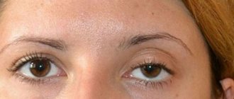
Ideally, both pupils should be the same size - and this is what they are for most people. However, if there is a slight “skew” in the diameter of one of the pupils, and it is slightly larger in size than the second, doctors advise not to sound the alarm. If the deviation is insignificant and the level of vision does not drop, this fact is considered normal and acceptable. However, if the size of the pupils differ greatly and noticeably, it is necessary to undergo an examination by an ophthalmologist.
Important: it is considered acceptable to exceed the diameter of one pupil by one millimeter, no more.
The causes of pathology can be various factors; below we will consider the most common of them.
Genetics
pupil reaction to light
One of the main factors determining the appearance of anicozoria in humans. In this case, the problem will manifest itself from an early age. Note that hereditary anicozoria does not cause any harm to health, and a drop in the level of vision does not occur in this case.
Medicines
The use of certain medications may cause temporary dilation of the pupils. Such medications are usually mydriatic drops, as well as drugs for the treatment of asthma in the form of inhalers.
Muscle work
If the eye muscles malfunction, the size of the pupil may be distorted when light rays enter the eyes.
Holmes-Eydie syndrome
With this disease, the pupil dilates in a person in adulthood, and, having dilated, it stops responding to light stimuli. The syndrome appears as an individual sensitivity of the body to pilocarpine. This expansion is detected randomly, since it does not cause any negative aspects in a person.
In addition, the causes of this pathology can also be:
- damage to the optic nerve;
- neurological pathologies;
- atrophy and aneurysm of the visual organs;
- taking drugs (opium, cocaine, etc.)
constriction and dilation of the pupil depending on external factors
Often the cause of this phenomenon is developed myopia - in this case, the pupil will be dilated in the eye that sees worse.
Pathological, dangerous causes of anicozoria are also possible, among them the following are especially likely:
- bleeding as a result of injuries, bruises of the head;
- tumor-like formations in the brain;
- infection of the lining of the brain, manifested as meningitis or encephalitis;
- pathological damage to the oculomotor nerve;
- glaucoma and migraine;
- cancer of the lung (upper part);
- lymph node cancer;
In addition, if there are pupils of different sizes with different colors of the iris or slightly drooping eyelids, we can talk about Horner's syndrome. This is a congenital pathology, but manifests itself only in adulthood. The “trigger” that triggers Horner’s syndrome is spinal injuries, osteochondrosis of the cervical area, injuries to the spinal and cervical muscles.
It is possible to accurately establish the cause of anicozoria only with a professional ophthalmological examination.
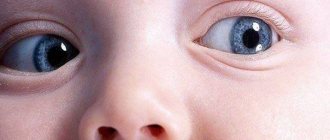
Anisocoria in children
If one pupil is larger than the other in a baby, this indicates a congenital pathology. Most often, the cause of such a defect is underdevelopment of the ANS or pathology of the iris. This disorder is often accompanied by the appearance of strabismus and ptosis, that is, drooping of the upper eyelid. If one pupil suddenly becomes larger, this may be a manifestation of the following pathologies:
- brain contusion;
- tumor process;
- aneurysm;
- encephalitis.
At an older age, one pupil larger than the other may appear for the following reasons:
- injuries;
- cerebral edema;
- inflammation of the iris;
- ophthalmic injuries;
- intoxication;
- aneurysm;
- tumor;
- overdose of medications.
In a child, one pupil larger than the other is most often the result of birth defects
Speaking about the reasons that can cause the appearance of pupils of different widths in a baby, it is worth paying attention to relatives. After all, if one of the parents had such a feature, then there is a high probability that such a phenomenon will be repeated in the newborn.
Treatment of anisocoria
Treatment of this disease depends entirely on the identified cause of the pathology. If this is a hereditary or physiological condition, then there is no need for treatment. If the cause is infectious or inflammatory processes, then treatment may be prescribed for a suitable nosology. Local or systemic antibiotics are used. For tumor processes, treatment requires surgical intervention. Due to the fact that the reasons for different pupil diameters can be completely different and can be of a different nature, it is better not to delay visiting a specialist.
And also about visual scotoma here.
Diagnostics
An ophthalmologist diagnoses anisocoria. To clarify the reasons for this phenomenon, the following studies will be required:
- ophthalmoscopy;
- measurement of intraocular pressure;
- electroencephalography;
- Brain MRI;
- X-ray of the lungs;
- cerebrospinal fluid analysis;
- Dopplerography of the blood vessels of the brain.
The photo shows the process of eye ophthalmoscopy
Treatment
The treatment process begins with a consultation with an ophthalmologist and a neurologist. Most often, anisocoria does not require treatment. But this largely depends on the main diagnosis and the provoking factor in the development of the phenomenon. Sometimes the use of anti-inflammatory drugs and antibiotics may be necessary. To relieve spasm and pupil dilation, anticholinergic drugs are used.
Important! The entire course of treatment is aimed exclusively at the underlying disease. If the phenomenon is not associated with pathologies of the iris, treatment is not required at all.
To combat inflammatory ophthalmological processes, you will need to take antibiotics, antipyretics and water-salt solutions. If anisocoria develops as a result of a stroke, doctors prescribe medications that thin the blood and dissolve blood clots.
Immediate surgical intervention is required if anisocoria occurs due to head injuries. For meningitis and encephalitis, leading to cerebral edema, complex treatment is required. If the reason lies in the tumor process, surgery will be required.
So, anisocoria is a condition in which the pupils differ in size. It can be physiological or congenital. In this case, treatment is not prescribed. Often, by the age of five or six, this feature goes away on its own. The acquired form can be a consequence of injuries, neoplasms, stroke, pathologies of the iris, and others. Treatment is aimed primarily at eliminating the underlying disease.
Anisocoria
Typically, a person's pupil size is 3-8 mm. If the diameter is different in the left and right eyes, they speak of anisocoria.
The physiological symptom is observed in 20% of the population. The difference in the size of the pupils in such people is the same in the light and in the dark, and the pupils themselves react normally to light. If you drop a special drug into an eye that has a constricted pupil to dilate it, they become the same. Physiological anisocoria is a normal variant (with a difference of up to 1 mm), does not affect visual acuity and does not require treatment.