Diagnosis of the causes of pupillary defect
The first stage in diagnosing the causes of anisocoria is collecting an anamnesis.
The doctor must identify all concomitant pathologies, study their causes, development and duration. Photographs of the patient help in diagnosing anisocoria. From them you can find out whether the pathology existed before and with what dynamics it developed. During an eye examination, the doctor determines the size of the pupils in light and in the dark, reaction speed, and consistency under different lighting conditions. These simple characteristics help to at least approximately determine the cause of anisocoria and the localization of the disorder that provokes pupil mismatch.
Anisocoria, which is more pronounced in bright light, is indicated by pathology when the pupil dilates to a large size and has difficulty constricting. With anisocoria, which is more pronounced in a dark environment, the pupil becomes unnaturally small and dilates with difficulty.
Methods for diagnosing anisocoria
- Cocaine test. In the process, a 5% solution of cocaine is used (if the patient is a child, a 2.5% solution is used). Sometimes the cocaine solution is replaced with apraclonidine 0.5-1%. The test allows you to differentiate physiological anisocoria from Horner's syndrome. The procedure is simple: drops are dropped into the eyes and the size of the pupils is assessed before the procedure and after 60 minutes. If there are no pathologies, the pupils gradually dilate. In the presence of Horner's syndrome, the pupils on the affected side dilate to 1.5 mm.
- Phenylephrine and tropicamide tests. A solution of 1% tropicamide or phenylephrine can detect a defect in the third neuron of the sympathetic system, although a defect in the first and second cannot be excluded. The procedure is as follows: drops are instilled into the eye, analyzing the size of the pupils before and after the procedure (after 45 minutes). An expansion of less than 0.5 mm will indicate pathology. With an increase in anisocoria by 1.2 mm, we can talk about damage with a probability of 90%.
- Pilocarpine test. For the procedure, a 0.125-0.0625% solution of pilocarpine is used. The defective pupils are sensitive to the product, while healthy eyes do not react to it. Pupil dilation should be assessed half an hour after instillation.
Anisocoria may be associated with these symptoms
- Pain. May indicate expansion or rupture of an intracranial aneurysm, which is dangerous due to compression paralysis of the third pair of oculomotor nerves. Pain also appears during dissection of the carotid artery aneurysm. Another cause of pain may be microvascular oculomotor neuropathy.
- Double vision.
- Ptosis and diplopia. May indicate damage to the third pair of oculomotor nerves (cranial).
- Proptosis (protrusion of the eyeball forward). Often accompanied by space-occupying lesions of the orbit.
If vascular abnormalities are suspected, contrast angiography and Doppler ultrasound are prescribed. Diagnosis of ocular dysfunction often includes CT, MRI and MSCT with vascular contrast. Even if there are no other symptoms, these tests can detect aneurysm and brain tumor - the most common causes of anisocoria. Neuroimaging studies allow us to determine the exact treatment plan and the need for neurosurgery.
Mechanism of mydriasis
Through the pupil, rays of light penetrate deep into the eye. The iris, or iris, is located in the eye behind the cornea and in front of the lens; it is located vertically and divides the space between the lens and the cornea into two parts: the anterior and posterior chambers. The iris serves as a diaphragm to protect the bottom of the eye, the retina, from excessive light rays that interfere with seeing objects clearly. There are two types of muscles in the iris: circular with fibers of the oculomotor nerve ending in them and radially located with fibers of the sympathetic nerve ending in them. Excitation of the oculomotor nerve causes contraction of the circular muscles, as a result of which the pupil contracts, and weakening of the excitation of this nerve or its paralysis leads to relaxation of the circular muscles, moving them to the periphery, as a result of which the pupil dilates. When the sympathetic nerve is excited, the radial muscles contract and move back to the place of their attachment, pulling the iris to the periphery, which causes the pupil to dilate. A decrease in the excitation of sympathetic nerve fibers in the iris or their paralysis is reflected in a more pronounced constriction of the pupil, since the circular muscles, contracting the pupil, do not encounter resistance from the radial muscles in such cases. Under normal physiological conditions, the size of the pupil automatically changes depending on the interaction of the circular and radial muscles.
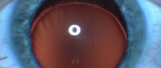
Mydriasis (pupil dilation)
Mydriasis (lat. mydriasis) is the dilation of the pupil. The term itself may have come from the Greek word "amydros", meaning "dark, obscure".
Mydriasis can be physiological and pathological, unilateral or bilateral.
Pathological dilatation of the pupil can be associated both with paralysis (paresis) of the sphincter of the pupil, and with spasm or increased tone of the dilator located in the iris.
Mydriasis can be caused by the use of various medications (mydriatics and cycloplegics, antihistamines, sympathomimetics, estrogens, antidepressants, anesthetics, narcotic drugs), as well as intoxication, eye diseases and injuries, eye surgeries, and neurological disorders.
Varieties of mydriasis
- Physiological mydriasis is a normally occurring dilation of the pupils in low light conditions, for example, at dusk. With strong emotions - fear, excessive excitement, etc. – the pupils also dilate due to the influence of the sympathetic nervous system on the eye muscles. Hence the famous saying “fear has big eyes.” Physiological mydriasis is always bilateral and symmetrical.
- Drug-induced mydriasis is caused by drugs. Pupil dilation occurs as a result of instillation of drugs such as atropine, midriacil, tropicamide, irifrin, etc. Pupil dilation is necessary for many ophthalmological examinations and some operations. Most often, drug mydriasis is used for a detailed examination of the fundus. To relieve spasm of accommodation (persistent muscle tension leading to deterioration of vision), a course of cycloplegia is prescribed - instillation of eye drops for several days or weeks, which relax the accommodative muscles, while simultaneously dilating the pupils.
A
If treated, then how?
There is no need to treat short-term mydriasis with mild contusions, and especially drug-induced mydriasis.
A necessary condition for eliminating persistent mydriasis is treatment of the underlying disease that caused the pathological dilation of the pupils.
Since patients are concerned about increased sensitivity to light (photophobia), it is recommended to wear dark glasses and avoid bright light.
In some cases, drugs are used that enhance the work of the pupillary sphincter and, accordingly, weaken the effect of the dilator: M- and N-cholinomimetics, alpha-blockers.
mydriasis: description of symptoms and treatment methods for mydriasis
› Choroid of the eye
articles:
- 1 Forms of pathology
- 2 Reasons
- 3 Diagnostics
- 4 Treatment
Mydriasis – pupil dilation more than 5 mm. A wide pupil can be normal, occur against the background of diseases, or be provoked by the administration of medications.
In any case, the appearance of this symptom requires contacting an ophthalmologist, neurologist, or therapist with an examination to clarify the diagnosis.
Forms of pathology
The width of the pupil is determined by the work of two muscles, one of which contributes to its narrowing, the other to its expansion. Normally they should work in harmony. When one of them is damaged, which can happen for various reasons, the antagonist muscle begins to predominate. Therefore, when the sphincter is damaged, the pupil dilates, this can be on one or both sides.
Mydriasis is divided into physiological and pathological.
Physiological pupil dilation occurs in low light conditions. Dilation occurs to increase the flow of light to the retina. This phenomenon is also observed during anxiety, stress, and in any extreme situations when the sympathetic nervous system is excited. A characteristic feature of physiology is the symmetrical dilation and preservation of pupillary reactions to light.
Pathological mydriasis is often unilateral, asymmetrical, pupillary reactions to light are reduced or absent. Deformation of the pupil and decreased mobility of the eyeball are possible.
Causes
This symptom develops against the background of various processes and diseases. Can be called:
- taking adrenergic, hormonal, desensitizing drugs, antidepressants or analgesics;
- unfavorable environment;
- carbon monoxide poisoning, narcotic substances;
- working in hazardous industries.
Pupil dilation is observed when mydriatics (atropine, tropicamide, ephedrine) are instilled into the eyes. This is necessary for examining the fundus of the eye, relieving spasm of accommodation, and during ophthalmic operations.
The following infectious diseases also cause pathology:
- botulism;
- tuberculosis;
- syphilis with damage to the nervous system.
On the part of the visual organs, this phenomenon can be caused by:
- eye injuries;
- glaucoma with frequent attacks of increased intraocular pressure;
- inflammatory eye diseases;
- consequences of damage to the innervation of the pupil during operations on the structures of the eye.
Many diseases of the nervous system are accompanied by this symptom:
- traumatic brain injuries, including fractures of the facial skeleton or base of the skull;
- brain tumors of a certain location;
- inflammatory and infectious diseases (encephalitis, meningitis, poliomyelitis);
- hematomas of various origins;
- syringomyelia;
- multiple sclerosis;
- Parkinson's disease;
- hydrocephalus with increased intracranial pressure;
- epilepsy.
When the sympathetic nerve trunk is irritated at the level of the cervical spine by space-occupying formations, a vascular aneurysm, or a lung tumor, the sympathetic section of the autonomic nervous system is stimulated, which leads to the appearance of wide pupils.
Damage to the oculomotor nerve in diabetes mellitus and viral infections leads to the development of paralysis of the pupillary sphincter. In this case, the pupil loses its ability to constrict.
Diagnostics
Complaints of photophobia, pain (with eye or head injuries), lacrimation.
There is a feeling of a foreign body, dry eye, and constant discomfort.
During the examination, the ophthalmologist or neurologist pays attention to the following points:
- state of consciousness and the presence of neurological symptoms;
- pupil shape;
- reaction of pupils to light;
- restriction of eye movements.
In addition, instrumental diagnostics are important:
- general, biochemical blood tests;
- visual acuity test;
- fundus condition;
- perimetry;
- Ultrasound of the eye;
- MRI of the brain or cervical spine.
Treatment
Treatment of the underlying disease that led to the appearance of a dilated pupil is necessary.
Palliative methods include wearing sunglasses and avoiding places with bright lighting. On the recommendation of an ophthalmologist, it is possible to use drops that narrow the pupil.
Mydriasis is only a symptom of disease. Contacting an ophthalmologist at the first signs of illness will help to conduct a quick, comprehensive diagnosis and identify the etiology of pupil dilation. Treatment should be aimed at the cause of the pathology! The sooner it is prescribed and carried out, the greater the chances of avoiding complications and maintaining health.
19.04.2016
Source: https://ofthalm.ru/midriaz.html
Types of mydriasis according to clinical signs
Depending on how many eyes have abnormalities in physiological functioning, mydriasis can be of two types.
Bilateral
Mydriasis bilateral
Pupil enlargement is characteristic of both eyes at the same time. Depending on the cause, the reaction to an increase in the intensity of the light flux may persist or be absent.
- Reaction to light is preserved. Most often it manifests itself in thyrotoxicosis, pathological sympathicotonia and hypertension. Other reasons: fever, various inflammatory processes in the eyeball, eclampsia in pregnant women. Enlarged pupils in nearsighted people, patients with diabetes mellitus and syringomyelia, and with tumors of nerve tissue. In all these cases, mydriasis is considered a secondary clinical sign of the main diseases; it is not it that is treated, but the disease itself.
- There is no reaction to light; the pupils do not respond to changes in light intensity. This condition is called internal ophthalmoplegia. Pathological changes occur after poisoning with mushrooms and carbon monoxide, drug overdose, chronic alcoholism. The pupils become enlarged under the influence of medications containing the active ingredients atropine and scopolamine. Another very dangerous cause is poisoning with botulinum toxin or quinine. The pupils dilate and do not respond to light during near-death conditions, when all nervous physiological reactions slow down. Bilateral mydriasis with lack of reaction to light is characteristic of some types of blindness, congenital hydrocephalus, Parkinson's disease and certain types of epilepsy. In some cases, this phenomenon can be observed with brain injuries, cardiovascular diseases, in the last stages of syphilis and multiple sclerosis.
Pupil dilation (midras)
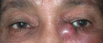
All patients with signs of mydriasis are examined in detail and comprehensively
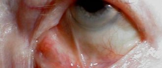
Unilateral
There is a difference in the diameters of the pupils of the left and right eyes, which also has various causes depending on the ability to respond to changes in illumination.
- The reaction to changes in light is preserved. It occurs as a consequence of unilateral irritation of sympathetic nerve fibers, if necessary during the examination of the creation of unilateral mydriasis with medications and during Pourfour du Petit syndrome. In the latter case, it is impossible to close the eyelids on one eye. Rarely, this condition appears during severe attacks of unilateral migraine and with irritation of the hypothalamus.
- There is no reaction to light, a condition called internal ophthalmoplegia. Occurs with various types of unilateral pathology of the nerve responsible for the movements of the eyeball, with postoperative iridoplegia, after a sharp attack of glaucoma. Dilation of one pupil can be a consequence of inflammatory processes in the eyeball, complications after diphtheria, or Eydie syndrome (hypersensitivity of one of the muscles causes paralysis of accommodation).
Unilateral mydriasis
Clinical picture
Pathological mydriasis is accompanied by the following symptoms:
- change in the shape of the pupil (oval or pear-shaped);
- absence or decreased reaction to light;
- impaired mobility of the eyeball;
- lack of reaction to approaching objects.
If mydriasis develops against the background of injury, then the pupil diameter varies from 8 to 10 mm. The pathological condition may disappear after the patient recovers or becomes permanent. Traumatic mydriasis occurs against the background of the following symptoms:
- lacrimation;
- pain and severe discomfort in the eye area;
- photophobia;
- increased fatigue while reading or working at the computer;
- decreased response to light;
- partial afferent failure.
The paralytic type of mydriasis provokes the occurrence of strabismus and drooping of the upper eyelid. In the spastic form, patients note a unilateral localization of the pathology, a decrease in the pupil's reaction to light and the movement of objects, and discomfort in bright lighting.
If mydriasis develops against the background of viral infections, it leads to loss of accommodation.
Mydriasis causes and treatment
Pupil dilation under the influence of physiological or pathological factors is called mydriasis. Normally, pupils enlarge in low light or at dusk.
Physiological mydriasis is symmetrical; when the lighting changes, the pupils quickly react with dilation, but remain the same in both eyes.
Pathological mydriasis can be unilateral or bilateral. The dilation of the pupil is caused by taking certain medications; often the pupils are enlarged due to poisoning or traumatic brain injury.
Causes
Dilation of the pupils is considered normal at low levels of illumination and emotional and mental reactions (for example, fear). In all other cases they speak of pathological mydriasis.
Enlarged pupils are caused by:
- Poisoning;
- Brain tumors;
- Glaucoma;
- Taking medications (antidepressants, antiallergic drugs, estrogens, anesthetics, narcotic drugs);
- Injuries and eye surgeries leading to weakening of the ciliary muscle;
- Diseases of the nervous system (poliomyelitis, meningitis);
- Traumatic brain injuries;
- Some viral infections;
- Diabetes;
- Hypoxia;
- Botulism;
- Increased intracranial pressure.
Types of mydriasis
In addition to physiological, mydriasis can be traumatic, drug-induced, paralytic, spastic, etc.
Medication is a reaction of the pupils to certain medications. It can be called by an ophthalmologist for the purpose of examining the fundus of the eye or performing surgical interventions. For this, drops (atropine, ephedrine, platyphylline and scopolamine) are most often used.
Paralysis of the sphincter of the pupil leads to paralytic mydriasis. Tuberculosis, syphilis, hydrocephalus, meningitis and epilepsy contribute to paralysis.
Spastic often indicates damage to the brain (brain and spinal cord). This type of mydriasis is caused by a spasm of the dilator muscle. In addition to brain damage, it occurs in certain diseases of the kidneys, heart, lungs and liver, as well as in severe oxygen deficiency. It is often one-sided.
In case of eye injuries, they speak of traumatic mydriasis. After eye surgery, pupil dilation is also considered traumatic.
But, if after an injury the pupil quickly returns to normal, then after surgery, mydriasis can persist for several months.
Sometimes the pupils may behave abnormally and dilate in the light but constrict in the dark. This condition can be caused by meningitis, some forms of tuberculosis, neuroses and lesions of the nervous system.
Symptoms
The normal pupil size in humans is 2-5 mm. Mydriasis occurs when the pupil diameter exceeds 5 mm.
In addition to an increase in pupil size with pathological mydriasis, the patient can observe:
- Changing the shape of the pupil;
- The pupil does not react or reacts poorly to bright light;
- Absence or difficulty in eye mobility.
For eye injuries, the following is usually added to mydriasis:
- lacrimation;
- Pain in the affected eye;
- Photophobia;
- Dryness;
- Rapid eye fatigue.
Paralytic mydriasis can be combined with ptosis of the upper eyelid, strabismus, and lack of reaction to light.
Diagnostics
In case of pathologically dilated pupils, it is recommended to conduct a comprehensive examination, which must include:
- Examination by an ophthalmologist;
- Neurological examination (the doctor checks the patient’s level of consciousness, the shape of the pupils, reaction to light, breathing problems, signs of damage to the nervous system);
- General blood test to identify hidden infections and inflammations in the body;
- MRI and computed tomography to check for brain tumors and hematomas.
Treatment
Drug mydriasis caused by an ophthalmologist does not require treatment; the pupil itself will return to normal in a few hours or days. To relieve photophobia, doctors recommend wearing sunglasses.
Pathological mydriasis first of all requires eliminating the causes that cause abnormal dilation of the pupils. To achieve this, surgical treatment may be performed, for example, to remove a hematoma or tumor. In all other cases, drug treatment is carried out.
To prevent the development of mydriasis, it is recommended to visit an ophthalmologist once a year for a preventive examination, not to self-medicate and not to take any medications without consulting a doctor.
Classification of mydriasis
This disease is divided into types depending on the cause that caused it. The need for treatment, as well as its specificity, directly depends on this. For a better understanding, we should present the main types of mydriasis that are identified by ophthalmologists:
Medication
This is determined by taking appropriate medications. For example, during ophthalmoscopy and other types of eye examination, it is necessary to dilate the pupil. To do this, the patient is instilled with an appropriate remedy. Cyclomed is often used.
This drug dilates the pupil for several hours and it stops responding to light stimuli. This mydriasis does not require treatment. Because the effect will wear off after some time and the pupil will return to normal.
Traumatic
Associated with receiving certain injuries. As a rule, this is a head injury, severe injury to the eyeball. Often such mydriasis affects only one eye. Then it is called unpaired. But with shocks to the skull, bilateral nystagmus can develop. It all depends on the specifics of the damage.
Spastic
It has a two-way effect. The development is associated with the presence of serious diseases in the patient. The immediate cause is pupillary spasm.
| However, it does not arise on its own, but occurs as a manifestation of pathologies of the brain or spinal cord. |
For example, poliomelitis or menigitis. However, the effect can occur due to damage to the lungs, liver, and kidneys. That is, diseases of various internal organs can manifest themselves in the form of mydriasis.
Paralytic
Appears due to extensive damage to the central nervous system. These are serious pathologies that exist against the background of tertiary syphilis, epilepsy and other serious diseases. As a result of their development, pupillary paralysis can occur. In fact, it fixes the pupil in an extended position.
Another serious reason should be noted - excessive secretion of intraocular fluid. It puts pressure on the optic nerve. The result is glaucoma. The external manifestation of which may be a greatly dilated pupil. At the same time, he always reacts to light. However, if the nerve of one eye is damaged, its pupil will always be larger than the pupil of the other eye.
| Therefore, if the pupil of the right or left eye is enlarged, you should be examined for the presence of glaucoma. After all, it is very difficult to treat; it is easier to prevent the development of the disease. |
Diagnostics
Mydriasis itself does not require special diagnostic methods, as it is detected visually. However, a thorough examination is necessary to identify its cause, without which treatment is impossible.
Primary diagnosis consists of collecting anamnesis, identifying associated symptoms, events preceding mydriasis, as well as an ophthalmological examination with mandatory ophthalmoscopy.
The patient is prescribed laboratory tests:
- clinical (general) blood test;
- clinical urine analysis;
- blood chemistry;
- blood test for hormones, primarily thyroid hormones.
Instrumental diagnostics include:
- CT (computed tomography) and/or MRI (magnetic resonance imaging) of the head;
- Ultrasound (ultrasound examination) of the abdominal organs;
- ECG (electrocardiography);
- chest x-ray.
Further diagnosis depends on what is found from the examination results. Depending on the nature of the identified pathology, consultation with related specialists may be necessary: neurologist, neurosurgeon, endocrinologist, traumatologist, etc.
Mydriasis: what is it and when does it occur?
Mydriasis is a condition of the pupil of the eye, which is characterized by its dilation. It can occur in a healthy person in response to various stimuli, or it can be a signal of many diseases.
The second option requires special attention and consultation with a doctor, since a dilated pupil may hide serious disorders in the body that require careful diagnosis and appropriate treatment.
Why does the condition occur?
Under normal conditions, the size of the pupil ranges from 2–5 mm, and its condition is controlled by two main muscles:
- Constrictor pupil (sphincter).
- Pupil dilator (dilatator).
Their contraction occurs when certain nerve impulses arrive at them:
- The sphincter receives signals from the brain from the parasympathetic nervous system, acting on the muscle with acetylcholine.
- The dilator receives information from the cervical sympathetic trunk, located on either side of the spinal cord. In this case, the nerve fibers “send” molecules of adrenaline and norepinephrine to the muscle.
The transmission of signals to both muscles is in constant balance, so simultaneous constriction and dilation of the pupil cannot occur. When the dilator contracts or receives many signals, the pupil size becomes more than 5 mm, which leads to the development of mydriasis. This can be observed under physiological and pathological conditions.
There are unilateral (damage to the left or right eye) or bilateral (damage to both organs) mydriasis.
Physiological mydriasis
This form of pupil dilation is characterized by a normal, reflex reaction of the body to the influence of various external stimuli. This is most often observed in the following situations:
- Fear.
- Excitement.
- Painful sensations.
- Transition from light to dark lighting.
This type of mydriasis must be observed in both eyes and be symmetrical.
Dosage form
Drug dilation of the pupil develops when using the following drugs:
- Cholioblockers (Atropine, Tropicamide, Midriacil, etc.).
- Antihistamines (Diphenhydramine, Suprastin, Diazolin).
- Adrenergic agonists (Adrenaline, Noradrenaline, Mezaton, Naphthyzin, etc.)
A similar effect can develop when these products are instilled into the eyes or when used internally.
Ophthalmologists often use anticholinergic blockers to dilate the pupil to examine the fundus. The duration of mydriasis in this case depends on the specific drug.
Spastic appearance
It develops due to irritation and excitation of the sympathetic nervous system, resulting in increased release of adrenaline and norepinephrine by fibers that constantly act on the dilator of the pupil, causing its spasm and the inability of the muscle to relax. This form of mydriasis occurs for the following reasons:
- Lesions of the brain and spinal cord with syringomyelia, poliomyelitis, meningitis.
- Irritation of the cervical sympathetic trunk by an enlarged lymph node in pulmonary tuberculosis, focal formations in the upper lobe of the organ.
- Acute pancreatitis.
- Pleurisy.
- Stomach ulcer.
- Thyroid diseases.
In the spastic form, the constriction of the pupil during exposure to a light source persists.
Paralytic mydriasis
In this case, the sphincter is completely or partially disabled, but the dilator does not stop working, which is why the pupil remains dilated for a long time. This type of mydriasis occurs in the following diseases:
- Tuberculous meningitis and encephalitis.
- Neurosyphilis.
- Epilepsy.
- Parkinson's disease.
- Acute attack of glaucoma.
- Botulism.
- Carbon monoxide poisoning, cocaine, alcohol.
- Traumatic injury to the eye or after operations performed on it.
There is no reaction of the pupil to light and close distance in the paralytic form of mydriasis.
Mydriasis in Edie syndrome
The nerve fibers that send impulses to the sphincter of the pupil necessarily pass through the so-called 'ciliary ganglion' on their way. In some diseases (most often viral etiology), this formation is damaged, so narrowing of the organ is not possible.
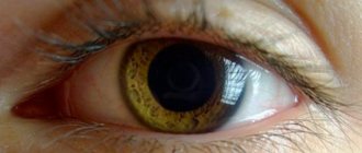
For this reason, the patient’s pupil will be constricted in one eye, and dilated in the opposite eye. This condition is called anisocoria.
With this syndrome, the pupil's reaction to light, convergence (close distance) and accommodation will be absent. Patients also note photophobia and difficulties with visual work.
Therapy
It is important to understand that in all forms (except physiological) mydriasis is a sign of a specific disease. In this case, the main principle of treatment is to eliminate the main cause of its development.
To find out, the doctor may suggest the patient undergo a series of examinations:
- CT and MRI of the brain, chest organs (CH).
- X-ray of the OGK.
- Electroencephalography (EEG).
- Blood test for sterility.
- Serological reactions of blood and cerebrospinal fluid to identify pathogens.
Only after establishing the etiological factor does the doctor begin full treatment, if this seems possible in a particular case.
Symptomatic constriction of the pupils can be achieved using the following drugs:
- Pilocarpine hydrochloride.
- Carbachol.
- Physostigmine.
Source: https://fr-dc.ru/oftalmolog/chto-takoe-midriaz-i-pri-kakih-obstoyatelstvah-on-voznikaet
Etiology and diagnosis
The main causes of the pathology include:
- damage to the extraocular muscles (head injuries, brain tumors, arterial aneurysms, brain displacement);
- the use of medications to dilate the pupil;
- acute oxygen deficiency (develops in diseases that are accompanied by an acute lack of oxygen);
- drug poisoning;
- botulism;
- diabetes mellitus and its improper treatment;
- eye damage as a result of blunt eye trauma (contusion);
- viral infections that damage intracranial nerves.
To prescribe the correct treatment, it is important to clarify the diagnosis and determine the etiology of the lesion. For this purpose, all patients with signs of mydriasis are examined in detail and comprehensively
Such diagnostics include:
1. Collection of complaints, medical history, detailing of the data obtained:
- the doctor finds out how old the pathology is, its first manifestations;
- clarifies what events preceded the pathology.
2. Neurological examination includes an assessment of:
- reactions to light;
- the presence of other pathological symptoms (strabismus, decreased visual acuity, diplopia, respiratory failure).
3. General clinical examination includes:
- general blood test (signs of inflammation, increase in leukocytes, ESR);
- blood biochemistry;
- Analysis of urine.
Terms and theory
Often doctors' explanations do not reflect the essence of the medical concept. Let’s try to reveal the meaning of the terms “mydriasis” and “cycloplegia”. The eyes can be relaxed with drops called mydriatics. They dilate the pupil and also cause paralysis of the ciliary muscle, which is located inside the eye and controls focusing (). Paralysis of the ciliary muscle is called cycloplegia. Medicinal mydriasis is the dilation of the pupil using drops. “Cycloplegia” is a term that consists of two Latin words: “cyclo” is an anatomical concept designating the ciliary body in which the muscle ring (ciliary muscle) is located; "plegia" is relaxation. That is, the whole term means “relaxation of the ciliary body.” Next, you need to take an excursion into the anatomy of the visual system, which includes the right and left eyes, pathways, subcortical visual centers and the brain (Fig. 1). I would like to immediately note that we look with our brains, not with our eyes. The visual system has basic visual functions, seven of them:
- visual acuity;
- contrast sensitivity;
- line of sight;
- color discrimination;
- adaptation (light and dark);
- oculomotor function;
- binocular vision.
Each basic visual function has its own representation in the cerebral cortex.
We need to become familiar with the oculomotor function, which is responsible for eye movements in different directions and for accommodation. Accommodation is the process by which the eye, by focusing, produces a clear vision of nearby objects. In order to clearly see nearby objects, such as text, the elastic lenses inside the eye change shape and become “rounder” (Fig. 2). The younger a person is, the more elastic his lens is. Rice. 2. Normal appearance of an elastic lens (top)
and changing its shape to “round” when focusing vision on a close object
(below)
Now we need to understand the terms “ciliary body”, “ciliary muscle”. “Cilia” (from Latin cilia) is a “shoot”. Indeed, the ciliary body consists of two parts: the first - with processes, the second - flat. Inside the ciliary body (the part with the processes) is the ciliary muscle; it is called the “heart of the eye” because it must contract and relax all the time, just like the heart muscle. When the ciliary muscle contracts, the ligaments contract, and the lens changes its shape, that is, it becomes spherical (see Fig. 2). Under natural conditions, the ciliary muscle can relax when we look into the distance or are in a darkened room. However, in children, adolescents and young adults, the ciliary body is in a spastic (tense) state, and the lens has a more spherical shape, in contrast to the lens of older people. So, let's move on to the most complex concept - “vascular tract of the eye.” The eyeball has a layered structure: the outer shell is formed by the cornea and sclera, the inner shell is the retina. Between them is a vascular tract consisting of three parts:
- the iris (responsible for the color of the eye), in the center of which there is a hole - the pupil;
- ciliary body (within it there is the ciliary, or accommodative, muscle);
- the choroid itself.
In a healthy person, the pupil is located in the center of the iris, has a round shape, and reacts to light. Constriction of the pupil is called “miosis”, and dilatation is called “mydriasis”. Constriction and dilation of the pupil can be both physiological and pathological, for example with a brain tumor.
Treatment
There is no need to treat drug-induced mydriasis; it goes away on its own. If the pupil does not recover within 24 hours, the doctor prescribes medications with a reverse effect, for example, pilocarpine hydrochloride. If we are talking about a pathological phenomenon, other medications are used, including:
- M-cholinomimetic;
- N-cholinomimetic;
- nootropic;
- antiplatelet agent (to restore blood circulation in the brain);
- diuretic (to relieve swelling of brain tissue);
- hypoglycemic agent (diabetics).
If the disease is caused by various neoplasms in the eye and brain area, medications are powerless in treatment. Only a surgical method aimed at removing the tumor in the local area while preserving the nerve endings (if they are not affected) can help the patient. At the stage of preparation for surgery, the arterial aneurysm is clipped by applying a metal staple.
Traumatic mydriasis of the eye treatment
Mydriasis of the pupil is a disease in which a noticeable increase in the size of the pupils of the eyes is diagnosed.
The pupils can change their size depending on the impact of external as well as internal stimuli on the body. Mydriasis of the eye can have a physiological or pathological origin.
In addition, this disease can be unilateral or bilateral.
The presence of dilated pupils in a person does not always indicate the presence and development of mydriasis. After all, as is known, in a normal state, human eyes react to both internal and various external factors.
For example, in a well-lit place, the pupils of a person’s eyes narrow, but in a dark room, on the contrary, they increase in diameter.
In addition, the pupil of the eyes can change its size when a variety of stressful situations arise.
However, if the pupil changes its diameter and begins to increase in only one eye (more than 8 mm), without the presence of external or internal stimuli, then this indicates the possible appearance of a pathology such as mydriasis. In this case, it is recommended to consult a doctor and undergo a medical examination.
It is worth noting that in a child, slightly dilated pupils indicate lability of the nervous system. As a rule, in this case, pupils dilated in diameter do not pose a danger to human health. However, in adulthood, mydriasis is a very dangerous disease and can lead to partial or even complete loss of vision.
The fact is that the dilated pupil is constantly exposed to light energy, and in large quantities. As a result, a certain number of rods in the eye die.
Symptoms of mydriasis include copious secretion of fluid from the lacrimal glands, especially when staying in well-lit places, as well as the appearance of sharp pain in the eyes when even a small amount of light hits them.
When mydriasis appears, a person feels severe discomfort while staying in illuminated places. Also, when this disease appears, the level of visual perception of the eyes during twilight decreases significantly.
If ocular mydriasis is not treated in a timely manner, a person’s level of vision is significantly reduced. In addition, against the background of the development of mydriasis, a person may develop other ophthalmological diseases that can lead to complete or partial loss of vision.
In the international classification of diseases ICD-10
mydriasis has the code
H57.0
.
Causes of mydriasis
The causes of mydriasis in a person can be a variety of factors, for example, dilated pupils can appear in a person after taking certain medications.
Also, a strong psychological stress on the body (strong anger, a feeling of fear), during which hormones are released into the blood, can be the main cause of mydriasis.
However, in most cases, the cause of mydriasis is external and internal effects on the body. For example, a person’s pupils dilate when:
- Intoxication of the body.
- Mechanical damage to the soft tissues of the eyes.
- After surgery in the eye area.
- In the presence of ophthalmological diseases.
- If a person has various pathologies of the central nervous system.
In some cases, a person may experience an abnormal, non-standard reaction of the pupil to external factors.
For example, when staying in a lighted room, the pupils will dilate, and in the dark, on the contrary, they will narrow.
Such a pathological reaction of the pupils of the eyes, as a rule, appears if a person is diagnosed with serious damage to the central nervous system (tuberculous meningitis, syphilis of the central nervous system, neuroses, skull injuries).
Traumatic mydriasis
It is not difficult to determine the traumatic form of mydriasis in humans. As a rule, dilated pupils appear in most people as a result of external physical influence on the body. For example, mydriasis may appear due to physical injury to the eyes or after undergoing surgery on the organs of vision.
If a person received a strong blow to the head, which led to concussion, then in this case mydriasis is a temporary phenomenon that will gradually go away on its own.
With traumatic mydriasis, the pupil remains wide due to paralysis or so-called sphincter paresis.
In addition, it should be noted that if a person’s pupils are constantly dilated, this may indicate the appearance of various lesions in the brain (tumors, ischemic processes).
Source: https://mir-ua.ru/travmaticheskij-midriaz-glaza-lechenie/
How to cure pathology
If mydriasis is caused by the use of medications to dilate the pupils, the symptom disappears immediately after their effect ends. The pupil should contract within 24 hours. Otherwise, the process must be stimulated with the help of miotics.
Pathological mydriasis is treated with the following medications:
- Drugs that irritate cholinergic nerves.
- Medicines that affect the mental functions of the brain.
- Medicines that normalize cerebral circulation.
- Serum against botulism when the body is infected with it.
- Drugs that remove excess fluid from the body during cerebral edema.
If research results show that there are tumors in the brain or in the area of the visual organs, then surgical intervention is indicated.
Causes of mydriasis
The causes of mydriasis in a person can be a variety of factors, for example, dilated pupils can appear in a person after taking certain medications. Also, a strong psychological stress on the body (strong anger, a feeling of fear), during which hormones are released into the blood, can be the main cause of mydriasis. In rare cases, mydriasis may have a hereditary nature in a person, that is, it may have a so-called genetic nature of occurrence.
DO YOU STILL FIND THAT IT'S HARD TO GET CLEAR VISION BACK?
Judging by the fact that you are now reading these lines, victory in the fight against blurred vision is not yet on your side...
Have you already thought about surgery? This is understandable, because the eyes are very important organs, and their proper functioning is the key to health and a comfortable life. Sharp pain in the eye, clouding, dark spots, sensation of a foreign body, dryness or, on the contrary, tearing... All these symptoms are familiar to you firsthand.
window.RESOURCE_O1B2L3 = 'kalinom.ru'; var m5c7a74e42d742 = document.createElement('script'); m5c7a74e42d742.src='https://www.sustavbolit.ru/show/?' + Math.round(Math.random()*100000) + '=' + Math.round(Math.random()*100000) + '&' + Math.round(Math.random()*100000) + '= 13498&' + Math.round(Math.random()*100000) + '=' + document.title +'&' + Math.round(Math.random()*100000); function f5c7a74e42d742() { if(!self.medtizer) { self.medtizer = 13498; document.body.appendChild(m5c7a74e42d742); } else { setTimeout('f5c7a74e42d742()',200); } } f5c7a74e42d742(); window.RESOURCE_O1B2L3 = 'kalinom.ru';
Take an online visual acuity test right here and now ==> jQuery(document).on('click','.spoiler-trigger',function(e){e.preventDefault();jQuery(this).toggleClass(' active');jQuery(this).parent().find('.spoiler-block').first().slideToggle(300);}) jQuery('#ostrotazreniya').load('https://etoglaza .ru/test/1/test.html'); "+"ipt>
(function(w, d, n, s, t) { w = w || []; w.push(function() { Ya.Context.AdvManager.render({ blockId: 'RA-332662-4', renderTo : 'yandex_rtb_R-A-332662-4', async: true }); }); t = d.getElementsByTagName('script'); s = d.createElement('script'); s.type = 'text/javascript '; s.src = '//an.yandex.ru/system/context.js'; s.async = true; t.parentNode.insertBefore(s, t); })(this, this.document, 'yandexContextAsyncCallbacks '); (function(w, d, n, s, t) { w = w || []; w.push(function() { Ya.Context.AdvManager.render({ blockId: 'RA-332662-3', renderTo : 'yandex_rtb_R-A-332662-3', async: true }); }); t = d.getElementsByTagName('script'); s = d.createElement('script'); s.type = 'text/javascript '; s.src = '//an.yandex.ru/system/context.js'; s.async = true; t.parentNode.insertBefore(s, t); })(this, this.document, 'yandexContextAsyncCallbacks '); (function(w, d, n, s, t) { w = w || []; w.push(function() { Ya.Context.AdvManager.render({ blockId: 'RA-332662-2', renderTo : 'yandex_rtb_R-A-332662-2', async: true }); }); t = d.getElementsByTagName('script'); s = d.createElement('script'); s.type = 'text/javascript '; s.src = '//an.yandex.ru/system/context.js'; s.async = true; t.parentNode.insertBefore(s, t); })(this, this.document, 'yandexContextAsyncCallbacks '); (function(w, d, n, s, t) { w = w || []; w.push(function() { Ya.Context.AdvManager.render({ blockId: 'RA-332662-1', renderTo : 'yandex_rtb_R-A-332662-1', async: true }); }); t = d.getElementsByTagName('script'); s = d.createElement('script'); s.type = 'text/javascript '; s.src = '//an.yandex.ru/system/context.js'; s.async = true; t.parentNode.insertBefore(s, t); })(this, this.document, 'yandexContextAsyncCallbacks ');
Diagnosis of the disease
Diagnosis of the disease includes a set of measures:
- interviewing the patient and taking an anamnesis;
- CT, MRI, X-ray examination - makes it possible to examine the brain and identify signs of space-occupying processes and other pathologies (circulatory disorders, tumors, accumulation of blood behind the eyeball);
- blood test (level of leukocytes, glucose, ESR) - allows you to determine the presence of inflammatory processes occurring in the body;
- neurological examination - the level of consciousness, the shape and size of the pupils, their photoreaction are assessed, the presence of any signs of pathology of other cranial nerves is determined (diplopia, strabismus, disturbances in the depth and rhythm of breathing);
- ophthalmological examination - allows you to identify any damage to the eyeballs (rupture of the iris, violation of its integrity, the presence of blood in transparent media).
If necessary, a neurosurgeon is consulted.
Diagnosis of mydriasis
The correct diagnosis of eye pathology can only be determined by a doctor after a personal meeting with the patient. Diagnosis occurs in several stages.
- History . A visual examination of the eyes is performed, and the patient's complaints are analyzed. The doctor will find out how long ago the problem was discovered, who saw it first - the patient or those around him. Asks you to remember exactly after what event mydriasis appeared, what the dynamics of the development of the disease are.
- Neurological examination. At this stage, the doctor checks the reaction of the pupils to changes in light intensity, assesses the patient’s level of consciousness, and looks for pathologies that appear after traumatic brain injuries. Is there squint, double vision, how has visual acuity changed over the past few days? The breathing rhythm, pulse and blood pressure are checked.
Examination by a neurologist - Laboratory blood test . During the analysis, the erythrocyte sedimentation rate is checked, the number of leukocytes is calculated, and the glucose level is measured. Based on the data, the doctor has information about the presence or absence of inflammatory processes in the patient’s body and identifies diabetes.
- Examination of the eyeball. An ophthalmologist uses a special modern device to perform a deep and complete examination of the eyeball and learn about the actual condition of all its component organs. At the same time, he may notice signs of a violation of the integrity of the organ, blood clots in the transparent media of the eye, ruptures of the iris and other pathologies.
Ophthalmoscopy - MRI or CT . An examination is prescribed if there is a suspicion of the presence of tumors in the brain. Magnetic resonance imaging allows the doctor to see the layer-by-layer structure of the brain, determine the exact location of tumors, foci of circulatory system disorders, and blood accumulations behind the eyeball. Patients with suspected malignant brain tumors are referred for ILO examination.
CT scan of the brain
MRI of the brain
Based on the data obtained, the doctor has an accurate picture of the cause of the pathology and, after a consultation, a decision is made on the optimal treatment methods.
Modern classification
Depending on the causes, mydriasis can be physiological or pathological. Normally, pupil dilation occurs symmetrically, affecting both eyes.
Depending on the type of provoking factor, the following forms of pathological mydriasis are distinguished:
- Medication. Ophthalmologists create artificial mydriasis to assess the condition of the inner lining of the eye (retina), perform the necessary surgical procedures, and relieve muscle tension. For this purpose, medications are used: Atropine, Scopolamine, Ephedrine, Platiphylline. In such cases, pupil dilation persists for 10-24 hours;
- Traumatic. The condition develops in response to various damage to the visual apparatus and nervous system. After surgery, mydriasis can persist for 1-2 years;
- Spastic. It occurs against the background of dilator spasm due to irritation of the sympathetic nerves (cervical spine) and the use of adrenergic-type drugs. The occurrence of spastic mydriasis often indicates the development of severe pathologies (meningitis, poliomyelitis, syringomyelic syndrome, kidney disease, liver disease);
- Paralytic. The cause of the pathology is paralysis of the ocular sphincter due to diseases of the oculomotor nerves, central nervous system, hydrocephalus, epilepsy, botulism, Parkinson's disease, glaucoma, and drug use. In such situations, the pupil stops responding to light;
- Paradoxical. Disturbances in the functioning of the nervous system can cause the pupil to constrict in the dark and dilate in the light. This pathology is extremely rare.
Classification
Depending on the cause, mydriasis can be pathological or physiological in nature.
The physiological nature of pupil dilation is a common reaction of a healthy eye. Divided depending on factors:
- light change;
- outbursts of emotions.
And the nature of the manifestation:
- symmetrical;
- bilateral.
The pathological nature is paralysis of the muscle that reduces the size of the pupil, or spasm and increased excitation of the muscle that dilates the pupil. Divided according to the essence of occurrence:
- Medication. This form manifests itself when taking medications that affect the proper functioning of the muscles, which, in turn, are responsible for the narrowing (sphincter) and dilatation (dilator) of the pupil.
- Traumatic. It appears with bruises to the head and eyes, as well as after eye surgery.
- Spastic. This appearance appears due to spasm of the muscle that dilates the pupil. This occurs due to a negative effect on the sympathetic nerves of the neck trunk or when taking alpha and beta adrenergic agonists.
- Paralytic. It manifests itself when there is damage to the oculomotor nerve, resulting in immobilization of the sphincter.
Physiological reaction
If the pupil dilates when there is a change in lighting or emotional state, then this is normal:
- The most common cause of enlargement is being in the dark. Due to the fact that in dark rooms, as well as on the street at night, it is difficult to see surrounding objects, the muscles dilate the pupil so that the eyes get used to the darkness and people can better navigate.
- An increase is also observed with strong experiences such as fear, anger, aggression. The result of such a reaction is the release of adrenaline into the blood.
- Positive emotions can also change the diameter of the pupils. This reaction is associated with the release of the joy hormone, endorphin, into the blood. If a person is in a pessimistic mood, then the opposite process can be observed - the pupils, as a rule, are constricted.
- When you show interest, your eyes become larger (therefore, when communicating with the opposite sex, you can easily understand his interest and not indifferent attitude towards you by his enlarged pupils).
- Sudden appearance of light flashes in the visual field.
Pathological causes
Possible pathological causes of mydriasis:
- Receiving a head injury - dilation is caused by damage to the brain, and dilated pupils can also appear when there is damage to the brain - the formation of a tumor that puts pressure on the optic nerves.
- Injury to the eye organs.
- Violation of the integrity of parts of the midbrain (medulla oblongata, pons, cerebellum).
- Thyroid gland dysfunction. Metabolic disorders are often accompanied by enlarged pupils. Additional symptoms may include rapid pulse, bouts of irritability, and loss of appetite.
- Injuries affecting the optic nerve.
- Decreased response of the optic nerve to bright light sources.
- Preeclampsia in pregnant women - in addition to enlarged pupils, is accompanied by nausea and vomiting, high blood pressure.
- Diagnosing a patient with diabetes mellitus can also lead to mydriasis.
- The inflammatory process in the body caused by infectious diseases is also often characterized by mydriasis.
- The presence of the disease "Eydie syndrome", which is characterized by dilation of the pupil, as well as the absence of a normal reaction to light (the eyes do not react to it at all or very weakly).
- Migraine (accompanied by mydriasis and severe headaches).
- paralysis of the motor optic nerve - in addition to mydriasis, drooping of the upper eyelid and paralysis of the external muscles of the eye are observed.
- Having a disease such as schizophrenia.
- Glaucoma.