As a result of impaired blood supply, ischemic optic neuropathy develops. The pathology is considered secondary and occurs against the background of diseases of the cardiovascular, nervous or endocrine systems. Symptoms manifest themselves actively, the patient’s vision sharply decreases. Males over 40 years of age are at risk. To confirm the diagnosis, it is necessary to study the condition of the optic nerves in detail, so a number of instrumental diagnostic procedures are prescribed. Treatment involves the use of conservative methods and physical therapy.
Due to the rapid development and characteristics of pathogenesis, ischemic optic neuropathy is often called disc infarction. This condition requires urgent hospitalization.
Etiology and pathogenesis
If the local blood supply is disrupted, the nerve tissues of the eyes do not receive the necessary substances for full functioning. As a result, optic nerve atrophy develops, which ultimately causes blindness. Ischemia occurs due to hemodynamic dysfunction. The following factors can trigger this process:
- vascular diseases (atherosclerosis, hypertension, Horton's disease, thrombosis);
- diabetes;
- neuritis;
- heavy bleeding;
- stress and frequent nervous overload.
In most cases, the pathology affects only one visual organ.
Pathology of the optic nerve is more often unilateral, only in 30% of cases ischemia of both eyes is observed. The disease is characterized by a sequence when the symptoms of the disease pass from one eye to the other after some time. This process is observed in a third of patients, but it is difficult to predict. The disease on the second organ can appear in a few days or over 10 years.
Ocular ischemic syndrome
Due to the presence of close anatomical and physiological cerebro-ophthalmic relationships, the clinic of vascular pathology of the eye with cerebrovascular lesions can be represented by a variety of disorders, with a common ischemic symptom complex, designated in clinical ophthalmology by the term “ocular ischemic syndrome” (OIS).
GIS is a set of symptoms of ischemic damage to the membranes of the eyeball and optic nerve, in which drug treatment remains ineffective, and surgical interventions aimed at improving ocular blood flow have not yet received proper use, although their clinical significance is beyond doubt.
The leading cause of HIS is primarily considered to be atherosclerotic lesions of the internal carotid artery (ICA), while at the same time, the participation in its development of hemodynamic disorders of other great vessels of the head and neck, as well as cardiovascular pathology, cannot be excluded. In addition to the stenotic pathology of the ICA, the pathophysiological mechanisms of GIS may be associated with an increase in total blood pressure, leading to disruption of autoregulation processes in the cerebral and ocular blood flow systems; with vertebral artery syndrome, which increases the tendency to vasospastic reactions; cardiovascular pathology, aggravating the severity of hemorheological disorders in the microvasculature of the brain and eye.
In recent years, a steady increase in GIS has been recorded, due to the widespread prevalence of systemic atherosclerosis, arterial hypertension (AH), coronary heart disease (CHD), and cervical osteochondrosis, which leads to an increase in the frequency of clinical manifestations and variations in the course of GIS.
It is extremely important for an ophthalmologist to know the symptoms and causes of GIS in order to promptly diagnose this disease with the participation of other specialists (neurologist, therapist, angiosurgeon), which is especially important for choosing the optimal treatment tactics and improving the visual prognosis. At the same time, many issues of clinical manifestations and early diagnosis of GIS still remain insufficiently studied, which affects the quality of medical care for this severe category of patients.
Clinical picture
Regardless of the degree of ICA stenosis, characteristic of patients with GIS is a decrease in the speed of blood flow in the ICA, which is combined with an increase in RI, which increases with increasing degree of stenosis.
It is known that in addition to stenotic processes caused by atherosclerosis in the ICA, other vascular diseases of the body can also form the prerequisites for the occurrence of GIS. These include arterial hypertension (AH), in which changes occur in the cytoarchitecture of the bloodstream in various organs, including the brain. With hypertension, the lumen of blood vessels decreases, vascular resistance increases due to hypertrophy of the vascular walls, occlusion of part of the arterioles occurs, and local ischemia and hypoxia occur.
Among neurological diseases accompanied by cerebro- and ophthalmovascular disorders, cervical osteochondrosis plays an important role. Characteristic of this pathology are spasms of small-caliber vessels in the ICA system against the background of sympathicotonia, including GA and CAS, due to the pathological influence of the sympathetic carotid plexus.
The likelihood of developing GIS increases with background vascular pathology, along with impaired hemodynamics of the ICA, damage is determined in the vascular systems of the body, indirectly affecting the blood flow of the brain and eye, including through: arterio-arterial embolism with atherosclerotic material, the source of which can be the heart; hemorheological microocclusions due to blood hypercoagulation and deficiency of anticoagulation factors characteristic of hypertension; hyperirritation of sympathetic formations in cervical osteochondrosis, which play an important role in the regulation of the entire circulatory system, primarily at the microcirculation level.
During GIS, two types are distinguished: acute and primary chronic. In both acute and chronic ocular symptoms, ischemia of the organ of vision is localized on the side of the affected vessel. However, the clinical features of visual disorders, the choice of optimal treatment tactics and visual prognosis depend on the type of course of GIS.
Among the manifestations of both acute and primary-chronic type of course, INOP predominates, predominantly unilateral anterior (28.3±1.1%) in the acute type of course of GIS, and bilateral posterior (50.0±2.7%) - in primary -chronic type (p<0.05). Also, with primary chronic GIS, clinical signs of CIR are diagnosed, which, as a rule, is observed with degrees of ICA stenosis of 60% or more.
At the same time, the clinical features of patients with chronic ischemic retinopathy (CIR) are: predominantly unilateral nature of the lesion, long-term high BCVA, aching ischemic pain behind the eyeball, no complaints about narrowing of the visual fields. The ophthalmoscopic picture was characterized as: dilation of venules, narrowing of arterioles, pinpoint intraretinal hemorrhages from the equator to the extreme periphery, areas of retinal non-perfusion. Fundus FA in patients with CID confirms the appearance of hypofluorescence zones, retinal hemorrhages, microaneurysms in the middle and extreme periphery of the fundus, and a late onset of the choroidal phase from 20 to 24 seconds. (at a norm of 7-15 seconds).
Compared to patients with CID, patients with INOP have different complaints and are characterized by bilateral lesions, concentric narrowing of visual fields, and decreased BCVA. In the fundus, a significant narrowing of the arterioles of the peripapillary region and retina is determined, and in the dynamics of the disease, partial atrophy of the optic nerve appears.
Possible symptoms
Signs depend on the location and scale of the process. In the first stages, partial or limited damage appears, then the disease develops into a total form. If the process develops in the intrabulbar region, anterior ischemic neuropathy is observed. This is an acute disorder of blood circulation, localized in the head. The main symptom is swelling of the organ, with the following symptoms occurring:
- rapid decrease in visual acuity;
- square loss from the field of view;
- dull headache;
- dilated retinal vessels;
- traces of pinpoint hemorrhage.
A person becomes completely blind in the total form of the pathology.
The acute form of the disease lasts for a month, then the swelling disappears. This means atrophy of nerve fibers begins. If swelling develops in the intraorbital region, posterior ischemic neuropathy is diagnosed. In this case, the damaged nerve is located immediately behind the eyeball. The symptoms are almost identical to the anterior type, but some changes in the fundus are observed. The danger is the total form, when all parts are damaged and optical neuropathy develops. This process leads to irreversible blindness. Ischemic neuroopticopathy is rare, but practically untreatable.
Ischemia is characterized by rapid development; sometimes the patient remembers the exact time when his vision began to decrease. But the first stages are completely asymptomatic.
Causes
Anterior ischemic neuropathy is expressed in impaired blood flow in the posterior ciliary arteries and, as a result, ischemia of the retinal, choroidal and scleral layers of the optic disc develops. Posterior ischemic neuropathy occurs in the process of circulatory disorders in the posterior parts of the optic nerve and stenosis of the carotid and vertebral arteries.
The disease is caused by various lesions and associated disorders, striking changes in the vascular bed, and microcirculation disorders. Neuropathy develops against the background of common vascular diseases: atherosclerosis, hypertension, temporal giant cell arteritis, periarteritis nodosa, obliterating arteritis, diabetes mellitus, cervical discopathy with disturbances in the vertebrobasilar system, thrombosis of the great vessels.
In individual cases, the disease can manifest itself as a result of serious blood loss during gastrointestinal bleeding, surgical interventions, injuries, anemia, arterial hypotension, after anesthesia, hemodialysis, and blood diseases.
Diagnostics
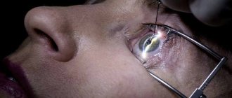
The study is carried out comprehensively, in addition to an ophthalmological examination, consultation with a neurologist, endocrinologist, hematologist and rheumatologist is necessary. Diagnostics involves a full range of procedures that allow you to determine the primary cause. First, the ophthalmologist examines the fundus of the eye for signs of swelling. Both eyes must be checked, and the emphasis is on studying the condition of the retinas. Then a number of additional procedures are prescribed:
An electroretinogram in such a situation is of an auxiliary nature.
- visual acuity test;
- X-ray of the head;
- angiography of the vascular circuit;
- electroretinogram;
- MRI and CT scan of the skull;
- kaogulogram.
During diagnosis, studies are performed to exclude retrobulbar neuritis. This disease is a formation in the orbital region of the central nervous system. The symptoms of the diseases are similar, in addition, active manifestations begin already at advanced stages of the process. The pathology is very life-threatening and requires urgent surgical intervention.
Treatment of the disease
It is very important to make a diagnosis on time. Treatment of ischemic neuropathy requires urgency; prolonged disruption of circulation provokes the rapid development of the disease and sudden death of neurons. First, emergency care is required; to slow down the process, the patient is injected with Eufillin intravenously and given a Nitroglycerin tablet. The patient must be admitted to a hospital, and then a treatment regimen is determined. It includes the following components:
- corticosteroid drugs to reduce tissue swelling;
- means for normalizing blood pressure;
- drugs to improve blood clotting;
- vasodilators;
- diuretics;
- nootropic drugs.
B vitamins in injection form will support the patient’s weakened immune system.
A full range of medications sharply reduces the level of immune defense. Vitamins C, B, and E are prescribed as supporting agents in the form of injections or tablets. The patient is in hospital under the watchful supervision of doctors. When the inflammatory process is stopped, physiotherapeutic procedures are prescribed to improve the condition of the optic nerve, such as:
- laser illumination;
- electrical stimulation;
- magnetotherapy.
Prevention and prognosis
Ischemic neuropathy develops against the background of cardiovascular and other diseases. Therefore, as a preventive measure, it is worth paying attention to timely treatment of the main ailments. It is very important to monitor the level of cholesterol in the blood; proper nutrition will help keep it normal. For patients with chronic forms of diseases, it is necessary to regularly see an ophthalmologist.
For patients who have previously suffered ischemic neuropathy in one eye, it is necessary to register with a doctor and undergo regular preventive examinations. Almost all patients experience a recurrence of the disease, but in the second eye. Therefore, a comprehensive examination of the right and left organs is required. The prognosis of such a disease is unfavorable: atrophy always leads to a decrease in visual acuity of varying degrees. And untimely treatment generally causes blindness.
Ischemic kidney disease (renal ischemia, ischemic nephropathy, vascular nephropathy)
The main signs to suspect ischemic nephropathy:
- end-stage renal failure that developed in individuals who did not have clinical manifestations of kidney damage (minimal or absent proteinuria, scanty urinary syndrome);
- age over 60 years;
- the presence of generalized atherosclerosis;
- frequent development of arterial hypertension.
It has now been proven that ischemic nephropathy is quite widespread, although it often remains undiagnosed.
In elderly and senile patients, ischemic nephropathy is one of the leading causes of renal dysfunction. Among patients with end-stage renal failure over the age of 50 years, ischemic nephropathy accounts for 15%.
There is currently no generally accepted approved modern classification of ischemic kidney diseases. Most often, ischemic nephropathy is classified according to 2 criteria:
- the rate of progression of renal failure, distinguishing acute (rapidly progressive) and chronic;
- localization of damage to the renal arteries (renal arteries of large, medium and small caliber).
Atherosclerosis is recognized as the most common cause of ischemic kidney disease, which has given grounds to distinguish separately the atherosclerotic form of ischemic kidney disease.
Ischemic nephropathy: three forms of the disease
The atherosclerotic process that damages the renal arteries most often affects large-caliber arteries. Damage to the renal arteries can be complete or partial.
What's going on? With almost complete damage to the arteries, acute renal failure develops with the actual cessation of blood supply to the kidneys. If the vessels are not completely affected, then the blood supply to the kidneys is partially restored and ischemic nephropathy does not progress so quickly.
There are three main variants of the course of ischemic nephropathy:
- acute renal failure;
- rapidly progressing renal failure;
- torpid (sluggish) chronic renal failure.
Acute renal failure is caused by a sudden obstruction of the renal vessels and depletion of renal blood flow. The following signs indicate this process: an acute rise in blood pressure, leukocytosis, fever, intense pain in the kidneys or back. The course of acute renal failure is usually asymptomatic.
Blockage of blood vessels by cholesterol crystals is the main cause of the development of rapidly progressive renal failure in ischemic nephropathy. In addition to the kidneys, the pathological process involves the skin, nervous system, and gastrointestinal tract. Signs that help recognize this type of renal failure are a rapid rise in blood creatinine, increased blood pressure, pain, nausea, vomiting, gastrointestinal bleeding, necrosis of the toes, erythema nodosum and others. Blockage of blood vessels by cholesterol crystals is confirmed by the detection of crystals in particles of the skin, muscles, and many internal organs, primarily the kidneys, as well as in the liver, pancreas and other organs.
Torpid (sluggish) renal failure is the most common clinical variant of ischemic nephropathy. As a rule, this clinical form is diagnosed only during special functional studies of blood vessels (arteriographic studies, duplex ultrasonography, etc.) or during autopsy. At the same time, it is important to remember that the reality of vascular kidney damage is high in elderly patients when they are diagnosed with generalized atherosclerosis.