Lens subluxation is a pathology characterized by a slight displacement of the lens, when part of it remains in place, but visual acuity does not decrease.
Despite this, lens subluxation requires treatment because over time, the likelihood of lens luxation and vision loss increases. Therefore, you should promptly consult a doctor at the first signs of a disorder.
general description
Lens luxation is a complete or partial (dislocation or subluxation) dislocation of the lens from its normal location.
Lens dislocation can be congenital or acquired. Congenital disorder of the location of the lens is a consequence of insufficient development, partial absence of the zonules of Zinn. An acquired disorder of the location of the lens develops as a result of rupture of the zonular ligaments due to blunt trauma to the eye or degenerative changes in the ligaments and vitreous body (cyanyl, high myopia, inflammatory processes). Congenital heterotopia lens is usually bilateral in nature and is often a stigma of connective tissue dysplasia.
Causes of pathology
The lens is an important microstructure of the eye, which is responsible for light refraction and occupies a clearly defined place in the optical system. The biological lens is held on all sides by thin fibers of the ciliary body.
The back surface of the lens is adjacent to the vitreous body, which is filled with intraocular fluid. Its front surface is covered by the pupil and iris, and the anterior and posterior chambers filled with ocular fluid are located right there. When the lens is dislocated, it is completely displaced from its point, and when it is subluxated, it is partially displaced.
Main reasons for the displacement:
- injury, rupture;
- some congenital genetic pathologies, such as Marfan syndrome;
- weakening or destruction of the ciliary ligaments in older people.
Clinical picture
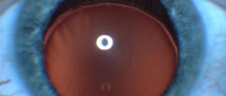
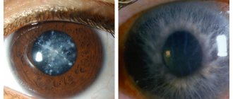
- Reduction of the anterior chamber on the side of displacement.
- Iridodonesis.
- Determination of the edge of the lens when examined in transmitted light in the form of an arcuate dark line.
- Double image of the fundus during ophthalmoscopy.
- When examined through a lens, the refraction changes towards myopia, and when examined through the area of the pupil devoid of a lens, towards hyperopia. Refractive errors are common.
- Visa depression.
- Aphakia due to dislocation of the lens into the vitreous body.
Dislocation of the lens into the vitreous body is fraught with such serious complications as iridocyclitis, secondary glaucoma, and retinal detachment. If the dislocation occurs after an eye injury, then complications such as inversion of the iris, uveitis, hemophthalmos, and retinal detachment are possible.
Why is deviation dangerous?
In severe cases of pathology or in case of untimely treatment, the following complications develop:
- Secondary glaucoma. It is expressed in a persistent increase in intraocular pressure.
- Iridocyclitis. Occurs when pathogenic microorganisms enter the iris and ciliary body. The danger of the disease is the spread of bacteria to all structures of the eye, as well as fusion of the pupil.
- Retinal detachment. With this pathology, the retina is rejected from the choroid and there is a danger of complete loss of vision.
A complication of lens subluxation is often a vitreous hernia, which is formed as a result of a defect in the structures located near the VT. May cause clouding of the cornea and decreased vision. Severe consequences often develop after surgery. These include intraocular lens displacement, postoperative astigmatism, and corneal edema.
Treatment of lens luxation
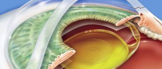
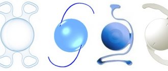
- In case of lens subluxation, phacoemulsification, surgical extraction of the lens with implantation of its artificial analogue.
- In case of dislocation in the anterior chamber, extraction of the lens with implantation of its artificial analogue is performed.
- In case of dislocation into the vitreous body, vitrectomy with extraction of the lens and implantation of its artificial analogue is performed.
The prognosis is favorable in most cases. The development of complications depends on the effectiveness of the treatment and the level of pathological changes in the retina and optic nerve.
Causes
The reason can be not only directly directed physical actions.
In most cases, dislocation of the eye lens is associated with existing diseases:
- Marfan syndrome is a hereditary disease. Caused by a mutation in the genes responsible for heart protein synthesis. The patient has elongated limbs and fingers, dystrophy, curvature of the spine, and decreased aortic tone. A characteristic feature of this type of disease is bipolar subluxation of the lens. The displacement usually occurs in the direction of the temporal cavities.
- Homocystinuria – occurs in patients with dementia. In humans, a pronounced form of skeletal deformation is visible. The body structure is identical to Morphan's syndrome, thromboembolism is observed, the risk of retinal detachment is increased, the lens is displaced downward.
- Weill-Marchesani – shortened phalanges, dwarfism. The patient experiences convulsive contractions, small pupils, underdeveloped limbs, and a shortened neck. The disease is caused by a mutation in an autosomal or recessive gene.
- Syphilis is an acquired infection, mainly transmitted through sexual contact. Damages soft tissues and the structure of cartilage and bones. The eye shell is damaged at an advanced stage.
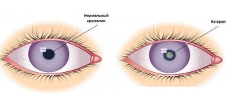
- Also, displacement of the lens can occur with cataracts, chronic processes of inflammation, high myopia, congenital ectopia of the eye.
Incidence (per 100,000 people)
| Men | Women | |||||||||||||
| Age, years | 0-1 | 1-3 | 3-14 | 14-25 | 25-40 | 40-60 | 60 + | 0-1 | 1-3 | 3-14 | 14-25 | 25-40 | 40-60 | 60 + |
| Number of sick people | 10 | 20 | 20 | 20 | 20 | 20 | 20 | 10 | 20 | 20 | 20 | 20 | 20 | 20 |
Lens luxation
Lens luxation is an eye pathology associated with displacement (luxation, dislocation, dislocation) of the lens from its normal anatomical position.
This pathology occurs more often in dogs than in cats.
Some dog breeds have a breed predisposition to lens displacement:
- Chinese Crested Dog
- Jack Russell Terrier
- Tibetan Terrier
- Wirehaired Fox Terrier, etc.
The disease is inherited and is called primary lens luxation (PLL). Affects both eyes. Most common at age 5 years.
Secondary luxation of the lens is the result of the presence of a concomitant pathology in the eye that causes lens displacement (cataracts, glaucoma, etc.). Thus, in cats, secondary luxation of the lens mainly occurs.
Causes
The reasons for the development of lens luxation in dogs and cats are associated with weakness and rupture of the zonules of Zinn, which hold the lens in a strict position along its entire circumference. As a result of tearing of these ligaments, the lens is displaced in different directions: into the anterior chamber, into the vitreous body, or pinched in the opening of the pupil.
If the tear of the ligaments is partial (not complete), then subluxation or subluxation of the lens is noted.
Symptoms
Clinical signs of lens luxation depend on the degree of displacement and the location of the lens. Possible development of edema and local clouding of the cornea, lacrimation, blepharospasm, pain syndrome . Specific signs include: vitreous hernia, iris tremors - iridodonesis, changes in the depth of the anterior chamber of the eye, the formation of an aphakic crescent. Animals may also experience a decrease in visual function.
Luxation of the lens in a dog
As a result of lens luxation, severe ocular pathologies may develop, such as uveitis, secondary phacotopic glaucoma. The development of secondary glaucoma is associated with impaired circulation of intraocular fluid due to blockage of outflow tracts by a displaced lens.
Diagnostics
Diagnosis is made by visual examination by a veterinary ophthalmologist, ophthalmoscopy, and slit lamp examination of the anterior segment of the eye. To identify the position of the lens, one of the informative methods of examining the eye is ultrasound, which allows one to assess the condition of the posterior segment of the eyeball. Tonometry is a mandatory method in the diagnosis of lens luxation in dogs and cats, for monitoring intraocular pressure and preventing glaucoma.
Treatment
Treatment of lens luxation in dogs and cats is possible with both medication and surgery. The essence of drug treatment comes down to the prescription of drugs that constrict the pupil (miotics) and retain the lens in the posterior segment of the eyeball. The surgical method of treatment depends on the location of the dislocated lens and the presence of concomitant ocular pathologies. Both intracapsular and extracapsular lens extraction are used. The most successful surgical treatment method is phacoemulsification with vitrectomy. In cases where there is a suspicion of the development of glaucoma, one-stage cyclophotocoagulation is performed.
It is important to understand that lens luxation in dogs and cats is a serious ophthalmopathy that requires urgent treatment. The outcome of treatment measures directly depends on the timing of animal owners contacting a veterinary ophthalmologist.
Possible complications
In cases of timely access to a medical institution and the selection of adequate therapy, the chances of complete preservation of vision are quite high. But in especially severe cases, it is almost impossible to restore visual functions. This is due to the fact that after injury there is a strong surge in pressure inside the eye, which destabilizes the internal structure of the eyeball. This pathology leads to damage to the iris, visual nerve, vascular layer and retinal detachment. Against the background of stress, they quickly progress and become irreversible.
General information
The lens is an outwardly colorless convex lens in both directions. It is held in a constant position by a ligamentous layer, consisting of a large number of connective fibers associated with the ciliary muscle. If, due to a rupture or absence of this layer, the lens moves freely, this is called luxation. Cases of partial damage to the connecting fibers are called subluxation. During luxation, the lens moves uncontrollably and may end up in the anterior chamber of the eyeball. If the lens is found on the back side of the iris, it is called a vitreous lens shift.
Causes of pathology
The most common factor is injury. Doctors also identify the following reasons:
- congenital underdevelopment of the ophthalmic apple;
- genetic predisposition;
- degenerative processes;
- long-term inflammatory processes.
Types of disease
There are 2 types of this disease.
There are the following 2 types of eye lens luxation:
- This type of disease most often manifests itself in incomplete separation from the ciliary muscle. The lens is displaced into the vitreous body, and incomplete dislocation occurs with one side leaving its normal state. In the case of partial dislocation, a significant decrease in vision is observed and retinal detachment occurs. Cases of complete displacement are extremely rare and occur due to serious injuries or underdevelopment of the eyeball in the fetus.
- Luxation of the lens into the anterior or posterior zone of the eyeball chamber is accompanied by an almost complete loss of the usual location of the lens. There is a strong impact on the iris, the hydrostatics of the eye is disrupted, and because of this, the pressure inside the eye increases. With such a dislocation, the lens occupies the entire posterior or anterior chamber of the eyeball, causing loss of visual functions and deformation of the pupil.
Luxation is often a factor in the development of glaucoma.