Visual pathway - what is it?
Visual path
- this is the path that nerve impulses take from the photoreceptors of the retina (the inner lining of the eyeball) to the nerve centers of the brain.
The main receptor of the eye is the retina, which contains rods and cones. They convert a beam of light into electrical impulses and transmit them to nerve cells. Nerve impulses, in turn, send information to the central section in the cerebral cortex, where the received characteristics are recognized and a real image of the surrounding world is formed.
That is, the visual pathway is the system of nerve cells that allows a person to see.
Diagnostic methods for diseases of the visual pathway
The visual pathway, under the influence of internal and external factors negatively affecting it, can change pathologically. This leads to various diseases. To identify lesions in the visual pathway, specialists can use various diagnostic techniques. The most common include:
- Visometry.
- Electroretinography.
- Perimetry.
- MRI and CT.
When examining a patient, ophthalmologists also check the lability of the optic fibers and nerves, as well as the potential of the cerebral cortex.
Structure of the visual pathway
The visual pathway begins with the retina of the eye. It is here that photoreceptors—rods and cones—translate light signals into nerve impulses. These nerve impulses are then transmitted to bipolar cells (connect one cone or rods to one ganglion cell (a nerve cell (neuron) in the retina of the eye that can generate nerve impulses, unlike other types of retinal neurons)) and retinal neurons.
Neurons have long processes called axons. They are responsible for collecting information from the entire surface of the retina. Millions of axons connected together form the optic nerve.
Groups of axons are arranged in a strictly defined order. Chief among these groups is the papillo-macular bundle, which transmits signals from the so-called macular zone of the retina.
The optic nerve then enters the skull through the optic canal. The fibers of the two optic nerves partially cross. This cross - chaism - is a particularly important part of our vision. Thus, this part of the ocular tract is associated with the fact that with lesions of the sella turcica (pathologies of the nervous or endocrine system), as well as with damage to the internal carotid arteries, a person experiences loss of parts of the visual field (internal and external).
Next, the bundle of nerve fibers (optic tract) bypasses the cerebral peduncles - its special paired structures - and enters the posterior part of the visual thalamus. The sensation of light that our brain experiences at this moment causes reflex reactions, manifested, for example, by turning the head towards a sharp flash.
In the same department, special groups of cells form visual radiance, which transmits information to the cells of the cerebral cortex, where nerve impulses are deciphered and an image of the surrounding world is created.
The structure of the visual pathway is complex and multifunctional. This is a whole mechanism that works every second and literally instantly performs all its tasks, thanks to which we see objects around us.
Conducting path of the visual analyzer
The conductive path of the visual analyzer is a complexly constructed organ. Only due to it we are able to perceive, transmit, analyze and synthesize light stimuli that are absorbed by light-sensitive cells at a speed of 720 m/s. Such cells can be divided into cones and rods. Initially, light rays visit the cornea, then continue their journey to the frontal and posterior chambers of the eye. After which they are processed on the lens and vitreous body. Only after going through all these stages do they reach the retina. Rods and cones act on light, thereby separating rhodopsin, forming energy that is taken up by the receptors of the first neuron, medically known as bipolar cells. After this stop, they move to ganglion neurocytes. The pathway of the visual analyzer consists of just such neurocytes that go to the disc and form the optic nerve. The latter has a vagina in its environment. The nerve originates from the eye socket, and then moves along the optic canal to the cranial cavity, thereby forming a visual chiasm on the lower part of the brain. If you do not pay attention to this, then you can confidently say that not all fibers related to the optic nerve are participants in this chiasm. It definitely involves only fibers starting from the middle region of the retina. In view of this state of affairs, the optic tract follows the chiasm, in which one can find nerve fibers of ganglion cells belonging to the lateral surface of the retina and also to the medial surface. Because of this, the slightest damage to the chiasm can deprive the path of impulses moving from the middle part of the retina. And damage to the optic tract will disrupt movement from the lateral and medial surfaces of the retina.
The conducting path of the visual analyzer includes nerve fibers that also belong to the optic tract. They move towards the subcortical optic nerves, which include the lateral geniculate body and the superior colliculus of the midbrain roof. In the geniculate body, the fibers representing the third neuron of the visual pathway end, but manage to create a message with the cells of the neuron following it. Axons belonging to neurocytes pass through the sublenticular part of the internal capsule. Thanks to this, visual radiance is formed. Their end point is located on the occipital lobe of the cortex, in the area of the calcarine sulcus. It is here that the highest analysis of visual perception is carried out. A certain proportion of axons do not have endings located in the lateral geniculate body; instead, they go as handles to the superior colliculus. Impulses emanating from the gray layer of the superior colliculus move into the nucleus of the oculomotor nerve, and at the same time to the accessory nucleus of Yakubovich. This is where innervation of the oculomotor muscles is produced, as well as constriction of the pupils and ciliary muscles. Along these fibers, a response to light stimulation occurs in the form of constriction of the pupil and rotation of the eyeballs in the required direction.
Conducting pathways of the visual analyzer (diagram 3)
The receptors of this analyzer are the rods and cones of the retina. The NI that appears in them under the influence of light is transmitted to the first neurons, which are bipolar cells, and from them they pass to the second neurons - ganglion cells (Fig. 9). Both neurons, like the receptors, are located in the retina. Ganglion cell axons within the optic disc, discus nervi optici
, are grouped into the optic nerve,
n.
opticus. Through the optic nerve canal,
canalis opticus,
the right and left optic nerves pass into the cranial cavity, where their medial fibers pass to opposite sides, forming the optic chiasma,
chiasma opticum,
and the laterally located fibers pass without crossing.
Medial fibers conduct NI from the medial sections (fields) of the retina of both eyeballs, and lateral fibers - from the lateral sections (fields). After decussation, these fibers form part of the optic tract, tractum optici
. Along each tract, NI are carried out from the lateral part of the retina of the eyeball on its side and from the medial part of the retina of the other eyeball. All fibers in the optic tracts reach three subcortical centers of vision, in which the bodies of third neurons are located, these are:
1) lateral geniculate bodies, corpora geniculata lateralеs;
2) superior colliculi of the roof of the midbrain, colliculi superiores tecti mesencephali;
3) pillows of the visual hillocks, pulvinares thalami dorsales.
The axons of the third neurons of the lateral geniculate body pass through the posterior part of the posterior limb of the internal capsule, after which they enter the optic radiation, radiatio optica
, and in its composition they reach the cortical nucleus of the analyzer - the calcarine sulcus. The nucleus of each hemisphere receives NI from the fields of the same name in the retinal membranes of both eyeballs.
Axons of the third neurons of the superior colliculus of the midbrain roof are directed to two groups of nuclei:
a) parasympathetic nuclei of Yakubovich of the oculomotor nerve, which participate in the innervation of mm. sphincter pupilli et ciliares,
ensuring, respectively, the implementation of the pupillary and accommodative reflexes;
b) the motor nuclei of the oculomotor (III), trochlear (IV) and abducens (VI) nerves, which innervate the skeletal muscles of the eyeballs, ensuring their movements.
The axons of the third neurons of the optic thalamus cushion reach both the cortical nucleus of the analyzer - the calcarine sulcus, and the structures of the midbrain and diencephalon to carry out somatic and visceral reflexes.
Function.
The visual analyzer provides the perception of light, color, determination of the degree of illumination, shape and size of objects, their distance and speed of movement, and also provides stereognosis perception of the surrounding world (binocular vision).
Rice. 9. Conducting pathways of the visual analyzer. 1 – receptors (rods and cones); 2 – bipolar cells; 3 – ganglion cells; 4 – n. opticus; 5 – chiasma opticum; 6 – tractus opticus; 7 – corpus geniculatum laterale; 8 – pulvinar; 9 – radiatio optica; 10 - sulcus calcarinus; 11 – colliculi superiores; 12 – nucleus oculomotorius accessorius; 13 – n. oculomotorius; 14 – fibrae parasympathica; 15 – ganglion ciliare; 16 – m. ciliaris; 17 – m. sphincter pupillae.
Symptoms of visual pathway diseases
The ophthalmologist, based on the patient’s complaints and during an external diagnostic examination, can name a presumptive diagnosis before conducting an instrumental examination. When the visual pathway is damaged, the following symptoms may often occur:
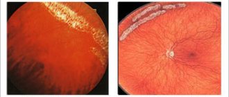
- Blindness on one side with complete preservation of the visual functions of the other eye often occurs when the optic nerve is damaged on the corresponding side.
- Damage to the chiasm in its central part will result in bitemporal hemianopsia.
- Damage to the external chiasm area causes binasal hemianopsia.
- If the optic tract or optic radiation is damaged, hemianopsia occurs on different sides.
- When parts of the optic radiation are damaged on a certain side, some areas of the visual fields fall out.
The main feature of damage to the visual pathway is that the patient does not feel pain in the structure of the visual organs. Now you know what the visual pathway is and what structure it has. We hope this information was useful and interesting.
Structure
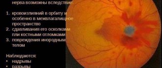
The optic nerve runs from the eyeballs to the brain. Its size is 5-6 cm. Adipose tissue is concentrated around the nerve formation, which is a protective layer from mechanical damage, bruises and the spread of infection.
Behind the eyeball, the optic nerve is formed in the form of a small disk, which accumulates information received from the photoreceptor of the retina. When it enters the orbit, it is surrounded by meninges. It goes into the eye socket along with them.
In the orbit, the right or left nerve plexuses intersect to form a chiasm. It is in this place that most pathologies of nervous tissue occur, which can reduce visual function. Under the weave is the pituitary gland. Because of this convergence, diseases that form in these structures can overlap with each other. The pituitary gland is an internal endocrine gland that produces substances that affect most human hormones.
After the chiasm, the nervous tissue enters the center of the visual analyzer. All information from the eyeballs is transmitted there, so a person can perceive surrounding objects and their colors. From the central nervous system, signals travel along the same pathways back to the eyeballs.
The nerve plexus is supplied with blood mainly through the internal carotid artery. The greatest blood flow is observed in the optic tract. The smallest part extends to the optic disc.
Thanks to knowledge of the structure of the optic nerve, an ophthalmologist and surgeon can identify the causes of the pathological condition, carry out diagnosis and treatment.