Causes
Impaired blood circulation in the brain leads to the development of a pathological condition. This occurs due to a malignant or benign neoplasm, head injury, or cerebral hemorrhage.
Less common causes of the disease include:
- infections;
- seizures or migraines;
- exposure to toxins;
- neurodegenerative disorders;
- non-ketotic hyperglycemia.
Common causes include stroke, tumors or complications from surgery. Also associated with high blood pressure or epileptic seizures.
The disease is caused by aneurysms. In most cases, the cause is a consequence of organic damage to the nerves of the optic pathway.
Causes of hemianopsia
The main factors provoking the development of the disease are damage to the optic tract or cerebral cortex. The cause of congenital pathology lies in developmental disorders of the fetus and in difficult childbearing (previous viral or infectious diseases, hypoxia, etc.). Acquired hemianopsia develops as a result of the following processes occurring in the body:
- Meningitis;
- Epilepsy. The disease is accompanied by swelling of the brain, resulting in impaired visual perception;
- Prolonged migraine. During headache attacks, the visual apparatus malfunctions because blood flow is disrupted;
- Formation of blood clots in the retinal vessels;
- Accumulation of fluid in the brain. Excess moisture puts pressure on the tissues of the main organ of the central nervous system, causing the development of various anomalies;
- Increased intracranial pressure;
- Inflammatory processes;
- Severe intoxication. Poisoning with methyl alcohol or certain medicinal drugs can cause visual impairment;
- Impaired functionality of the central nervous system;
- Brain injury. Most often, damage is accompanied by the formation of hematomas, which put pressure on the tissues responsible for visual perception. This leads to partial loss of vision;
- Poor circulation;
- Anorexia (due to it there is a lack of nutrients and vitamins);
- Neoplasms in the brain. They compress the tissues responsible for visual perception. As a result, fields disappear.
Neurological diseases can also cause the formation of “blind spots”. Return to contents
Symptoms
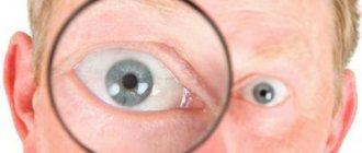
Hemianopia is manifested by narrowing of peripheral vision or the appearance of blind spots in the central part of visual perception. This symptomatology causes the patient difficulty in moving and performing minor work.
The clinical picture depends entirely on the type of disease. Pain may occur due to eye strain. There is no redness.
The heteronymous form differs from the homonymous one in that with the first there is a clear boundary between the visible area and the one that is outside the field of view.
Classification
Homonymous
With this type, the patient is not able to immediately identify two left or two right halves of the visual field.
The pathological process is characterized by loss of the temporal half of the visual field. The disease is caused by damage to the optical tract on the side opposite to loss of peripheral vision.
The homonymous form progresses rapidly. It practically never disappears.
Heteronymous
Bitemporal
The lesion is located in the optic chiasm, compressing the dissecting fibers. Most often caused by abnormal growth of the pituitary gland, which gives way to the optic chiasm.
Partial blindness. The perception of the outer half of the visual fields of both eyes is lost. Most often, the bitemporal form is the result of tumors that are located in the central region of the optic nerve intersection.
The nerve fibers from the nasal halves of the retina are the only ones that cross to the other side of the brain, resulting in loss of vision only on the temporal side.
Some of these changes are associated with a pituitary adenoma increasing the production of any of the following hormones: LCH, PSH, TTX, ACTH, GC, prolactin, vasopressin, oxytocin, and Alpha-MCX.
Binasal
This form is also associated with damage to the uveal tract and central nervous system. Binasal hemianopsia develops when the lateral parts of the chiasm fibers are affected on both sides.
This form of the disease is found in patients with sclerotic lesions of the internal carotid artery on both sides.
Quadrant
Left superior quadrant hemianopsia is a visual defect that occurs when the inferior optic emitting fibers (Meyer's loop) in the temporal lobe of the brain are damaged. Strokes involving the middle cerebral artery can lead to the development of this form of blindness.
Temporal lobe lesions cause other neurological manifestations, including aphasia, memory deficits (if hemisphere dominant), seizures, and auditory and visual hallucinations.
The left lower homonymous form of pathology is damage to the upper fibers in the parietal lobe. Because the parietal lobe is considered the primary sensory area of the cerebral cortex, these lesions often cause sensory deficits.
Gerstmann's syndrome (finger agnosia, agraphia, acalculia and right-sided disorientation) accompanies this defect if the dominant angular lobe of the gyrus is involved. Contralateral heminglect (inability to recognize the visual field opposite the area of traumatic brain injury) is observed with lesions of the parietal lobe of the non-dominant hemisphere, which is difficult to distinguish from this defect.
One-sided
Right-sided blindness occurs when the lesion is located in front of the optic chiasm. Has a wide differential, including corneal diseases or cataracts to optic neuritis. If the site of the stroke is the optic nerve, patients will usually have a relative afferent pupillary defect.
The left-sided form of the disease results from damage to the optic tract en route to the lateral genital body of the thalamus, and damage just after the radiating fibers leave the lateral genital body.
The damage is often caused by strokes or neoplasms.
Double-sided
A type of pathology in which defects are found in both corner spaces visible to the eye with a fixed gaze. The pathological condition develops in the presence of several foci in the optical pathways, tracts, and occipital lobe.
Scotoma
This is a blind spot in the visual field. Scotoma takes different shapes - oval, round, arched. It can form in different parts of the visual field and is not associated with peripheral boundaries.
General signs and classifications of hemianopsia
It happens that such visual impairment is not detected by the patient and is diagnosed only during a medical examination. Homonymous types of hemianopsia always limit a person’s movement, and he or she experiences visual difficulties. These defects may be temporary and may disappear after some period. The main symptom from the fundus of the eye is nerve atrophy of varying degrees.
With heteronymous hemianopia, the boundary between the visible and invisible parts of the visual field passes through its center. This happens if the middle parts of the chiasm are affected. Such damage is observed much less frequently. Sometimes, with a significant increase in intracranial pressure, a person experiences characteristic signs of congestion in the optic nerve.
Homonymous hemianopsia occurs when the right or left portion of the visual field disappears. When scalene zones fall out, they speak of heteronymous hemianopsia. There may be incomplete, quadrant hemianopsia and, finally, scotoma. Bilateral pathology is observed if both halves of vision are lost.
Bitemporal hemianopsia
Doctors distinguish the following types of hemianopsia:
- Different types of homonymous vision loss. All of them indicate a specific location of the pathogenic focus.
- Heteronymous hemianopsia is the exclusion of the nasal and temporal halves of the visual field. In bitemporal hemianopsia, the superior or temporal visual fields are lost.
- Binasal hemianopsia is loss of the lower portion of vision (usually diagnosed in both eyes).
- Bilateral - the appearance of visual perception defects on two halves.
- A scotoma is a dark spot in the visual field that has a variety of shapes. For example, it could be an oval, circle, square, arc, etc. Such a defect can occur in any part of the field.
It is necessary to consider these types of violations in more detail.
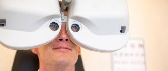
Diagnostics
The disease does not affect the clarity of visual perception. Hemianopsia does not change the condition of the fundus. Therefore, to determine the correct diagnosis, you will need to perform the following tests:
- perimetry;
- computer campimetry;
- computed or magnetic resonance imaging;
- ultrasound examination of the head;
- vascular angiography.
Diagnostics makes it possible to establish the cause of the pathological condition, to detect a cyst or tumor that presses on the optic tract. A thorough examination gives a complete picture of the arteries of the brain.
Treatment
For hemianopia, two therapies are possible - both approaches are neuropsychological in nature. The goal of the first method is to restore vision functions. Method 2 is called compensation training.
This treatment approach is designed to compensate for impairment as effectively as possible. However, visual field defects are not compensated for in 95% of cases. Visual perception is not fully restored.
The first method is carried out by visual stimuli - light or color. They are designed to stimulate the areas between the working part and the defective part. Thus, partial restoration of the visual field is possible.
This approach to therapy is controversial, but doctors actively use treatment with visual stimuli. For this reason, treatment is not covered by health insurance.
The exercises consist of targeted eye movements that can be easily done at home or on the go after training - while shopping or reading a newspaper. Hemianopsia deprives sufferers of the enjoyment of life. With correctly applied compensatory training, deterioration decreases relatively quickly.
Exercises are used to teach children to find necessary objects and read. The saccade exercise is a technical term that implies rapid and strictly coordinated eye movements occurring simultaneously in one direction.
Training is carried out using additional electronic programs developed for this purpose. They are designed to encourage patients to look for specific visual stimuli on a screen—with just one eye movement.
Learning to read is an important part of compensation training. People with hemianopia have difficulty reading due to a severely limited field of vision.
The goal is to adapt the reading movement to the boundaries of the visual field. There are specially designed programs. But even at home they study effectively - short texts and lines are suitable. It is important that the head remains straight during the exercise - the search for lines and words should occur precisely with the help of eye movements.
Useful video
Irina Sergeevna Mironova’s story about the causes and features of the development of the pathological condition:
Author's rating
Author of the article
Alexandrova O.M.
Articles written
2031
about the author
Was the article helpful?
Rate the material on a five-point scale!
( 1 ratings, average: 2.00 out of 5)
If you have any questions or want to share your opinion or experience, write a comment below.
Prevention
It is almost impossible to prevent the development of homonymous hemianopia. However, you can reduce the risk of the problem occurring by following these preventive measures:
- avoid head injuries;
- undergo regular preventive medical examination;
- promptly and correctly treat existing pathologies;
- avoid any intoxication of the body;
- take only medications prescribed by a doctor;
- monitor the condition of the circulatory system;
- try to avoid stressful situations.
Symmetrical loss of half of the visual fields usually occurs against the background of severe brain diseases.
The development of this pathological condition can be avoided if you consult a doctor in a timely manner and carry out competent therapy for the underlying disease.
Author of the article: Kvasha Anastasia Pavlovna, specialist for the website glazalik.ru Share your experience and opinion in the comments.