Morphology of brain gliosis
The destruction of functional brain tissue leads to the replacement of damaged areas with neuroglia. The process is necessary to ensure cerebral nutrition and provide necessary substances.
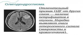
MRI of brain gliosis
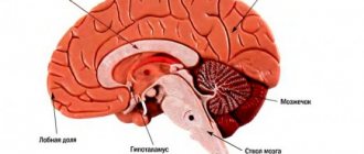
The main anatomical structures of the central nervous system:
- The cell layers of the lining of the ventricles are ependymal;
- Functional elements of nerve signal transmission are neurons;
- Tissues that provide framework, construction, metabolic, secretory functions are neuroglia.
Most of the cerebral structure consists of glial elements. The complex provides many functions, but signal transmission is achieved through interconnections between neurons. The fewer nerve fibers, the slower the processes proceed. Trophic functions are performed by glia, which fills about forty percent of the brain.
Inflammation, infections, and traumatic brain injuries can lead to the destruction of neurons and ependyma. The sites of destruction are filled with neuroglia, creating scars that are similar to scars on the skin (overgrowth of connective tissue fibers).
Treatment methods
Treatment of gliosis is aimed at eliminating the causes of the pathology - first of all, treatment is carried out for the primary disease that affects the structures of the brain. There is no specific treatment for gliosis changes. Depending on the indications, medications are prescribed, and surgery is performed less frequently. Nutrition for cerebral gliosis should be complete and balanced.
Multiple foci of gliosis detected during the study of brain structures, regardless of size, require dynamic monitoring. The frequency of control studies is prescribed by the attending physician.
Drug therapy
The attending physician will tell you how to treat cerebral gliosis, based on the results of a diagnostic examination, taking into account the primary pathology, the patient’s age and symptoms. Essential medicines:
- Nootropic. Protect neurons from damage, stimulate metabolic processes in nerve cells.
- Regulating cerebral blood flow. Drugs that normalize the activity of the circulatory system that feeds the brain.
- Antioxidant. Drugs that prevent oxidative reactions in nervous tissue.
- Antihypertensive. Medicines that normalize blood pressure.
In parallel, depending on the type of primary disease and symptoms, medications are prescribed to eliminate neurological symptoms. This group includes anticonvulsants, antiepileptics, painkillers, and antiemetics.
Surgical intervention
Surgical treatment is resorted to when it is impossible to maintain the patient’s well-being with medications. Surgical treatment of gliosis is indicated under certain conditions:
- Single, large lesions.
- The presence of severe neurological symptoms - convulsive syndrome, epileptic seizures, serious impairment of motor activity and mental activity.
- The patient's age is not older than 60 years.
- Mass effect (negative impact of education on surrounding healthy brain structures).
During the operation, areas of cystic glial tissue that cause symptoms and disorders are removed. Treatment must be comprehensive and timely.
Where do gliosis foci appear?
If you have a headache due to nosology, MRI will detect multiple areas of glial tissue. The lesions affect different parts of the brain. They are located with equal probability both inside the white matter and superficially.
Forms of gliosis according to the structure of the foci:
- Fibrous – morphological defects contain most of the fibers;
- Marginal - zones are localized superficially under the shell;
- Vascular - the proliferation of small capillaries predominates;
- Anisomorphic – chaotic arrangement of glial elements predominant over the fibers;
- Perivascular – neuroglia are located around the affected vessels (with vasculitis);
- Focal - a limited area at the site of inflammation;
- Diffuse – spread of the process to the brain and spinal cord;
- Supratentorial - single foci of gliosis of vascular origin in the brain are formed during human aging, after childbirth. The course is asymptomatic.
Magnetic resonance imaging of the brain allows one to count foci of gliosis. Tomograms show the structure of the areas. The wider the focus, the more clinical symptoms of central nervous system damage.
Dynamic examination (repeated MRI) allows us to evaluate the increase in the size of brain lesions.
Symptoms
A few, small lesions may not appear for a long time. They are often discovered accidentally during a diagnostic test ordered for another reason. Symptoms are often associated with manifestations of the primary disease. Main general symptoms:
- Pain in the head area, dizziness.
- Impaired motor coordination, changes in gait (unsteadiness, uncertainty, expansion of the support base).
- Amplitude jumps in blood pressure indicators.
- Deterioration of cognitive abilities (memory, attention, mental activity).
- Sleep disorder.
- Increased fatigue, decreased performance.
- Convulsive, epileptic seizures.
- Visual and auditory dysfunction.
Gliosis localized in the frontal lobe is typical for elderly patients, which is associated with previous diseases and various destructive processes occurring in the brain. A single focus of gliosis, located in the left or right frontal lobe, is small in size and may not appear throughout life. Focal lesions of the frontal lobes are often associated with disorders such as inability to concentrate, memory impairment, and the development of dementia.
The appearance of lesions in the white matter of the frontal lobes may be accompanied by contralateral (located on the side opposite to the lesion) paresis and aphasia (impaired speech function). Neurogliosis in epilepsy is a secondary type of scar-forming process initiated in response to damage and death of neurons. Scientists have not come to a consensus on whether this process is an epileptogenic (causing epileptic seizures) factor or a consequence of epilepsy.
Causes of glial growths
Any pathological processes with destruction of the anatomical structures of the white matter of the brain lead to glial growths. The most common causes:
- Infectious processes – tuberculosis, cytomegalovirus, chlamydial vasculitis;
- Autoimmune processes with destruction of nerve fiber sheaths (demyelination, multiple sclerosis);
- Inflammatory diseases – encephalitis, meningitis;
- Open and closed traumatic brain injuries (TBI);
- Genetic defects of fat metabolism;
- Hypoxic conditions, cerebral ischemia;
- Lack of oxygen supply to the brain during childbirth;
- Hypertensive crisis;
- Consequences of stroke;
- Surgical interventions;
- Encephalopathy;
- Intracranial hypertension;
- Hematoma.
In addition to etiological factors, there are two provoking mechanisms of gliosis:
- The introduction of narcotic drugs leads to gradual atrophy of the white matter. Clinical studies have identified isolated glial foci in patients with drug addiction;
- Alcohol abuse – excessive intake of alcohol metabolites into the brain leads to the death of neurons. Moderate consumption improves cerebral blood circulation.
Detection of single foci of gliosis requires treatment. It is impossible to restore dead neurons, but with the help of therapy it is possible to prevent the progression of the disease.
Definition of pathology
When the neurons that make up the nervous tissue are damaged, glial cells form in their place. Gliotic changes are a process occurring in the brain, which is characterized by an increase in the number of glial cells, which, when the scale of replacement expands, leads to a deterioration in the functioning of the central nervous system. The more glial foci in the medulla, the worse the brain performs its functions.
Disorders are most often associated with a deterioration in the transmission of nerve impulses, with the help of which the nervous system controls the organs and systems of the body. Proliferation of glial cells most often occurs in the form of diffuse distribution of astrocytes. As areas consisting of glial cells grow, signs of pathology appear, such as impaired motor coordination, memory impairment, slowness of movements and reactions. Gliotic transformation of the brain, depending on the type of primary pathology, occurs with characteristic features.
In diabetes mellitus, large-scale infiltration of macrophages and hypertrophy (pathological increase in size) of astrocytes is observed. Gliotic changes in drug addicts are accompanied by an increase in the number of oligodendrocytes of drainage forms. In multiple sclerosis, hypertrophy of astrocytes and changes in the glial formula are detected (astrocytes - 46%, oligodendrocytes - 40%, other cells - 14%).
Normally, the glial formula looks like this: astrocytes - 8.5%, oligodendrocytes - 85%, other cells - 7.5%. In epilepsy, there is a decrease in the number of oligodendrocytes by 20% and microgliocytes by 6%. Glial cells are the most numerous and active components of brain tissue. They retain the ability to divide throughout their lives. Due to their high activity, glia cells immediately respond to any changes in the functioning conditions of the brain.
The average number of cells in 1 mm2 of brain tissue varies depending on the location of the site. For example, in the parietal lobe the number of cells is 2 times greater than in the frontal region. When diagnosed with multiple sclerosis, the number of glial cells in areas not affected by the demyelination process may increase by about 3 times. In patients with drug addiction, the proportion of glial cells increases approximately 2 times.
With dyscirculatory encephalopathy, this indicator increases slightly. Foci of gliosis arise as a reaction of neuroglia to damage to nervous tissue or changes in the functioning conditions of the brain. The mitotic activity of gliocytes increases in response to the development of pathological processes in the tissues of the central nervous system. Gliotic foci are a consequence of the process of restoration of destroyed brain tissue. However, the functions of areas of restored tissue are not always adequate to normal physiological processes.
The reason for the incomplete correspondence of functions lies in the underdevelopment of glial cells, which, having not reached maturity and a normal level of functioning, undergo apoptosis (a regulated process of cell death). Similar phenomena are most clearly observed in multiple sclerosis. What is dangerous for humans is not the process of glial transformations itself, but rather its scale and incompleteness associated with the interruption of the normal development of gliocytes.
Signs of neuroglial accumulation in the brain
Pathological manifestations of the accumulation of neuroglia inside the brain are only in rare cases provoked by serious diseases. In older people, foci of gliosis gradually increase, which leads to the progression of neurological symptoms:
- Impaired coordination of movements;
- Memory loss;
- Prolonged pain syndrome of the head;
- Frequent changes in blood pressure;
- Muscle cramps;
- Epileptic seizures;
- Dizziness.
Using MRI and CT, it is possible to calculate the number of neuroglial proliferations per unit volume of cerebral tissue. The value is used to assess the progression of the disease and evaluate the effectiveness of treatment.
Interesting fact
A study by epidemiologists has proven the influence of neuroglia on the aging process. Experts added blood taken from an elderly person to the morphological material of glia. The consequences of the experiment turned out to be impressive - neuroglial layers began to actively multiply, destroying neurons. The results suggest mechanisms for the development of senile dementia.
A similar process does not occur in children. The proliferation of neuroglia in the white matter is observed only after the death of neurons. The mechanism is local in nature. The lesions cover the formed defects.
Symptoms of focal white matter gliosis
Clinical symptoms are determined by the size, location, and progression of neuroglial accumulations:
- Severe headaches - diffuse type;
- Instability of blood pressure, frequent hypertensive crises;
- Unsteady gait, lack of coordination;
- Decreased mnestic functions;
- Memory impairment.
Therapeutic techniques are ineffective in the presence of diffuse periventricular lesions. Large accumulations in a child are rarely observed, so symptoms do not occur.
What is dangerous about gliosis - prognosis for life and health
Life prognosis depends on the underlying cause of the disease. Clinical symptoms are determined by the severity of white matter damage. Affecting the functional centers of breathing and blood circulation can lead to death. Encephalitis and meningitis are dangerous, which often lead to an unfavorable prognosis and death.
Minor symptoms, favorable outcome for gliosis in the frontal lobes of the brain. Subcortical species are dangerous if they spread diffusely.
Signs of gliosis
Quite often, the patient learns about the presence of a single focus of gliosis in his brain, by chance, during a routine examination. At the same time, nothing bothers the person. This situation requires special attention.
The patient must be carefully examined and the reason why the lesion formed is identified, that is, the disease that provoked the proliferation of glial fibers. In the case of multiple foci of glia, the situation is different; unpleasant symptoms cannot be avoided.
Symptoms:
- Constant headaches.
- Blood pressure surges
- Dizziness.
- Violation of intellectual activity.
- Loss or impairment of coordination.
- Changes in speech functions.
- Paresis and paralysis.
- impairment .
- Changes in the mental sphere.
- Dementia.
The larger the area of brain damage, the more pronounced the symptoms of this disease.
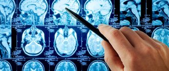
Diagnosis of cerebral gliosis
In most cases, the disease is verified by chance during an MRI or CT scan of the brain. The form is not dangerous. A repeat examination after a few months is carried out in order to study old glial areas. The absence of an increase in size indicates a favorable prognosis.
MR and CT angiography with a contrast agent helps to identify the vascular genesis of the nosology. Electroencephalography (EEG) determines or excludes changes in cerebral activity. The procedure is prescribed for the occurrence of epileptic seizures and muscle cramps.
Practice shows a predominant transformation of neuroglia during aging in the frontal lobes.