Brain tumors and their symptoms
Brain tumors refer to a whole group of diseases that develop in the brain or internal structures of the cranial cavity. They account for about 6% of all types of neoplasms in the human body.
The risk of developing a brain tumor increases with age; according to statistics, the majority of patients with this pathology are people who have crossed the 40-year mark. Although there are types of tumor-like formations that are diagnosed only in children.
In addition to age, other risk factors for developing brain tumors include:
- presence of traumatic brain injuries;
- bad ecology;
- radiation exposure and exposure to toxic substances in the workplace;
- disruption of intrauterine development leading to congenital tumors;
- family predisposition to genetic mutations.
Tumor-like formations in the brain are usually divided into benign and malignant.
Distinctive features of benign tumors are slow development without germination into neighboring tissue structures and organs, and the absence of metastasis. After their removal there is no relapse.
Malignant tumors are more aggressive and tend to return even after treatment. In addition, brain tumors are also usually classified as primary or secondary tumors. The development of the primary begins from brain tissue, and the secondary is the result of metastasis from any organ.
Signs of brain tumors are very diverse, their manifestations are determined by the size, location and speed of development of the pathology.
Let us dwell on the general symptoms that are characteristic of many tumor-like formations:
- Headache. Almost all patients complain of periodic headaches, although they do not assume that it may be the result of tumor growth.
- Muscle spasms and cramps . This is the most common phenomenon in cases of dysfunction of the central nervous system due to a tumor process in the brain.
- Feeling of nausea, vomiting . Due to increased intracranial pressure, the patient experiences nausea in the morning, which is not associated with indigestion.
- Visual impairment. Brain tumors can affect the optic nerves, so the patient experiences a gradual deterioration of vision.
- Personal changes. The location of a tumor-like formation in a certain part of the brain leads to mental disorders.
- Problems with speech and hearing . Loss of communication skills is associated with tumors in the frontal and parietal lobes of the brain.
- Memory impairment . Patients complain of problems with remembering and memory loss.
- Weakness and fatigue . Brain tumors lead to exhaustion of the body, the patient experiences constant physical fatigue.
- Difficulty maintaining coordination of movements . With the development of tumor-like formations in the brain, the patient’s balance when walking and coordination of arms and legs is impaired.
- Endocrine disorders . Tumors affecting the pituitary gland lead to hormonal imbalances.
Even at the slightest suspicion of the development of a tumor-like formation, it is necessary to immediately undergo examination, and if the diagnosis is confirmed, begin therapeutic procedures. Treatment of malignant brain tumors cannot be delayed, as this may negatively affect the results of therapy. Modern foreign clinics have advanced technological resources to successfully deal with various types of intracranial formations. It is necessary to take into account: any brain surgery is a complex process that requires high professionalism of a doctor, even with ultra-modern equipment. If we compare the cost of cancer treatment abroad, then in Israel the prices for medical services compare favorably with the prices of other countries.
Symptoms of brain cancer
There are many symptoms and signs of brain tumors. Their manifestation depends on the size of the tumor, location and speed of its growth. The most common symptoms of brain cancer include:
- the occurrence of frequent, uncharacteristic headaches, a change in the nature of the pain or its intensification;
- unexplained nausea or vomiting;
- vision problems such as blurred and double vision or loss of peripheral vision;
- gradual loss of sensation or movement in an arm or leg;
- difficulty with balance;
- speech problems;
- confusion in daily activities, feelings of confusion or disorientation;
- changes in personality or behavior;
- convulsions, especially if not previously observed;
- hearing problems;
- memory impairment and difficulty concentrating;
- drowsiness, sleep problems.
In order to understand how brain tumors manifest themselves, we have listed below the symptoms of the most common ones:
- meningioma (headaches, weakness in an arm or leg, personality changes, vision problems, seizures);
- glioblastoma (nausea and vomiting, headaches that may be worse in the morning, weakness in the body (eg, arm, leg, or face), memory problems, seizures, difficulty with balance);
- astrocytomas (headaches, memory loss, seizures, changes in behavior);
- pituitary tumors (headaches, vision problems, changes in behavior, changes in hormone levels);
- metastatic tumors (headaches, seizures, loss of short-term memory, changes in personality and behavior, weakness on one side of the body, difficulty with balance).
These symptoms do not always indicate the presence of a brain tumor. To confirm or refute suspicions, at the first signs of brain cancer, doctors conduct the necessary diagnostic examinations.
Diagnostics
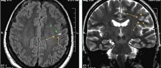
Instrumental imaging methods - computed tomography (CT) and magnetic resonance imaging (MRI) - allow one to accurately determine a brain tumor. MRI is considered the best method for detecting tumors. The images obtained with this method are more detailed than those obtained with CT.
Magnetic resonance imaging scanner Siemens Magnetom Sola at the Spizhenko Clinic
In general, MRI is used more often when diagnosing brain tumors. A CT scan is indicated in cases where MRI is not appropriate (for example, in overweight people or people with a fear of closed spaces). Also, CT, unlike MRI, allows you to evaluate the bone structures in the area of the tumor.
Instrumental imaging methods show the location and extent of the tumor, but they cannot always accurately determine the type of tumor. In most cases, this can only be done with a biopsy. It is performed as an independent procedure or during an operation to remove a tumor. Based on the selected tissue sample, the pathologist determines whether the tumor is benign or malignant and classifies it.
The results of the diagnostic examinations allow the doctor to establish a diagnosis, describe the tumor and plan future treatment.
How to detect a brain tumor?
A multidisciplinary approach to the treatment of brain tumors abroad involves high-quality diagnostics, correctly selected therapeutic procedures, a rehabilitation course, social adaptation of the patient, and qualified psychotherapeutic assistance.
The main methods for detecting tumor formations in the brain today are:
- CT scan . This study is carried out for suspicious pathology in the early stages of the disease.
- Nuclear magnetic resonance . The procedure allows you to obtain an image of the tumor and determine its characteristics.
- Whole body PET . The examination helps diagnose the primary tumor, as well as assess the spread of abnormal cells if there are metastases. This method is often used in the diagnosis and treatment of brain cancer abroad.
In addition to these methods, to clarify the diagnosis after a consultation of specialists, other additional diagnostic procedures may be required, including ventriculoscopy and stereotactic biopsy, which are related to complex procedures.
Causes of brain cancer development
The occurrence of a tumor is caused by abnormal cell division. For what reason the pathological process is formed cannot be determined. There are several risk factors that increase the risk of cancer. Namely:
- Radiation. According to statistics, in people regularly exposed to ionizing radiation at nuclear plants, brain cancer is detected much more often. At the same time, the influence of electromagnetic radiation from modern gadgets does not increase the risk of developing cancer.
- Genetic predisposition. A history of cancer in one of the family members increases the risk of developing this pathology, especially when it comes to glioma. Heredity is aggravated by genetic pathologies: tuberculous sclerosis, neurofibromatosis type 2.
- Immune system disorders. The high-risk group includes people with weakened immune systems, since lymphoma forms in the cells of the immune system - lymphocytes.
Risk factors for the formation of pathologies in the brain also include: bad habits, previous head injuries, taking certain medications, and the presence of serious diseases, including HIV.
Symptoms and stages of low-quality brain tumors
The development of low-quality brain tumors occurs quite quickly, so it is necessary to react sensitively to any failures and disturbances in the body so as not to miss alarming symptoms.
- Stage 1 of tumor formation in the brain
At this stage, cancer can be detected using special diagnostics. Symptoms of the disease coincide with those of other diseases. The patient usually associates weakness, drowsiness, dizziness and pain in different areas of the head with climate change or weakened immunity.
- Stage 2 of tumor formation in the brain
As the pathological process moves to the next stage, further growth of abnormal cells occurs, they begin to put pressure on the brain and disrupt its normal functioning. The patient may have problems with the gastrointestinal tract, vomiting, and seizures.
- Stage 3 of tumor formation in the brain
At stage 3, the tumor affects surrounding healthy structures, spreading very quickly. The patient's symptoms become even more pronounced. Problems with memory, hearing, speech, vision, and vestibular apparatus are noticeable. Horizontal nystagmus (involuntary movements of the eyeball) at this stage is a hallmark sign of a low-grade brain tumor.
- Stage 4 of tumor formation in the brain
The abnormal cells in this case are in vital areas of the brain, so symptoms are associated with loss of related basic functions. Palliative techniques, radio irradiation, and the use of painkillers are used to treat cancer abroad.
Prognosis for brain tumors
Until now, in people’s minds, any pathological neoplasm of the brain is associated with an incurable disease, but the latest methods for diagnosing and treating brain tumors in Israel prove that this is not the case at all.
A favorable prognosis largely depends on the complex of therapeutic procedures started in the early stages of the disease, as well as the degree of benignity of the tumor process. The key to effective treatment of a malignant brain tumor is timely detection and obtaining complete information about its size, location, and stage of development.
Patients from other countries often turn to cancer centers in Israel because domestic doctors have declared the tumor inoperable. And in this case, Israeli specialists, who have mastered the most advanced treatment technologies, work a real miracle, saving the patient, preserving his brain function and improving his quality of life.
Classification
Depending on the type of brain cells that degenerated and gave rise to pathogenesis, the following types of tumors are distinguished:
- astrocytomas are the most common type, their share in the general population is about 50%;
- oligodendrogliomas – the share in the general population of tumors of this type is up to 10%;
- Ependymomas are the rarest forms (occurrence less than 7%).
According to the WHO classification (which is generally accepted), neoplasia is ranked depending on the degree of malignancy.
Benign gliomas
These are tumors of the first degree of malignancy, for example, astrocytomas: giant cell, pilocytic, juvenile subependymal. They are benign because they grow slowly, have no signs of cancer, and are easy to treat. The prognosis for life with benign glioma is favorable. After tumor removal, patients live 10 years or longer.
Low grade glioma
Second degree of malignancy. It is also classified as a benign neoplasm, but with a borderline degree of malignancy. The growth of pathological tissue is slow (low-grade), they are well differentiated, as a rule, there is only one sign of cancer (cell atypia). Tumors of this type can degenerate into cancer and easily progress to the third and fourth degree of malignancy. These include diffuse and fibrillary astrocytomas. Treatment is complex: surgery to remove atypical tissues, additionally radio and chemotherapy.
Malignant gliomas
These include gliomas of grade 3 and 4:
- Third degree of malignancy . There are all signs of malignancy, except for tissue necrosis. Tissues lose clear differentiation, tumor growth accelerates (high-grade), the boundaries are unclear, and growth into nearby tissues is typical. The most striking example is anaplastic astrocytoma, which most often develops in middle-aged and older people. Treatment is hampered by the lack of clear boundaries of the tumor; life expectancy depends on how much atypical tissue is removed, as well as the stage of carcinogenesis at the beginning of treatment.
- The most dangerous is the fourth degree of malignancy (glioblastoma). It develops between the ages of 40 and 70 years. In this case, all the signs of malignancy are present, including necrosis. The tumor grows quickly, penetrates other tissues and has no clear boundaries. This significantly complicates therapy. The prognosis is unfavorable.
Neoplasia is divided into two types, depending on growth characteristics:
- Tumors of nodular growth . As a rule, these are benign neoplasms with clear contours. They are characterized by formation anywhere in the brain and the presence of cysts. Examples: pleomorphic xanthoastrocytoma and piloid astrocytoma.
- Diffuse type formations . There are no obvious boundaries, the size can reach significant sizes, neoplasia grows into adjacent brain tissue, which complicates the removal of dysplasia. These are often malignant tumors, such as glioblastoma or anaplastic astrocytoma, or those that can quickly degenerate.


