Dear colleagues, the material offered to you was once prepared by the author for the chapter of the manual on neuraxial anesthesia, which, for a number of reasons, was not completed and was not published. We believe that the information presented below will be of interest not only to novice anesthesiologists, but also to experienced specialists, since it reflects the most modern ideas about the anatomy of the spine, epidural and subarachnoid spaces from the point of view of an anesthesiologist.
Anatomy of the spine
As you know, the spinal column consists of 7 cervical, 12 thoracic and 5 lumbar vertebrae with the adjacent sacrum and coccyx. It has several clinically significant bends. The greatest anterior curves (lordosis) are located at the C5 and L4-5 levels, and posteriorly at the Th5 and S5 levels. These anatomical features, together with the baricity of local anesthetics, play an important role in the segmental distribution of the level of spinal block.
The characteristics of individual vertebrae influence the technique, primarily of epidural puncture. The spinous processes extend at different angles at different levels of the spine. In the cervical and lumbar regions, they are located almost horizontally in relation to the plate, which facilitates median access when the needle is perpendicular to the axis of the spine. At the mid-thoracic level (Th5-9), the spinous processes extend at fairly acute angles, which makes the paramedian approach preferable. The processes of the upper thoracic (Th1-4) and lower thoracic (Th10-12) vertebrae are oriented intermediately compared to the above two features. At these levels, neither access has advantages over the other.
Access to the epidural (ED) and subarachnoid space (SP) is carried out between the plates (interlaminar). The superior and inferior articular processes form the facet joints, which play an important role in correctly positioning the patient before ED puncture. The correct positioning of the patient before ES puncture is determined by the orientation of the facet joints. Because the facet joints of the lumbar vertebrae are oriented in the sagittal plane and allow anterior-posterior flexion, maximum spinal flexion (fetal position) increases the interlaminar spaces between the lumbar vertebrae.
The facet joints of the thoracic vertebrae are oriented horizontally and provide rotational movements of the spine. Therefore, excessive spinal flexion does not provide additional benefit when performing EC puncture at the thoracic level.
Anatomical bony landmarks
Identification of the required intervertebral space is the key to the success of epidural and spinal anesthesia, as well as a necessary condition for patient safety.
In a clinical setting, the choice of puncture level is made by the anesthesiologist through palpation in order to identify certain bony landmarks. It is known that the 7th cervical vertebra has the most pronounced spinous process. At the same time, it must be taken into account that in patients with scoliosis, the most protruding process may be the spinous process of the 1st thoracic vertebra (in approximately ⅓ of patients).
The line connecting the inferior angles of the shoulder blades passes through the spinous process of the 7th thoracic vertebra, and the line connecting the iliac crests (Tuffier's line) passes through the 4th lumbar vertebra (L4).
Identification of the required intervertebral space using bone landmarks is not always correct. The results of the study by Broadbent et al. (2000), in which one of the anesthesiologists used a marker to mark a certain intervertebral space at the lumbar level and tried to identify its level with the patient in a sitting position, the second made the same attempt with the patient in the lateral position. A contrast marker was then attached over the mark made and magnetic resonance imaging was performed.
Most often, the true level at which the mark was made was one to four segments lower than the values reported by the anesthesiologists participating in the study. It was possible to correctly identify the intervertebral space only in 29% of cases. The accuracy of the determination did not depend on the patient's position, but worsened in overweight patients. By the way, the spinal cord ended at the L1 level in only 19% of patients (in the rest at the L2 level), which created a risk of damage to it if a high puncture level was incorrectly chosen. What makes it difficult to choose the intervertebral space correctly?
There is evidence that Tuffier's line corresponds to the L4 level in only 35% of people (Reynolds F., 2000). For the remaining 65%, this line is located at the level from L3-4 to L5-S1.
It should be noted that an error of 1-2 segments when choosing the level of puncture of the epidural space, as a rule, does not affect the effectiveness of epidural anesthesia and analgesia.
Spinal ligaments
The anterior longitudinal ligament runs along the anterior surface of the vertebral bodies from the skull to the sacrum, which is rigidly fixed to the intervertebral discs and the edges of the vertebral bodies. The posterior longitudinal ligament connects the posterior surfaces of the vertebral bodies and forms the anterior wall of the spinal canal.
The vertebral plates are connected by the ligamentum flavum, and the posterior spinous processes by the interspinous ligaments. The supraspinous ligament runs along the outer surface of the spinous processes of C7-S1. The pedicles of the vertebrae are not connected by ligaments, resulting in the formation of intervertebral foramina through which the spinal nerves exit.
The ligamentum flavum consists of two layers fused along the midline at an acute angle. In this regard, it is, as it were, stretched in the form of an “awning”. In the cervical and thoracic regions, the ligamentum flavum may not be fused in the midline, causing problems in identifying ES using a loss of resistance test. The ligamentum flavum is thinner in the midline (2-3 mm) and thicker at the edges (5-6 mm). In general, it has the greatest thickness and density at the lumbar (5-6 mm) and thoracic levels (3-6 mm), and the smallest in the cervical region (1.53 mm). Together with the vertebral arches, the ligamentum flavum forms the posterior wall of the spinal canal.
When passing the needle through the median approach, it must pass through the supraspinous and interspinous ligaments, and then through the ligamentum flavum. With the paramedian approach, the needle bypasses the supraspinous and interspinous ligaments, immediately reaching the ligamentum flavum. The ligamentum flavum is denser than the others (80% consists of elastic fibers), therefore, an increase in resistance when passing it with a needle, followed by its loss, is known to be used to identify ES.
The distance between the ligamentum flavum and the dura mater in the lumbar region does not exceed 5-6 mm and depends on factors such as arterial and venous pressure, pressure in the spinal canal, pressure in the abdominal cavity (pregnancy, abdominal compartment syndrome, etc. ) and the chest cavity (ventilator).
With age, the ligamentum flavum thickens (ossifies), making it difficult to pass a needle through it. This process is most pronounced at the level of the lower thoracic segments.
Symptoms and signs
A wide variety of clinical manifestations can indicate the presence of the disease. So, warning signs are symptoms such as:
- The child's increased sensitivity to extraneous sounds, even quiet ones.
- Photophobia.
- After each feeding, the child regurgitates profusely, sometimes the regurgitation turns into vomiting.
- The baby does not sleep well and often wakes up.
- Visual impairment, manifested in the form of asymmetrical pupil size, strabismus.
- Increase in head size.
- Weather sensitivity (the child becomes more restless when weather conditions change).
- Disruption of the process of overgrowth of the fontanelle, increased tension in its area.
- Tremor of the limbs, slight trembling of the chin.
- Severe crying, which is caused by headaches that bother the child, mainly in the morning (after waking up).
- Drowsiness, lethargy, decreased motor activity.
From the editor: Doppler ultrasound of the vessels of the head and neck
Spinal cord membranes
The spinal canal has three connective tissue membranes that protect the spinal cord: the dura mater, the arachnoid mater, and the pia mater. These membranes participate in the formation of three spaces: epidural, subdural and subarachnoid. The spinal cord (SC) and roots are covered by a well-vascularized pia mater; the subarachnoid space is limited by two adjacent membranes - the arachnoid and dura mater.
All three SC shells continue in the lateral direction, forming the connective tissue covering of the spinal roots and mixed spinal nerves (endoneurium, perineurium and epineurium). The subarachnoid space also extends for a short distance along the roots and spinal nerves, ending at the level of the intervertebral foramina.
In some cases, the cuffs formed by the dura mater lengthen by a centimeter or more (in rare cases by 6-7 cm) along the mixed spinal nerves and extend significantly beyond the intervertebral foramina. This fact must be taken into account when performing a brachial plexus block from supraclavicular approaches, since in these cases, even with the correct orientation of the needle, intrathecal injection of a local anesthetic is possible with the development of a total spinal block.
The dura mater (DRM) is a sheet of connective tissue consisting of collagen fibers oriented both transversely and longitudinally, as well as a certain amount of elastic fibers oriented in the longitudinal direction.
For a long time, it was believed that dura fibers have a predominantly longitudinal orientation. In this regard, it was recommended that when puncturing the subarachnoid space, the cut of the spinal needle with the cutting tip should be oriented vertically so that it does not cross the fibers, but rather pushes them apart. Later, using electron microscopy, a rather random arrangement of dura fibers was revealed - longitudinal, transverse and partially circular. The thickness of the dura mater is variable (from 0.5 to 2 mm) and may differ at different levels in the same patient. The thicker the dura mater, the higher its ability to retract (constrict) the defect.
The dura mater, the thickest of all SC membranes, has long been considered as the most significant barrier between the EP and the underlying tissues. In reality this is not the case. Experimental studies with morphine and alfentanil performed on animals showed that the dura mater is the most permeable membrane of the SC (Bernards C., Hill H., 1990).
A false conclusion about the leading barrier function of the dura mater on the diffusion path has led to an incorrect interpretation of its role in the genesis of post-puncture headache (PDPH). If we assume that PDPH is caused by a leak of cerebrospinal fluid (CSF) through a puncture defect in the membranes of the spinal cord, we must correctly conclude which of them is responsible for this leak.
Since the CSF is located under the arachnoid membrane, it is the defect of this membrane, and not the dura mater, that plays a role in the mechanisms of PDPH. Currently, there is no evidence indicating that it is the defect in the membranes of the spinal cord, and therefore its shape and size, as well as the rate of CSF loss (and therefore the size and shape of the needle tip) that influence the development of PDPH.
This does not mean that clinical observations indicating that the use of thin needles, pencil-point needles, and the vertical orientation of the cut of Quincke-type needles reduce the incidence of PDPH are incorrect. However, explanations for this effect are incorrect, in particular, statements that when the cut is vertically oriented, the needle does not intersect the fibers. These statements completely ignore modern understanding of the anatomy of the dura mater, which consists of randomly arranged fibers rather than oriented vertically. At the same time, the cells of the arachnoid membrane have a cephalo-caudal orientation. In this regard, when the cut is oriented longitudinally, the needle leaves a narrow slit-like hole in it, damaging fewer cells than when oriented perpendicularly. However, this is only an assumption that requires serious experimental confirmation.
Arachnoid
The arachnoid membrane consists of 6-8 layers of flat epithelial-like cells located in the same plane and overlapping each other, tightly connected to each other and having a longitudinal orientation. The arachnoid membrane is not just a passive reservoir for CSF; it actively participates in the transport of various substances.
It was recently discovered that the arachnoid membrane produces metabolic enzymes that can affect the metabolism of certain substances (for example, adrenaline) and neurotransmitters (acetylcholine), which are important for the implementation of the mechanisms of spinal anesthesia. Active transport of substances through the arachnoid membrane occurs in the area of the cuffs of the spinal roots. Here, a unilateral movement of substances from the CSF into the EP occurs, which increases the clearance of local anesthetics introduced into the EP. The lamellar structure of the arachnoid membrane facilitates its easy separation from the dura mater during spinal puncture.
The thin arachnoid membrane, in fact, provides more than 90% of the resistance to drug diffusion from the EP into the CSF. The fact is that the distance between the randomly oriented collagen fibers of the dura mater is large enough to create a barrier to the path of drug molecules. The cellular architecture of the arachnoid membrane, on the contrary, provides the greatest obstacle to diffusion and explains the fact that CSF is located in the subarachnoid space, but is absent in the subdural.
Understanding the role of the arachnoid membrane as the main barrier to diffusion from the EP into the CSF allows us to take a new look at the dependence of the diffusion ability of drugs on their ability to dissolve in fats. It is traditionally believed that more lipophilic drugs are characterized by greater diffusion ability. This is the basis for recommendations for the preferential use of lipophilic opioids (fentanyl) for EA, which provide rapidly developing segmental analgesia. At the same time, experimental studies have established that the permeability of hydrophilic morphine through the membranes of the spinal cord does not differ significantly from that of fentanyl (Bernards C., Hill H., 1992). It was found that 60 minutes after an epidural injection, 5 mg of morphine at the L3-4 level is detected in the cerebrospinal fluid already at the level of the cervical segments (Angst M. et al., 2000).
The explanation for this is the fact that diffusion from the epidural to the subarachnoid space occurs directly through the cells of the arachnoid membrane, since the intercellular connections are so dense that they exclude the possibility of penetration of molecules between cells. During the process of diffusion, the drug must penetrate into the cell through the double lipid membrane, and then, once again breaking through the membrane, enter the SP. The arachnoid membrane consists of 6-8 layers of cells. Thus, during the diffusion process, the above process is repeated 12-16 times.
Drugs with high lipid solubility are thermodynamically more stable in the lipid bilayer than in the aqueous intra- or extracellular space, and therefore, it is more difficult for them to leave the cell membrane and move into the extracellular space. Thus, their diffusion through the arachnoid membrane slows down. Drugs with poor lipid solubility have the opposite problem - they are stable in an aqueous environment, but have difficulty penetrating the lipid membrane, which also slows down their diffusion.
Drugs with intermediate lipid solubility are least susceptible to the above-mentioned water-lipid interactions.
At the same time, the ability to penetrate through the membranes of the SM is not the only factor determining the pharmacokinetics of drugs introduced into the EP. Another important factor (which is often ignored) is the volume of their absorption (sequestration) by the fatty tissue of the EP. In particular, it has been established that the duration of stay of opioids in the EP linearly depends on their ability to dissolve in fats, since this ability determines the amount of sequestration of the drug in fatty tissue. Due to this, the penetration of lipophilic opioids (fentanyl, sufentanil) into the SM is difficult. There is good reason to believe that with continuous epidural infusion of these drugs, the analgesic effect is achieved primarily due to their absorption into the bloodstream and suprasegmental (central) action. In contrast, when administered as a bolus, the analgesic effect of fentanyl is primarily due to its action at the segmental level.
Thus, the widespread idea that drugs with a greater ability to dissolve in fats after epidural administration penetrate into the SM faster and more easily is not entirely correct.
Structural features of the brain
To understand the essence of this pathology, it is important to know what membranes cover the brain. There are three of them:
- arachnoid;
- hard;
- soft.
The subarachnoid space is located between the arachnoid and soft membranes. The first covers the entire surface of the brain, which in turn is enveloped by the endometrium. To communicate with other tissues, plexuses under the arachnoid membrane are used - membranes. The system of ventricles of the spinal cord and brain consists of the subarachnoid vascular plexuses. It consists of 4 reservoirs in which cerebrospinal fluid constantly circulates.
Subarachnoid spaces are small cavities in the brain filled with a special fluid (CSF). Their job is to nourish and protect the brain. The cerebrospinal fluid contains nutrients that are used to maintain the vital activity of nerve cells and the ventricles of the brain. Tissue waste products are also removed through the cerebrospinal fluid. If the subarachnoid space is expanded, it begins to compress adjacent tissues and blood vessels. Brain cells that do not receive proper nutrition suffer.
Liquor continuously circulates in the cavities of the brain. This is ensured by heart contractions, breathing, and body position. Normally, the volume of liquid filling the cerebrospinal fluid spaces should not exceed 140 ml.
Epidural space
The ES is part of the spinal canal between its outer wall and the dura mater, extending from the foramen magnum to the sacrococcygeal ligament. The dura mater is attached to the foramen magnum, as well as to the 1st and 2nd cervical vertebrae, and therefore solutions injected into the EP cannot rise above this level. The ES is located anterior to the plate, laterally limited by the pedicles, and anteriorly by the vertebral body.
EP contains:
- fatty tissue,
- spinal nerves exiting the spinal canal through the intervertebral foramina,
- blood vessels supplying the vertebrae and spinal cord.
The vessels of the EP are mainly represented by epidural veins, forming powerful venous plexuses with a predominantly longitudinal arrangement of vessels in the lateral parts of the EP and many anastomotic branches. The EP has minimal filling in the cervical and thoracic spine, maximum in the lumbar spine, where the epidural veins have their maximum diameter.
Descriptions of the anatomy of the ES in most manuals on regional anesthesia present fatty tissue as a homogeneous layer adjacent to the dura mater and filling the ES. The veins of the EP are usually depicted as a continuous network (Batson's venous plexus) adjacent to the SM along its entire length. Although back in 1982, data from studies performed using CT and contrasting veins of the EP were published (Meijenghorst G., 1982). According to these data, the epidural veins are located predominantly in the anterior and partly in the lateral sections of the ED. Later, this information was confirmed in the works of Hogan Q. (1991), who showed, in addition, that the fatty tissue in the EP is arranged in the form of separate “packets”, located mainly in the posterior and lateral sections of the EP, i.e. it does not have the character of a continuous layer.
The anteroposterior size of the ES progressively narrows from the lumbar level (5-6 mm) to the thoracic level (3-4 mm) and becomes minimal at the level of C3-6.
Under normal conditions, the pressure in the EA is negative. It is lowest in the cervical and thoracic regions. An increase in pressure in the chest during coughing and the Valsalva maneuver leads to an increase in pressure in the ES. The introduction of liquid into the ED increases the pressure in it, the magnitude of this increase depends on the speed and volume of the injected solution. At the same time, the pressure in the joint increases.
The pressure in the ED becomes positive in late pregnancy due to an increase in intra-abdominal pressure (transmitted to the ED through the intervertebral foramina) and dilation of the epidural veins. Reducing the volume of EN promotes wider distribution of the local anesthetic.
The immutable fact is that the drug injected into the ED enters the CSF and SM. A less studied question is how does it get there? A number of manuals on regional anesthesia describe the lateral spread of drugs injected into the ED followed by their diffusion through the spinal root cuff into the CSF (Cousins M., Bridenbaugh P., 1998).
This concept is logically justified by several facts. Firstly, in the cuffs of the spinal roots there are arachnoid granulations (villi), similar to those in the brain. Through these villi, CSF is secreted into the subarachnoid space. Secondly, back at the end of the 19th century. in experimental studies by Key and Retzius, it was found that substances introduced into the SP of animals were later found in the EP. Thirdly, it was found that red blood cells are removed from the CSF by passage through the same arachnoid villi. These three facts were logically combined, and it was concluded that drug molecules smaller than the size of red blood cells can also penetrate from the ER into the subarachnoid through the arachnoid villi. This conclusion is, of course, attractive, but it is false, based on speculative conclusions and not supported by any experimental or clinical research.
Meanwhile, with the help of experimental neurophysiological studies, it has been established that the transport of any substances through the arachnoid villi is carried out by micropinocytosis and only in one direction - from the CSF to the outside (Yamashima T. et al., 1988, etc.). If this were not so, then any molecule from the venous bloodstream (most villi are washed by venous blood) could easily enter the CSF, thus bypassing the blood-brain barrier.
There is another common theory that explains the penetration of drugs from the EP into the SM. According to this theory, drugs with a high ability to dissolve in fats (or rather, non-ionized forms of their molecules) diffuse through the wall of the radicular artery passing through the ER and enter the SM through the bloodstream. This mechanism also does not have any supporting data.
In experimental studies on animals, the rate of penetration into the SM of fentanyl introduced into the ES was studied with intact radicular arteries and after applying a clamp to the aorta, blocking blood flow in these arteries (Bernards S., Sorkin L., 1994). There were no differences in the rate of penetration of fentanyl into the SM, but delayed elimination of fentanyl from the SM was detected in the absence of blood flow through the radicular arteries. Thus, the radicular arteries play an important role only in “washing out” drugs from the SM. Nevertheless, the refuted “arterial” theory of drug transport from the EP to the SM continues to be mentioned in special guidelines.
Thus, at present, only one mechanism for the penetration of drugs from the EP into the CSF/SM has been experimentally confirmed - diffusion through the membranes of the SM (see above).
New data on the anatomy of the epidural space
Most early studies of the anatomy of the ES were performed using radiopaque solutions or at autopsy. In all these cases, researchers were faced with distortion of normal anatomical relationships caused by displacement of the EC components relative to each other.
Interesting data have been obtained in recent years using computed tomography and epiduroscopic techniques, which make it possible to study the functional anatomy of the esophagus in direct connection with the technique of epidural anesthesia. For example, using computed tomography it was confirmed that the spinal canal above the lumbar region has an oval shape, and in the lower segments it is triangular.
Using a 0.7 mm endoscope inserted through a 16G Tuohy needle, it was found that the volume of the ES increases with deep breathing, which may facilitate its catheterization (Igarashi, 1999). According to CT data, adipose tissue is predominantly concentrated under the ligamentum flavum and in the area of the intervertebral foramina. Adipose tissue is almost completely absent at the C7-Th1 levels, while the hard shell is in direct contact with the ligamentum flavum. The fat of the epidural space is arranged in cells covered with a thin membrane. At the level of the thoracic segments, the fat is fixed to the canal wall only along the posterior midline, and in some cases it is loosely attached to the hard shell. This observation may partially explain the cases of asymmetric distribution of MA solutions.
In the absence of degenerative diseases of the spine, the intervertebral foramina are usually open, regardless of age, which allows the injected solutions to freely leave the ED.
Using magnetic resonance imaging, new data were obtained on the anatomy of the caudal (sacral) part of the ES. Calculations performed on the bone skeleton indicated that its average volume was 30 ml (12-65 ml). MRI studies have taken into account the volume of tissue filling the caudal space and found that its true volume does not exceed 14.4 ml (9.5-26.6 ml) (Crighton, 1997). The same work confirmed that the dural sac ends at the level of the middle third of the S2 segment.
Inflammatory diseases and previous operations distort the normal anatomy of the esophagus.
Expansion of the external cerebrospinal fluid spaces of the brain in adults
Cerebrospinal fluid continuously circulates in a closed system, maintaining stable intracranial pressure. The expansion of the external cerebrospinal fluid spaces of the brain in adults is caused by the accumulation of fluid in the subarachnoid space, which leads to disruption of the functionality of the central nervous system.
- 1 What is it
- 2 Causes and symptoms
- 3 Treatment
What it is
The brain contains billions of nerve cells that interact with each other, supporting the functioning of all organs and systems. External protection for the organ is provided by a strong bone frame – the skull. Internal protection is provided by cerebrospinal fluid.
What is cerebrospinal fluid - it is a biological fluid that acts as a shock absorber between the membranes of the brain and spinal cord, protecting the central nervous system from mechanical stress.
Liquor performs many functions:
- Maintains intracranial pressure balance;
- Supports metabolic processes in the central nervous system;
- Maintains oncotic and osmotic pressure at the tissue level;
- Supports cellular immunity;
- Delivers nutrients.
The fluid is formed from glandular cells in the ventricles of the brain and circulates in a closed liquor system, being renewed several times a day (up to 4). The spaces in the central nervous system filled with biological fluid are called liquor spaces.
External expansion of the cerebrospinal fluid spaces of the brain in adults refers to neurological pathology. The accumulation of excess secretion occurs in the ventricles of the organ and in the cavity between the soft membrane and the arachnoid. Obstructed outflow leads to compression of the meninges and increased intracranial pressure.
Causes and symptoms
Hydrocephalus in adults is an acquired pathology and is divided into forms and degrees. Clinical manifestations depend on the speed of the pathological process.
Root causes of pathology:
- The secretion of cerebrospinal fluid and its absorption by the villi of the arachnoid membrane are impaired. The circulation of secretions is not impaired;
- Violation in the cerebrospinal fluid spaces;
- The disorder is caused by changes in the brain parenchyma (senile age, central nervous system pathologies);
- Hypersecretion of cerebrospinal fluid.
From the editor: What to do if you have headaches with dizziness
The liquor system in the brain fills all free cavities (ventricles, cisterns, gaps between the membranes). A closed system allows the liquor fluid to circulate, as a continuous cycle of secretion formation and absorption occurs.
Impaired cerebrospinal fluid dynamics may be caused by concomitant diseases.
Secondary causes:
- Malignant or benign neoplasm, cyst;
- Inflammation of the meninges;
- Vascular pathology;
- Traumatic brain injuries;
- Intracranial hemorrhage;
- Intoxication due to poisoning (food, heavy metals, poisons).
Acquired external hydrocephalus is a fairly rare pathology in adults. A small accumulation of cerebrospinal fluid in the subarachnoid space has a blurred clinical picture. The patient experiences only minor headaches and does not seek medical help.
Symptoms increase as the subarachnoid cavity expands (circulation in the ventricles of the brain is not impaired). An increase in intracranial pressure leads to damage or death of nerve cells, which can have serious consequences.
Common clinical manifestations:
- Jet lag;
- Fatigue and drowsiness immediately after rest;
- Continuous headaches;
- Memory disorder;
- Disorientation in space;
- Change in gait (elementary purposeful actions are impossible, movements are uncertain and shaky);
- Development of strabismus;
- Attacks of vomiting, followed by a decrease in the intensity of the headache;
- Meteosensitivity.
The increase in fluid leads to pressure from the membranes on vital centers in the brain. The chronic form is manifested by additional symptoms:
- Disturbance in the respiratory center (from difficulty breathing to complete stop);
- Enuresis (urinary incontinence);
- Convulsive syndrome;
- Paresis or paralysis (lower limbs);
- Development of dementia;
- Heart rhythm failure.
For children, this symptomatology is characteristic of the expansion of the subarachnoid convexital spaces (the area of the central sulcus). This pathology is not typical for adults.
Subarachnoid space
It starts from the foramen magnum (where it passes into the intracranial subarachnoid space) and continues approximately to the level of the second sacral segment, limited by the arachnoid and pia mater. It includes the spinal cord, spinal roots, and cerebrospinal fluid.
The width of the spinal canal is about 25 mm at the cervical level, at the thoracic level it narrows to 17 mm, at the lumbar (L1) it widens to 22 mm, and even lower - to 27 mm. The anteroposterior size throughout is 15-16 mm.
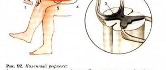
Inside the spinal canal are the spinal cord and cauda equina, CSF, as well as blood vessels supplying the spinal cord. The end of the CM (conus medullaris) is located at the level of L1-2. Below the cone, the SC is transformed into a bundle of nerve roots (cauda equina), freely “floating” in the CSF within the dural sac. Currently, it is recommended to puncture the subarachnoid space in the L3-4 intervertebral space to minimize the likelihood of injury from the SM needle. The roots of the cauda equina are quite mobile, and the risk of injury to them by a needle is extremely low.
Symptoms of expansion of subarachnoid spaces
In children and adults, the pathology manifests itself differently. So, in children of the first year of life, the following signs help to suspect something is wrong:
- Negative reaction to noise, medium-level sounds, light;
- Frequent regurgitation;
- Poor sleep, anxiety;
- Strabismus, different pupil diameters;
- Head growth that does not correspond to age;
- Bulging and slower overgrowth of the fontanel;
- Weather sensitivity, anxiety when weather changes;
- Trembling, tremors of the chin, arms or legs.
As you can see, the symptoms are quite non-specific. They may more likely indicate hydrocephalus with intracranial hypertension secondary to expansion of the subarachnoid space. These signs cannot make an accurate diagnosis, but they should prompt an immediate visit to a neurologist or pediatrician.
In adults, one of the main symptoms of expansion of the subarachnoid space is headache, which can be quite intense and prolonged. Patients often complain of severe dizziness and nausea, inability to perform work duties, weakness, restlessness, and anxiety.
Cranialgia is especially pronounced early in the morning and may be accompanied by vomiting, dizziness, hypersensitivity to light and sounds. During the day, patients are often drowsy, at night they cannot sleep, or their sleep is restless and intermittent. An increase in headache, its pulsating nature, and the appearance of vomiting at the height of pain are characteristic symptoms of increasing intracranial pressure.
Diffuse expansion of the subarachnoid space and an increase in the volume of the lateral ventricles will sooner or later cause atrophic and dystrophic changes in the cerebral cortex, which will manifest itself as symptoms of encephalopathy - decreased memory and attention, visual impairment, and intelligence. Autonomic symptoms are often associated in the form of tachycardia, pressure surges, unmotivated anxiety and even fainting.
Movement disorders are more typical for cerebellar localization of pathology. In this case, symptoms may include unsteadiness and uncertainty in gait, dizziness, loss of coordination and fine motor skills.
Constant fatigue, repeated attacks of severe headaches, and the inability to get a good night's sleep disrupt the emotional state of patients, who can become irritable, anxious, and prone to depression. Secondary changes in the brain impair the ability to work and limit active life.
compression of the brain due to subarachnoid hemorrhage
Uneven expansion of the subarachnoid space is possible after trauma, brain surgery, or with tumors that locally compress the membranes of the brain and the cerebrospinal fluid pathways. In this case, focal neurological symptoms are observed in the form of disturbances in sensitivity, speech, hearing, etc. A severe course of the pathology is indicated by convulsive syndrome or epilepsy.
Depending on the width of the intershell space, there are light (1-2 mm expansion), moderate (up to 4 mm) and severe (over 4 mm) degrees of expansion. To a certain extent, such a division is arbitrary, especially with uneven or local changes in the liquor spaces.
The described symptoms usually occur with severe variants of the pathology, severe hydrocephalus and intracranial hypertension. Most often, mild and even moderate degrees of impairment are manifested only by periodic headaches, fatigue and autonomic dysfunction.
Blood supply to the spinal cord
The spinal cord is supplied by the spinal branches of the vertebral, deep cervical, intercostal and lumbar arteries. The anterior radicular arteries enter the spinal cord alternately - sometimes on the right, sometimes on the left (usually on the left). The posterior spinal arteries are upward and downward oriented continuations of the posterior radicular arteries. The branches of the posterior spinal arteries are connected by anastomosis with similar branches of the anterior spinal artery, forming numerous choroid plexuses in the pia mater (pial vasculature).
The type of blood supply to the spinal cord depends on the level of entry into the spinal canal of the largest diameter radicular (radiculomedullary) artery - the so-called artery of Adamkiewicz. Various anatomical options for the blood supply to the spinal cord are possible, including one in which all segments below Th2-3 are fed from one artery of Adamkiewicz (option a, about 21% of all people).
In other cases the following are possible:
b) the inferior accessory radiculomedullary artery, accompanying one of the lumbar or 1st sacral roots,
c) superior accessory artery accompanying one of the thoracic roots,
d) loose type of feeding of the SM (three or more anterior radiculomedullary arteries).
In both option a and option c, the lower half of the SC is supplied by only one artery of Adamkiewicz. Damage to this artery, compression by an epidural hematoma or an epidural abscess can cause serious and irreversible neurological consequences.
Blood flows from the SM through the tortuous venous plexus, which is also located in the soft membrane and consists of six longitudinally oriented vessels. This plexus communicates with the internal vertebral plexus of the EP from which blood flows through the intervertebral veins into the azygos and semi-gypsy vein systems.
The entire venous system of the ES does not have valves, so it can serve as an additional system for the outflow of venous blood, for example, in pregnant women with aortocaval compression. Overfilling of the epidural veins with blood increases the risk of their damage during puncture and catheterization of the EP, including the likelihood of accidental intravascular injection of local anesthetics.
Cerebrospinal fluid
The spinal cord is bathed in CSF, which plays a shock-absorbing role, protecting it from injury. CSF is an ultrafiltrate of blood (a clear, colorless fluid) that is produced by the choroid plexus in the lateral, third, and fourth ventricles of the brain. The rate of CSF production is about 500 ml per day, so even the loss of a significant volume is quickly compensated.
CSF contains proteins and electrolytes (mainly Na+ and Cl-) and at 37°C has a specific gravity of 1.003-1.009.
Arachnoid (Pachionian) granulations, located in the cerebral venous sinuses, drain most of the CSF. The rate of CSF absorption depends on the pressure in the joint. When this pressure exceeds the pressure in the venous sinus, thin tubes in the pachyonic granulations open, allowing CSF to flow into the sinus. After the pressure equalizes, the lumen of the tubes closes. Thus, there is a slow circulation of CSF from the ventricles into the ventricle and further into the venous sinuses. A small portion of CSF is absorbed by the veins of the joint and lymphatic vessels, so there is some local circulation of CSF in the vertebral subarachnoid space. CSF absorption is equivalent to its production, so the total CSF volume is usually in the range of 130-150 ml.
There may be individual differences in the volume of CSF in the lumbosacral parts of the spinal canal, which may influence the distribution of arterial fluid. NMR studies have revealed variability in lumbosacral CSF volumes ranging from 42 to 81 ml (Carpenter R., 1998). It is interesting to note that overweight people have lower CSF volume. There is a clear correlation between CSF volume and the effect of spinal anesthesia, in particular, the maximum extent of the block and the rate of its regression.