The human spinal cord is a complex mechanism that consists of many different “parts”. And each of them is important for our full life. It is thanks to the fact that we have a spine that we move. The reflex function of the spinal cord is one of the main ones it performs. In addition to it, there is also a conductor. It is worth considering these functions in more detail and finding out what each of them is responsible for.
The formation of the spinal cord in the fetus occurs in the mother's womb, and at a time when she is not yet aware of her pregnancy. By the end of the first month, the first foundation of the spinal column is laid. However, it will take some more time for its complete formation after the birth of the child. Some parts of the brain are fully formed when the baby is 2 years old.
What does the spinal cord look like?
Not everyone knows what the spinal cord looks like. Moreover, not all people have an idea of what its role is in the life of every person. It is therefore worthwhile to fill this knowledge gap. In addition, many people mistakenly believe that the brain and spinal cord are separate parts.
To find out what the reflex function of the spinal cord is for, let's try to determine what it looks like. It is impossible to clearly understand where the spinal cord begins and ends. It starts from the first vertebra just below the skull, smoothly connecting to the brain in this area. The division into the spinal cord and the brain is formal, but in reality the spinal cord smoothly passes into the brain. Thus, we can conclude that these two parts are a single whole.
Location of the spinal cord and its membrane
The brain is protected by the cranium, and the spinal cord is hidden in the spine and surrounded by three membranes. The first of them is the most delicate, thin and soft. It contains blood vessels that deliver nutrients to the brain. In other words, the spinal cord is a kind of “courier” for delivering food.
Continuing to talk about how the reflex function of the spinal cord works, we cannot ignore the analysis of the structure of the second arachnoid membrane. There is a special space here called the subarachnoid space. Along the entire length of the spine it is filled with cerebrospinal fluid (CSF). It is this that is taken during puncturing for analysis in order to determine the health status of the spinal cord.
The last shell is located on the outside and has a harder surface, which allows it to provide protective functions against various types of external damage.
Characteristics of the spinal cord
In adults, the spinal cord reaches 45 cm in length and 1.5 cm in thickness. Its weight, by the most conservative standards, is no more than 35 grams. The entire brain is divided into several sections, from which various roots extend:
- cervical;
- chest;
- lumbar;
- cross;
- coccygeal
Since the reflex function of the spinal cord is carried out, the cervical and lumbosacral regions are the most important parts of the spine. In this regard, they are best protected - nature itself took care of this, making them significantly thicker and denser. It is in these places that important nerve endings are located, the defeat of which can lead to serious consequences. In the cervical region there is a cluster of roots responsible for the movement of the arms. The roots of the lower section are responsible for the movement of the lower extremities.
The human spinal cord controls the activity of all internal organs. Each of them is associated with a specific department. In addition, the entire spinal canal is divided into segments and each of the listed sections has its own number. There are 8 of them in the cervical, 12 in the thoracic, 5 in the lumbar and sacral, and one or two in the coccygeal.
Gray matter
Gray matter or substantia grisea is represented by several columns connected to each other by two plates (anterior and inferior), called commissures. On a cross-section of one of these pillars, you can see that the gray matter in its shape resembles a butterfly with spread wings or the Latin letter H.
In addition, you can also notice that there are projections extending from the substance, which are otherwise called horns. They can be either front, located on the front wall, or rear, running along the back wall. Both the first and second pairs, and have a narrow and wide shape. But in addition to the rear and anterior ones, there are also lateral horns, which contain the centers of the autonomic nervous system.
What is the reflex function of the spinal cord? The fact is that in the anterior horns there is a special type of motor neurons, the processes of which form nerve roots.
In the middle of the gray matter there is a central canal, which is also filled with cerebrospinal fluid. In the upper part, the canal is connected to the ventricles of the brain. In this case, all sections: the ventricles, the central canal and the subarachnoid space take an active part in the circulation of cerebrospinal fluid.
Internal structure of the spinal cord
The central sections of the spinal cord are made of gray matter. On a preparation of a section of an organ, this substance resembles a butterfly in outline. This component of the brain consists of nerve cell bodies (intercalary and motor type). This part of the nervous system is divided into functional zones: anterior and posterior horns. The former contain motor-type neurons, the latter have intercalary nerve cells. Along the segment of the spinal cord from the 7th cervical segment to the 2nd lumbar segment there are additional lateral horns. It contains centers responsible for the functioning of the autonomic nervous system (nervous system).
Internal structure of the spinal cord
The posterior horns are characterized by the heterogeneity of their structure. These zones of the spinal cord contain special nuclei made of interneurons.
The outer part of the spinal cord is formed by white matter, made of axons of butterfly neurons. The spinal grooves conventionally divide the white matter into 3 pairs of cords, known as: lateral, posterior and anterior. Axons unite into several conducting tracts:
- associative fibers (short) - provide connection between different spinal segments;
- ascending fibers, or sensory fibers, transmit nerve signals to the head section of the central nervous system;
- descending fibers, or motor fibers, transmit impulse signals from the cerebral cortex to the anterior horns that control the executive organs.
From the editor: Forms, causes and symptoms of encephalitis
The posterior cords contain only ascending conductors, and the remaining two pairs are characterized by the presence of descending and ascending pathways. The number of conducting tracts in the cords varies. The table below shows the location of the conduction tracts in the dorsal part of the central nervous system.
Lateral cord of conductors:
- spinocerebellar tract (posterior) – transmits proprioceptive impulse signals to the cerebellum;
- spino-cerebellar tract (anterior) - responsible for communication with the cerebellar cortex, where it transmits impulse signals;
- spinothalamic tract (external lateral) - responsible for transmitting impulse signals to the brain from receptors that respond to pain and temperature changes;
- pyramidal tract (outer lateral) – conducts motor impulse signals from the cortex of the large hemispheres to the spinal cord;
- red nuclear spinal tract - controls the maintenance of skeletal muscle tone and regulates the performance of subconscious (automatic) motor functions.
Front cord of conductors:
- pyramidal tract (anterior) – transmits the motor signal from the cortex of the upper parts of the central nervous system to the lower ones;
- spinothalamic tract (anterior) – transmits impulse signals from tactile receptors;
- vestibulospinal - carries out coordination of conscious movements and balance, and is also characterized by the presence of a connection with the medulla oblongata.
Posterior cord of conductors:
- thin bundle of Gaulle fibers - responsible for transmitting impulse signals from proprioceptors, interoreceptors and skin receptors of the lower torso and legs to the brain;
- wedge-shaped bundle of Burdach fibers - is responsible for transmitting the same receptors to the brain from the arms and upper torso.
White matter
White matter - substantia alba, envelops the gray matter, is formed by a collection of nerve fibers, which also come in three types:
- front;
- rear;
- lateral.
Moreover, all the roots have a different direction, and some of them are connected directly to the brain and central nervous system (hereinafter simply the CNS). And if the reflex function of the spinal cord is to transmit signals from the motor neurons of the gray matter, then the task of the white matter neurons is the prompt delivery of impulses from the muscles and joints to the medulla oblongata. Thus, the transmission of all commands along the entire spinal cord is realized.
Here are the paths through which all information regarding sensitivity and pain is transmitted. Only before entering the cerebral cortex, the information first reaches the diencephalon, and only then rushes further to its destination.
Functions of the spinal cord: the main thing
The spinal cord is a highly complex system of nerve fibers that perform two important tasks in the life of the body:
- reflex;
- conductive.
Conductive function
Any movement originates initially in the brain. Impulses come to it from the mucous membranes, skin or internal organs, after which it processes them and sends a signal to the spinal cord, and then to the peripheral nervous system. That, in turn, transmits signals along nerve endings that cause your muscles to contract.
When performing a certain movement, a person does not even think about which muscles need to be used at the moment - this function is automatically performed by the spinal cord.
Serious injuries, for example, rupture of an organ, lead to partial or complete loss of a person’s ability to move. In this case, the information simply does not reach the nerve endings that would cause the muscles to contract.
Reflex function
The reflex function is not only the withdrawal of a hand upon contact with fire or the flexion of limbs. The reflex also includes coughing during illness, closing the eyes during contact with ultraviolet light, and many other uncontrolled defensive reactions. At the same time, a certain segment is responsible for each reflex, and its damage causes the loss of a certain skill.
The brain does not take any part in the reflex function. The reflex itself is a natural protective reaction of the body, which a person is not able to control.
It has been scientifically proven that if reflexes were also processed by the head, human survival rate would be much lower. He would react much more slowly to irritation, which would increase the size of the damage.
How our brain works
Ascending and descending pathways are responsible for the fast and correct functioning of our body. The latter flows are formed using the red nuclear and lateral pathways. It is thanks to these pathways that the reflex and conduction functions of the spinal cord are carried out. Thanks to the red nuclear spinal tract, involuntary motor impulses are produced. While the lateral corticospinal tracts are responsible for voluntary impulses.
All roots are supplied by personal veins and arteries, which as a result form neurovascular bundles. Each such beam is responsible only for its own segment and works autonomously, analyzing incoming information and transmitting the necessary impulses.
Damage to these bundles leads to serious pathological and sometimes irreversible changes in the human body. And so that specialists can determine which particular bundle is damaged and localize the pain, it is necessary to conduct a whole range of studies.
Reflex function
Everything in our body is thought out to the smallest detail, and our body reacts differently to every external stimulus. The defense mechanism is based on reflexes. We sneeze, cough, get burns, flinch from a sharp sound, or react in our own way to gusts of wind. These are all examples of the reflex function of the spinal cord and such actions occur outside of our control.
So that we can respond in a timely manner to any irritant, including critical situations, pain receptors are located throughout the surface of our skin. As a striking example: after touching a hot kettle or any surface, we almost instantly withdraw our hand. The reaction speed is so fast that it is impossible to understand the time frame. In a split second, a reflex ring is formed, which causes the muscles to contract.
Another common case can be cited. If you accidentally swallow a portion of smoke or sniff dust particles with your nose, you will start sneezing or coughing. Thus, it became clear that in such a short time the information was received, processed, and our “defenders” received instructions to free the body from the presence of foreign bodies.
Reflex
The reflex brain function is to organize reflexes. This allows the body, for example, to instantly respond to a signal of pain. The action of reflexes is striking in its efficiency. A person withdraws his hand from a hot object in a split second. During this time, information from the receptors to the brain and back has traveled a long way along the reflex arc.
When the sensitive nerve endings of the skin, muscle fibers, tendons, and joints receive irritation, this means that a nerve impulse has been sent to them. Such signals travel along the dorsal roots of nerve fibers and enter the spinal cord. Receiving a signal, motor and intercalary cells are excited. Then, along the motor fibers of the anterior roots, impulses are sent to the muscles. Having received such a signal, the muscle fibers contract. Simple reflexes occur through this mechanism.
A reflex is a reaction of the body in response to received irritation. All reflexes are provided by the work of the central nervous system. One of the functions of the spinal cord is reflex. It is provided by the so-called reflex arc. This is a complex path that nerve impulses travel from the peripheral components of the body to its spinal cord, and from there directly to the muscles. This is a difficult but vital process.
The simplest reflexes can save a person’s life and health. Retracting the hand that touched the hot item, we do not even suspect that the signal from the skin was transmitted with lightning speed along the nerve fibers to the brain, and then to the spinal cord. In response, an impulse was sent that contracted the muscles of the arm to avoid being burned. This is a clear manifestation of the reflex function.
Neurophysiologists have studied in detail almost all reflexes and the neural arches that ensure their implementation. These data allow for effective rehabilitation after injuries and a number of diseases, and also help in their diagnosis.
From the editor: Treatment and symptoms of mixed type dystonia
It is on this reflex that a diagnosis by a neurologist is based, in which the doctor lightly hits the patient’s patella tendon with a hammer. This is how the knee reflex is studied, by which one can judge the state of a certain part of the spinal cord.
However, the spinal cord is not an independent reflex system. Its functions are constantly controlled by the brain. They are closely connected by special bundles of nerve fibers. The fibers are very long, thin, and consist of white matter. Some signals are transmitted upward to the brain, and others - to the spinal cord.
The entire central nervous system is involved in the formation of coordinated complex movements. Each movement is a continuous flow of impulses from the brain to the spinal cord, and from it to the muscle fibers.
Conductor function
So, it is now clear what the reflex function of the spinal cord is expressed in, we can move on to another, also significant task - conduction. It involves transmitting signals along ascending pathways to the main brain. From it, depending on the situation, the impulse is directed along descending pathways to some organ.
The conductor function allows us to perform meaningful actions:
- take or throw;
- stand up or sit down;
- walk slowly or run;
- draw;
- cut off.
We perform all these actions in everyday life: at home or at work, and usually we simply do not notice.
This whole connection of the brain, spinal cord, the entire central nervous system, internal organs and all limbs makes the human body unique in nature. Even the most modern robot cannot boast of the number of movements that any biological organism can perform.
Conducting function of the spinal cord
Bundles of axons form anterior, posterior and lateral cords around the gray matter, in which ascending and descending pathways are located. Descending tracts are located in the anterior funiculi :
1) the anterior corticospinal (pyramidal) tract, which is straight and uncrossed;
2) posterior longitudinal fasciculus; 3) tectospinal tract; 4) vestibulospinal tract. In the posterior cords there are ascending pathways :
1) thin bundle (Gaull's bundle);
2) wedge-shaped bundle (Burdach bundle). In the lateral cords there are descending and ascending tracts : 1) descending
tracts: - lateral corticospinal (pyramidal) tract, which is crossed;
— red nucleus-spinal cord (rubrospinal) tract; - reticular-spinal (reticulospinal) tract. 2) ascending
pathways: - spinothalamic tract;
- lateral and anterior spinocerebellar (Flexig and Gowers bundles). Associative ( propriospinal ) pathways. Associative pathways connect neurons of the same or different segments of the spinal cord. They start from the neurons of the gray matter of the intermediate zone, go to the white matter of the lateral or anterior cord of the spinal cord and end in the gray matter of the intermediate zone or on the motor neurons of the anterior horns of other segments. These connections perform an associative function , which consists in coordinating posture, muscle tone, and muscle contractions in various metameres of the torso. The propriospinal tracts also include commissural fibers that connect functionally homogeneous symmetrical and asymmetrical areas of the spinal cord. Descending paths. These pathways connect various parts of the brain with motor or autonomic efferent neurons of the spinal cord. They start from neurons of various brain structures and end on motor neurons of the anterior horns of the spinal cord or on interneurons. The pyramidal tract is crossed and consists of two bundles - lateral and direct. The lateral bundle starts from the neurons of the cerebral cortex, at the level of the medulla oblongata passes to the other side, forming a decussation, and descends along the opposite side of the spinal cord. The direct fasciculus descends to its segment and only there passes to the motor neurons of the opposite side. The rubrospinal tract consists of the axons of neurons in the red nucleus. Immediately after leaving the nucleus, these axons move to the opposite side ( Forel's decussation ) and divide into three bundles. One goes to the spinal cord, the other to the cerebellum, the third to the reticular formation of the brain stem. The neurons that give rise to this pathway are involved in controlling muscle tone. The rubrocerebellar and rubroreticular pathways ensure coordination of the activity of pyramidal neurons of the cerebral cortex and cerebellar neurons involved in the organization of voluntary movements. The vestibulospinal tract the
neurons of the lateral vestibular nucleus (
Deiters nucleus , which lies in the medulla oblongata. This nucleus regulates the activity of spinal cord motor neurons, ensures muscle tone, coordination of movements, and balance. The reticular-spinal cord ( reticulospinal ) pathway
goes from the reticular formation of the brain stem to the motor neurons of the spinal cord.
Through it, the reticular formation regulates muscle tone. Damage to the conduction apparatus of the spinal cord leads to disorders of the motor or sensory system below the site of damage. Crossing the pyramidal tract causes muscle hypertonicity below the transection (motoneurons of the spinal cord are freed from the inhibitory influence of the pyramidal cells of the cortex) and, as a consequence, spastic paralysis. When sensitive pathways are crossed, muscle, joint, pain and other sensitivity below the site of transection of the spinal cord is completely lost. Ascending paths. The spinocerebral ascending tracts connect segments of the spinal cord with structures of the brain. Their function is to transmit information to the brain from extero-, intero- and proprioceptors. The proprioceptive pathway ( thin and wedge-shaped fascicles ) originates from the receptors of deep sensitivity of the muscles of the tendons, periosteum, and joint membranes. The thin bundle ( Gaulle's bundle ) starts from the spinal ganglia, which collect information from the caudal parts of the body, pelvis, and lower extremities. The wedge-shaped fasciculus ( Burdach's fasciculus ) arises from the dorsal ganglia, which collect information from the muscles of the chest, neck and upper extremities. From the spinal ganglion, axons go to the dorsal roots of the spinal cord, into the white matter of the posterior funiculi, and rise into the thin and wedge-shaped nuclei of the medulla oblongata. Here the first switch to a new neuron occurs, and then the path goes to the lateral nuclei of the thalamus of the opposite hemisphere of the cerebrum, where the second switch occurs. From the thalamus, the path rises to the neurons of layer IV of the somatosensory area of the cortex. The fibers of these pathways give off collaterals in each segment of the spinal cord, which makes it possible to correct the posture of the entire body. The speed of excitation along the fibers reaches 60-100 m/s. The spinothalamic tract is
the main pathway of cutaneous sensitivity, which begins from pain, temperature, tactile and pressure receptors.
Signals from skin receptors go to the spinal ganglion, then through the dorsal roots to the dorsal horns of the spinal cord, where the first switch occurs. Sensory neurons in the dorsal horn send axons to the opposite side of the spinal cord and ascend along the lateral funiculus to the thalamus (second switch), from where impulses are sent to the sensory area of the cerebral cortex. Some of the fibers of the skin receptors go to the thalamus along the anterior cord of the spinal cord. The speed of excitation along the fibers of this path reaches 110-120 m/s. The spinocerebellar tracts lie in the lateral cords of the spinal cord and are represented by the non-crossing anterior spinocerebellar tract ( Gowers' bundle ) and the double-crossing posterior spinocerebellar tract ( Flexig's bundle ). Information along these pathways comes from Golgi tendon receptors, proprioceptors, pressure and touch receptors. Along the dorsal roots, nerve impulses are sent to the gray matter of the spinal cord, where they are switched to neurons of the thoracic nucleus (Flexig's bundle), or neurons adjacent to it on the lateral side (Gowers' bundle).
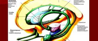
Along the processes of these neurons, impulses are sent to the cerebellum. The speed of excitation along these paths reaches 110-120 m/s. 3) Morpho - functional features of the medulla oblongata and pons. Sensory functions of the medulla oblongata and pons. Medulla oblongata ( bulbus, medulla oblangata
) is a continuation of the spinal cord.
Its length is approximately 2.5 cm. This part of the brain does not have a clear segmental structure, although segmental and suprasegmental levels are distinguished. The olives are located in the medulla oblongata - these are the thin (Gaull) and wedge-shaped (Burdach) nuclei of proprioceptive sensitivity. Here are the intersections of the pyramidal tracts and Gaulle and Burdach bundles, as well as neurons of the reticular formation. The upper part of the medulla oblongata is somewhat thicker than the lower part, which gives it the appearance of an onion. The bridge has the form of a transversely located roller. In the central part of the section there is a transversely running bundle of fibers - the trapezoidal body; between these fibers the anterior and posterior trapezoidal bodies are located. In the anterior part, longitudinal fibers are visible; they belong to the pyramidal tract. Sensory function of the medulla oblongata. In the sensory nuclei located in the medulla oblongata, the strength and quality of stimulation of the following types of sensitivity are analyzed: 1) primary sensitivity of the facial skin (trigeminal nerve nucleus); 2) primary reception of sound signals (nucleus of the cochlear nerve); 3) primary reception of taste (nucleus of the glossopharyngeal nerve); 4) primary reception of vestibular stimuli (superior vestibular nucleus). Next, from the listed nuclei, nerve impulses are transmitted to the subcortical nuclei to determine the biological significance of the stimulation. 4) Reflex function of the medulla oblongata. Bulbar mechanisms for maintaining human posture . This function is provided by the nuclei of 5-10 pairs of cranial nerves. We can say that the medulla oblongata performs the main (vital) reflex functions: 1) vital reflexes on the heart, blood vessels, breathing, gastrointestinal tract; 2) protective reflexes: sneezing, blinking, coughing, vomiting, lacrimation, etc.; 3) complexly coordinated reflexes of chewing, swallowing, sucking; 4) reflexes associated with maintaining posture, straightening and changing the position of the body in space when a person moves. The respiratory (medial parts of the reticular formation) and cardiovascular are located in the medulla oblongata . They function together with all neurons of the reticular formation, with the hypothalamus and other overlying brain structures. Therefore, when the cardiovascular center is excited, breathing and the tone of the muscles of the intestines, bladder, bronchi, etc. change. If these centers are damaged, for example, when the brain is herniated, a person may die. Protective reflexes are realized from the receptors of the mucous membranes of the nasopharynx, oral cavity, larynx, and eyes through the afferent branches of the trigeminal and glossopharyngeal nerves, going to the corresponding sensory nuclei of the medulla oblongata. From these nuclei, nerve impulses go to the motor nuclei of the trigeminal, facial, vagus, glossopharyngeal, accessory and hypoglossal nerves, from which impulses travel along the efferent nerves to the corresponding effectors that implement protective reflexes. Complexly coordinated reflexes are implemented in exactly the same way as protective ones, due to sequentially activated muscle groups. Thus, when the lip receptors are stimulated, a sucking . At the same time, excitation spreads along the afferent fibers of the trigeminal nerve to the medulla oblongata, where it switches to the efferent neurons of the facial and hypoglossal nerves. In newborns, sucking is an involuntary reflex. With age, due to the formation of associative connections with the cerebral cortex, it comes under its influence and can be voluntarily controlled. Chewing as an involuntary process can only be observed in bulbar animals (animals with the medulla oblongata and spinal cord preserved and other parts of the central nervous system removed). When the receptors of the oral mucosa are irritated, nerve impulses along the sensory fibers of the trigeminal nerve are directed to its sensory nuclei, and then switch to motor neurons of the motor nuclei of the trigeminal and hypoglossal nerves, from which impulses are sent to the masticatory muscles and muscles of the tongue. Swallowing begins from the receptors of the oral mucosa and soft palate. Excitation from these receptors through the afferent fibers of the trigeminal, glossopharyngeal and vagus nerves enters the swallowing center of the medulla oblongata, which provides a strictly coordinated sequence of reflex contraction of the muscles involved in this act. The swallowing center is closely connected with the respiratory center - when swallowing, the activity of the respiratory muscles is inhibited. Classification of reflexes that support a person’s posture according to Magnus: 1) static
(postural and straightening);
2) statokinetic
(nystagmus, elevator reflexes).
Static reflexes ensure that a person’s posture in space is maintained at rest. They begin from the vestibular apparatus, proprioceptors of the deep muscles of the neck, as well as from receptors of the torso with unilateral stimulation. Postural reflexes (positional reflexes) are responsible for maintaining a person’s horizontal, sitting and vertical posture in a calm state. When the labyrinths of the inner ear are destroyed or a plaster cast is applied to the neck, these reflexes are not realized. Rectifying reflexes are activated when the body is in an uncomfortable position. Thanks to them, a person assumes a posture of average physiological rest. To carry out these reflexes, in addition to the nuclei of the medulla oblongata, the nuclei of the midbrain are needed. For example, if you throw a cat with its back down, then from the receptors of the semicircular canals impulses are transmitted through the medulla oblongata to the muscles of the neck, and the head turns down, the receptors of the deep muscles of the neck are excited, from which impulses go to the Deiters nucleus of the medulla oblongata, and from it along the vestibulospinal tracts to spinal cord extensor motor neurons, which causes the extensor muscles to contract, causing the cat to flip in the air and land on its feet. This righting reflex is controlled by γ neurons in the spinal cord. Statokinetic reflexes provide redistribution of muscle tone in the trunk and neck to organize a posture corresponding to the moment of linear or rotational movement. Nystagmus ( nystagmos,
gr. - blinking) is the movement of the eyes (eye nystagmus) and head (head nystagmus) in the direction opposite to the movement, and then their return to their original position.
For example, if a person is traveling on a train and at the same time looking out the window, then his eyes and head involuntarily make these movements. If nystagmus appears in a person in the absence of linear or rotational movement, then this is a serious neurological symptom. Elevator reflexes occur when moving up or down on a high-speed elevator. When going up, the tone of the leg flexor muscles increases, and the person squats. When going down, the tone of the extensors increases. To carry out these reflexes, the nuclei of the medulla oblongata and midbrain are necessary . 5) Structural and functional organization of the midbrain. Motor functions of the midbrain. The midbrain consists of the quadrigeminal plate, the red nucleus, the substantia nigra, the oculomotor nerve nucleus and the trochlear nerve nucleus. Functions of the midbrain: 1) sensory function (analysis of the biological significance of visual and sound information); 2) conduction function (conduction of nerve impulses along ascending pathways to the thalamus, cerebellum and cerebrum and descending pathways to the medulla oblongata and spinal cord); 3) motor function (implemented by the nuclei of the trochlear, oculomotor nerves, red nucleus and substantia nigra); 4) reflex function (implemented through the quadrigeminal structures, which are functionally independent). The quadrigeminal plate includes the superior and inferior colliculus. The superior colliculus is the primary center of vision. The inferior colliculus carries out orienting reactions to sound, i.e. the primary hearing centers are located here . In the substantia nigra there are neurons that coordinate the reflexes of chewing and swallowing, coordinate small movements of the fingers (playing the piano, violin), ensure the plastic tone of a person, and participate in the contraction of facial muscles. When the neurons of the substantia nigra are damaged (for example, with atherosclerosis of the cerebral vessels), parkinsonism develops (tremor; amia - mask-like face; increased salivation, etc.), and the emotional sphere also suffers. The red nucleus receives impulses from the cerebellum, the motor cortex (anterior central gyrus) and the subcortical nuclei. They, in turn, through the vestibular nucleus of Deiters and the adjacent reticular formation, inhibit the a-motoneurons of the extensors of the anterior horns of the spinal cord. When the red nuclei decerebrate rigidity occurs ( rigidus,
lat. - stiff, inflexible).
Decerebration is an operation of cutting between the upper and lower tubercles of the quadrigeminal tubercles, then the red nucleus remains above the cutting. This phenomenon consists of rigidity of the extensor muscles. In this case, the animal’s tail is raised, its head is thrown back, all its limbs are extended, and an attempt to bend them can lead to broken limbs. A person has opisthotonus, i.e. the person lies leaning on the back of his head and heels, but since the person’s flexors are stronger than the extensors, his arms will be bent at the elbows. The mechanism of this phenomenon is as follows: the red nucleus, as well as the cerebellum and overlying structures inhibit the Deiters nucleus and the adjacent reticular formation. This determines the normal distribution of muscle tone between flexor and extensor neurons. When the red nucleus is destroyed, its inhibition on the Deiters nucleus and reticular formation is removed, and the excitability of these structures increases sharply. As a result of this, an increased number of nerve impulses go to the a-motoneurons of the extensor muscles, and the tone of the extensor muscles increases. Thus, the red nucleus, together with the vestibular nuclei, regulates the distribution of tone between the flexors and extensors, and also carries out rectifying and statokinetic reflexes.